ISSN 0378-6471 (Print)⋅ISSN 2092-9374 (Online)
https://doi.org/10.3341/jkos.2019.60.4.340
Original Article
시신경유두오목 황반병증에 대한 유리체절제술의 장기 결과
Long-term Clinical Outcome of Vitrectomy for the Treatment of Optic Disc Pit Maculopathy
박종호1,2⋅박성후3,4,5⋅이지은3,4,5⋅변익수1,2,5
Jong Ho Park, MD1,2, Sung Who Park, MD3,4,5,Ji Eun Lee, MD, PhD3,4,5, Ik Soo Byon, MD, PhD1,2,5 양산부산대학교병원 안과1, 양산부산대학교병원 의생명융합연구소2, 부산대학교병원 안과3, 부산대학교병원 의생명연구소4,
부산대학교 의과대학 안과학교실5
Department of Ophthalmology, Pusan National University Yangsan Hospital1, Yangsan, Korea
Research Institute for Convergence of Biomedical Science and Technology, Pusan National University Yangsan Hospital2, Yangsan, Korea Department of Ophthalmology, Pusan National University Hospital3, Busan, Korea
Medical Research Institute, Pusan National University Hospital4, Busan, Korea Department of Ophthalmology, Pusan National University School of Medicine5, Yangsan, Korea
Purpose: To evaluate the long-term outcomes of optic disc pit maculopathy after vitrectomy.
Methods: We evaluated retrospectively the medical records of eight patients with macular retinal detachment or retinoschisis who underwent vitrectomy due to optic disc pit maculopathy. The best-corrected visual acuity and optical coherence tomography findings were analyzed after surgery.
Results: Eight eyes of eight patients (two male and six female) were enrolled. The mean best-corrected visual acuity was 0.76 log MAR, the mean age was 42.8, and the mean follow-up period was 56 months (range: 8-120 months). At baseline, retinoschisis was observed in all eight eyes. Six eyes had serous retinal detachment of the macula. Vitrectomy for a complete posterior vitre- ous detachment was performed. Additional internal limiting membrane peeling and tamponade were performed in six and four eyes, respectively. After surgery, serous retinal detachment was gone in all eyes (100%) at a mean of 22.8 months (range: 18 days-60 months). Three of eight eyes (37.5%) showed the disappearance of retinoschisis at a mean of 6.8 months (range:
1.7-21 months), but the remaining patients still had retinoschisis at the final visit. Ocular complications were full-thickness mac- ular hole and iatrogenic retinal detachment in each case. The final corrected visual acuity improved to 0.29 logMAR.
Conclusions: Vitrectomy is an effective treatment for patients with optic disc pit maculopathy. It achieved anatomical and visual improvements over a long period of time. However, retinoschisis due to inner retinal fluid remained in many patients.
J Korean Ophthalmol Soc 2019;60(4):340-347 Keywords: Maculopathy, Optic disc pit, Vitrectomy
■Received: 2018. 9. 6. ■ Revised: 2018. 9. 21.
■Accepted: 2019. 3. 18.
■Address reprint requests to Ik Soo Byon, MD, PhD
Research Institute for Convergence of Biomedical Science and Technology, Pusan National University Yangsan Hospital, #20 Geumo-ro, Mulgeum-eup, Yangsan 50612, Korea
Tel: 82-55-360-2592, Fax: 82-55-360-2161 E-mail: [email protected]
*Conflicts of Interest: The authors have no conflicts to disclose.
ⓒ2019 The Korean Ophthalmological Society
This is an Open Access article distributed under the terms of the Creative Commons Attribution Non-Commercial License (http://creativecommons.org/licenses/by-nc/3.0/) which permits unrestricted non-commercial use, distribution, and reproduction in any medium, provided the original work is properly cited.
시신경유두오목은 선천성 질환으로 11,000명당 1명 정도 의 발병률을 보인다.1 주로 단안에 원형 혹은 타원형의 함 몰이 시신경유두의 하이측에 존재한다.2 시신경유두오목 자체로는 시력에 영향을 주는 경우는 드물지만, 시신경유 두오목 환자의 약 25-75%에서는 낭포황반부종, 망막층간 분리, 장액망막박리 등 황반부의 변화로 인해 시력저하가 발생할 수 있다.2 Halbertsma1는 황반부 이상을 보이는 증 례를 시신경유두오목 황반병증이라고 하였다. 시신경유두
Case Age (years) Sex
Preop.
Vac.
(Snellen) Pit location
Prev.
Tx.
(laser)
Surgical treatment Maculopathy
Final Vac.
(Snellen) Time to resolution
of SRF (months)
Complic ations
Follow up (months)
Baseline Final
Px. Vx. ILM
P Tamponade IRF (schisis)
SRF (serous
RD) IRF (schisis)
SRF (serous
RD)
1 41 F 0.02 T + + + + + - 0.2 19 NA 27
2 19 M 0.2 T + + + SF6 18% + + - - 1.0 21 NA 72
3 36 F 0.16 IT + + + + + - 0.2 26 FTMH 60
4 31 F 0.06 I + + Air + + + - 0.32 8 NA 8
5 53 F 0.4 T + + + Silicone oil + + - - 0.63 0.6 RD 14
6 29 M 0.16 T + + C3F8 12% + + + - 0.8 63 NA 102
7 66 F 0.32 IT + + + + - - - 0.8 11 NA 46
8 68 F 1.0 T + + + + - + - 1.0 1 NA 120
Preop. Vac. = preoperative visucal acuity; Prev. Tx. = previous treatment; Px. = phacoemulsification; Vx. = vitrectomy; ILMP = internal limiting membrane peeling; IRF = intraretinal fluid; SRF = subretinal fluid; RD = retinal detachment; Final Vac. = final visucal acuity; F = female; T = temporal; NA = not avail- able; M = male; SF6 = sulfur hexafluoride; IT = inferotemporal; FTMH = full-thickness macular hole; I = inferior; C3F8 = octafluoropropane.
Table 1. Clinical characteristics of patients
오목 환자에서 망막내액 및 망막하액의 발생기전은 아직 정확하게 알려진 바는 없으나, 시신경유두오목과 지주막하 공간 사이의 직접적인 교통, 뒤유리체와 시신경유두오목 부위의 비정상적인 강한 유착 등이 원인으로 추측되고 있 다.3,4
시신경유두오목 황반병증에 대해서 시신경유두 주변 레 이저광응고술, 유리체절제술, 황반부 공막돌륭술 등 다양한 치료 방법들이 시도되었으나 질환 자체가 발생률이 낮고, 이에 대한 치료에 있어 전향적 다기관 연구 결과가 없어 아 직까지 합의된 치료 방법은 없다. 그중에서 유리체절제술 은 Hirakata et al5이 시신경유두오목 황반병증환자에서 처 음 시도하여 우수한 치료 효과가 있었다고 보고한 이후로 국내외 연구가 보고되고 있으나, 대상 환자 수가 적으며, 술 후 장기 결과에 대한 보고는 거의 없는 실정이다. 이에 저자들은 시신경유두오목 황반병증으로 유리체절제술을 시행받은 환자들의 술 후 장기 결과에 대해 알아보고자 하 였다.
대상과 방법
2007년 1월부터 2017년 12월까지 시신경유두오목 황반 병증으로 진단받고 유리체절제술을 시행받은 환자 8명, 8안 의 환자의 의무기록을 후향적으로 분석하였다. 본 연구는 헬싱키선언에 입각하여 시행되었으며, 부산대학교병원 임 상시험심사위원회(Institutional Review Board, 1808-015-070) 의 승인을 받았다. 안저사진과 빛간섭단층촬영을 이용하여 시신경유두에 오목과 망막층간분리, 장액망막박리 등의 황 반병증이 동반된 경우 시신경유두오목 황반병증으로 진단 하였으며, 최대교정시력과 빛간섭단층촬영을 이용한 해부 학적 변화를 조사하였다. 고도근시, 눈 수술의 기왕력 등
백내장을 제외한 시력에 영향을 줄 수 있는 기타 눈 질환을 가진 환자는 제외하였다. 안저사진은 안저카메라(Canon digital retinal camera, Canon Inc., Tokyo, Japan)를 이용하 여 시신경유두와 중심와를 중심으로 하여 30° 영역을 촬영 하였고, 빛간섭단층촬영은 공간영역 빛간섭단층촬영 장비 (Cirrus HD-OCT, Carl-Zeiss Meditec Inc., Dublin, CA)의 512 × 128 macular cube scan과 파장가변 빛간섭단층촬영 장비(Atlantis DRI-OCT, Topcon, Tokyo, Japan)의 5 line cross 영상을 이용하여 촬영하였다.
국소마취하에 23게이지 유리체절제술이 시행되었다. 완 전한 뒤유리체박리를 만드는 것을 목표로 하였다. 트리암시 놀론(Tamceton injection, Hanall biopharma., Seoul, Korea) 으로 시신경유두 위의 유리체를 가시화한 뒤 유리체절제침 으로 흡인 후 당겨서 유도하였다. 술자의 판단에 따라 추가 적인 백내장수술, 내경계막제거술, 눈속충전물주입술을 시 행하였으며, 충전물을 삽입한 경우에는 1주일간 엎드린 자세 를 유지하였다. 내경계막제거는 0.25%로 희석한 indocyanine green (Diagnogreen® Injection, Daiichi Pharmaceutical, Tokyo, Japan)를 이용하여 내경계막을 염색한 후 중심와를 중심으로 3-4 유두직경 크기로 벗겨내었다. 백내장을 동시 에 수술하는 경우에는 유리체절제술에 앞서 투명각막절개 창을 만들고 수정체유화술로 수정체를 제거한 뒤 인공수정 체를 수정체낭에 삽입하였다.
최대교정시력은 스넬렌 시력표를 이용하여 측정한 뒤 통 계를 위해 logarithm of the minimum angle of resolution (logMAR)으로 변환하여 계산하였다. 수술 전후의 시력 변 화는 Wilcoxon signed rank test를 사용하여 비교하였으며 p 값이 0.05 미만인 경우를 통계적으로 유의하다고 판단하였 다.
A B
C D
E F
G H
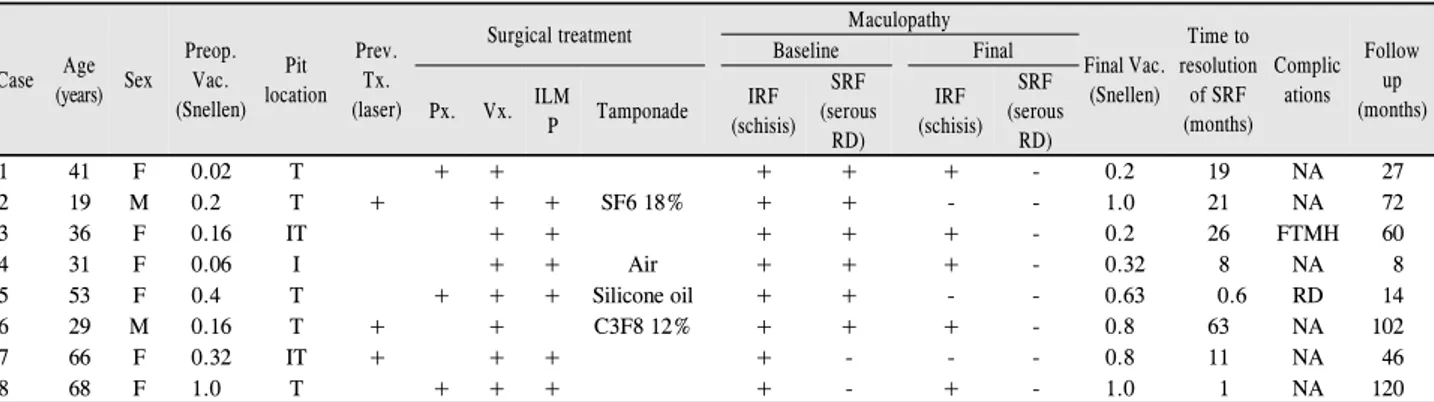
Figure 1. Composite of the representative images from patient 1. (A) fundus photograph showed a round optic pit (white arrows) and
serous retinal detachment (yellow arrowheads) at the initial visit. (B) The baseline optical coherence tomographic (OCT) image showed an excavation of the optic disc (asterisk). (C) Macular OCT image demonstrated schisis-like intraretinal fluid in the outer plexiform layer and serous retinal detachment involving macula. (D-H) Serial OCT images showed a gradual reduction of serous ret- inal detachment and outer retinoschisis at 1, 3, 8, 18, and 27 months, respectively. However, the retinoschisis still remained at the final visit. Snellen visual acuity improved from 0.02 to 0.2.결 과
총 8명, 8안 남자 2명, 여자 6명이 포함되었다. 평균 경과 관찰 기간은 56개월(8-120개월)이었다. 평균 나이는 42.9세 (19-68세)였으며, 술 전 최대교정시력은 0.76 ± 0.52 logMAR 였다. 신경유두오목의 위치는 하측 1안, 하이측 2안, 이측 5안이었다. 빛간섭단층촬영에서 망막층간분리는 8안 모두 에서 관찰되었으며, 6안에서 황반부 장액망막박리가 있었 으며, 2안은 망막앞막으로 인한 표층황반원공이 있는 상태 였다. 3안은 수술 전 시신경유두주변으로 레이저광응고술 을 받은 병력이 있었으나 시술 후 호전이 없는 상태였다.
백내장 동시수술은 3안에서 시행되었다. 6안에서는 내경계 막을 동시에 제거하였다(Table 1).
수술 과정에서 8안 모두 시신경유두와 유리체 사이에 유 착이 있는 것이 확인되어, 유리체절제침으로 완전한 뒤유 리체박리를 만들었다. 유리체절제술 후 황반부 장액망막박 리가 있던 6안은 모두 평균 22개월(18일-60개월)에 걸쳐 망 막하액이 완전히 소실되어 망막이 유착되었다. 망막하액은 수술 후 경과 관찰 기간 동안 재발한 증례는 없었다. 망막 내액이 동반된 망막층간분리는 8안 중 3안에서만 평균 6.8개 월(2-21개월)만에 망막내액이 완전히 소실되었고, 나머지 5안 도 감소하였으나, 최종 경과 관찰 시기까지 일부 남았다.
A B
C
D E
F G
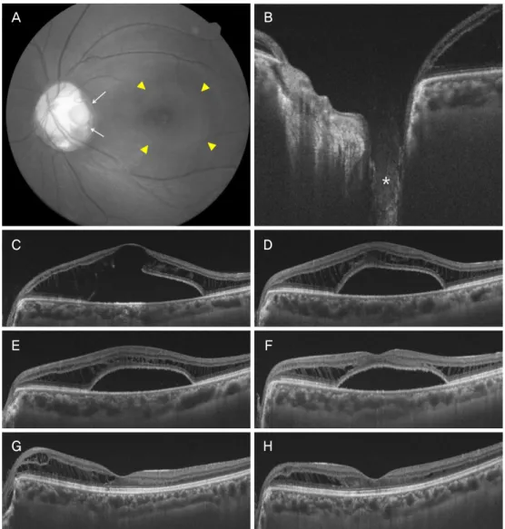
Figure 2. Composite of the representative images from patient 2. (A, B) Fundus photograph and optical coherence tomographic
(OCT) images showed an optic pit (white arrows), retinoschisis and serous retinal detachment (yellow arrowheads) at the baseline.The patient underwent vitrectomy, internal limiting membrane peeling and 18% SF6 tamponade. (C-G) A gradual absorption of in- traretinal and subretinal fluid was seen in longitudinal OCT scan images at 3, 9, 16, 24, and 72 months, respectively. The Snellen visual acuity improved 0.2 to 1.0 after vitrectomy.
장액망막박리가 있었던 6안 중에 4안에서는 내경계막제 거술이 시행되었는데, 내경계막을 제거한 경우에는 망막하 액이 소실되기까지 평균 13.8개월(0.6-25개월)이 소요되었 으나, 내경계막을 보존한 2안은 평균 40개월(18, 62개월)이 걸렸다. 하지만 이러한 차이는 통계적으로는 의미가 없었 다.
눈속충전물은 8안 중 3안에서 시행되었는데, 각각 공기 1안, 18% 육불화황 1안, 12% 과불화프로판 1안이었다. 눈 속충전물을 사용한 3안은 평균 30개월(8-63개월)에 망막하 액이 소실되었는데, 눈속충전물을 사용하지 않은 증례의 23개월(19, 26개월)과 같이 오랜 시간에 걸쳐 소실되었다.
총 8안의 최종교정시력(평균 56개월)은 0.29 logMAR로 수 술 전에 비해 유의하게 호전되었다(p=0.017).
시신경유두 황반병증에 대한 유리체절제술과 관련하여 2안 에서 합병증이 발생하였다. 내경계막을 제거했던 4안 중 1안 에서 술 후 2년째 전체층황반원공이 발생하여 추가적인 내 경계막 절편삽입술을 시행하였으며, 1안에서 수술 중 의인 성 망막박리가 발생하여 망막의 재유착을 위해 망막열공
부위로 망막하액을 배액하고 실리콘오일을 주입하였다. 의 인성 망막박리의 회복을 위한 의도적인 망막하액 배액술로 인해 황반부 장액망막박리가 18일 만에 완전히 소실되었 다.
증례 1
41세 여자 환자가 6개월 전부터 시작된 좌안 시력저하로 내원하였다. 최대교정시력은 Snellen 시력 0.02였다. 안저 검사에서 시신경유두 이측에 시신경유두오목이 관찰되었 으며, 빛간섭단층촬영에서 시신경유두오목과 함께 황반부 장액망막박리와 망막층간분리가 확인되었다. 백내장수술과 함께 유리체절제술이 시행되었다. 18개월 뒤 장액망막박리 는 완전히 소실되었으나, 망막층간분리는 수술 후 27개월 까지 일부 남았다. 최종 시력은 Snellen 시력 0.2로 호전되 었다(Fig. 1).
증례 2
19세 남자 환자가 3일 전부터 시작된 우안 시야 가림 증
A B
C
D E
F
G H
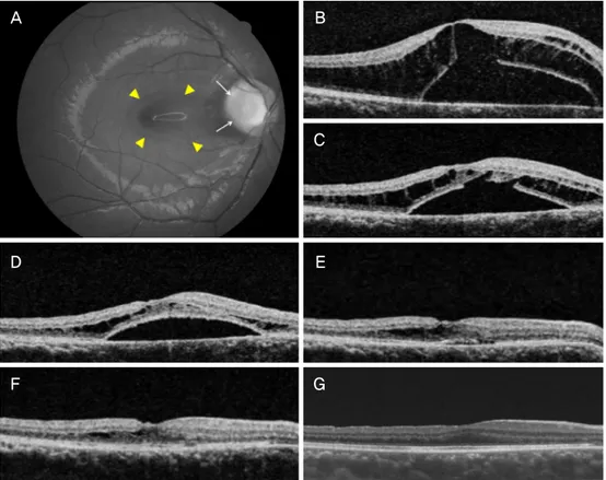
Figure 3. Composite of the representative images from patient 5. (A, B) Fundus photograph and optical coherence tomography
(OCT) images showed the optic pit (white arrows), retinoschisis and and serous retinal detachment (yellow arrowheads) at the initial visit. The patient underwent vitrectomy and internal limiting membrane peeling. (C) After 2 weeks, OCT showed small reduction of intra and subretinal fluid. (D, E) After 2 years, full-thickness macula hole was detected in OCT (white arrowheads). The patient underwent (F-H) OCT images and they showed successful closure of macular hole 3, 12, and 36 months after vitrectomy, autologous internal limiting membrane transplantation and gas tamponade. But retinoschisis remained through the follow-up period. The Snellen visual acuity improved 0.16 to 0.2 at the final visit.세로 내원하였다. 최대교정시력은 Snellen 시력 0.2로 측정 되었다. 안저검사에서 시신경유두 이측에 시신경유두오목 이 관찰되었으며, 빛간섭단층촬영에서 망막층간분리와 황 반부 장액망막박리가 확인되었다. 신경유두 주변으로 2-3줄 의 레이저광응고술을 시행하였으나, 변화가 없어 유리체절 제술과 함께 내경계막제거술, 18% 육불화황주입술을 시행 하였다. 망막내액과 망막하액은 천천히 흡수되어 수술 후 3년 째 완전히 소실되었고 최종시력은 Snellen 시력 1.0으로 호 전되었다(Fig. 2).
증례 3
36세 여자 환자가 1년 전부터 좌안 중심시야가 흐린 증
상으로 내원하였다. 안저검사에서 하이측에 시신경유두오 목에 동반된 망막층간분리와 황반부 장액망막박리가 관찰 되어 시신경유두오목 황반병증으로 진단되었다. 유리체절 제술 및 내경계막제거술이 시행되었으나, 1개월까지 호전 되지 않은 채 경과 관찰이 되지 않았다. 수술 후 2년 뒤 환 자는 좌안 시력의 저하로 내원하였다. 망막하액은 완전히 흡수되었으나 전체층황반원공이 발생한 상태였다. 내경계 막 절편을 원공 속에 삽입하는 내경계막 자가 이식술을 시 행하였다. 수술 후 원공은 폐쇄되었으나, 망막층간분리는 60개월까지 남았다. 교정시력은 Snellen 시력 0.16에서 0.2로 상승하였다(Fig. 3).
고 찰
시신경유두오목 황반병증에 대한 정확한 병태생리는 아 직까지 알려져 있지는 않지만, 여러 연구를 통해 유리체 견 인6,7 및 시신경유두와의 비정상적인 유착3,4이 중요한 요인 으로 알려져 있다. 망막내액 및 망막하액의 병태 생리에 대 해 명확하게 규명되어 있지는 않지만, 시신경유두오목과 지주막하 공간의 직접적인 연결이나 비정상적으로 강한 뒤 유리체막과 시신경유두오목의 유착이 망막내 혹은 아래로 액체 유입을 일으키는 것으로 설명되고 있다.3,4
이러한 가설을 근거로 Hirakata et al5은 유리체절제술을 이용한 유리체 견인의 제거는 시신경유두오목 황반병증의 효과적인 치료 방법이 될 수 있다고 처음 소개하였다. 시신 경유두 주변의 유리체 견인을 제거하면, 시신경유두오목을 통한 망막하액의 유입을 억제할 수 있어, 황반병증이 있는 환자의 약 80%에서 호전을 얻을 수 있다고 보고된 바 있 다.7,8 이후 Bottoni et al9과 Hirakata et al7은 유리체절제술 을 통해 시신경유두오목 황반병증환자의 약 80%에서 평균 12-14개월에 걸쳐 망막하액이 소실되며, 시력도 황반병증 의 호전과 함께 상승하였다고 보고하였다. 본 연구에서도 유리체절제술 후 8안 모두 망막하액 및 내액의 감소를 보 였으며 시력도 호전되었다. 이러한 연구결과들을 통해 시 신경유두오목 황반병증에서 유리체절제술은 효과적인 수 술적 치료 방법이라고 생각된다.
유리체절제술과 함께 부가적인 내경계막의 제거나 눈속 충전물의 사용에 대한 효과에 대해서는 다양한 결과들이 보고되었다. 내경계막제거술에 대해서는 일부 증례보고가 되었는데, 내경계막을 제거하여 해부학적인 성공을 얻었음 을 보고하였다.10-12 하지만 Shukla et al8은 표층황반원공을 동반한 시신경유두오목환자에서 내경계막제거술 후 7안 중 4안(57%)에서 전체층황반원공이 발생하였다고 보고한 바 있다. 본 연구에서도 6안에서 내경계막제거술을 시행하였 는데, 이 중 1안(16.7%)에서 24개월 뒤 전체층황반원공이 발생하였다. 시신경유두오목 황반병증환자에서 내경계막을 제거한 뒤 황반원공이 발생하게 된 기전으로는 장액망막박 리가 감소되면서 얇아져 있는 바깥망막에 접선 방향의 견 인력이 작용하여 원공이 유발될 수 있으며, 또한 내경계막 을 제거할 때 망막내층에 기계적 손상을 유발하여 원공이 발생할 수도 있다.8,13 하지만 시신경유두오목에 동반된 망 막층간분리환자의 자연 경과에서도 전체층황반원공이 발 생할 수 있다고 보고된 바 있고,14,15 전체층황반원공에서 내 경계막의 제거가 접선 방향 견인력을 제거하여 원공 폐쇄 성공률을 높인다는 점을 생각해보면, 시신경유두오목 황반 병증에서 내경계막 제거술의 효과 및 합병증에 대해서는
추가적인 연구가 필요하다고 생각된다.
본 연구에서는 장액망막박리가 있었던 6안 중 3안에서 눈속가스와 공기가 사용되었는데, 눈속충전물을 사용하지 않은 2안과 망막하액의 소실 여부와 기간에는 차이가 없었 다. Hirakata et al7도 눈속가스를 사용하지 않아도 눈속가스 를 충전한 눈과 수술 성공률에는 차이가 없다고 보고하였 다. Bottoni et al9은 눈속가스의 사용 여부는 회복 기간과의 상관성은 보이지 않는다고 하였다. 가스 충전물은 그 효과 가 종류에 따라 약 2-4주 이상 지속되지 않을 뿐만 아니라 시신경유두오목 황반병증에서는 해부학적 호전이 유리체 절제술 후 수개월에 걸쳐 나타나므로, 눈속충전물이 황반 병증의 호전에 큰 영향을 미치지 않는다고 생각된다.
시신경유두오목 황반병증에서 실리콘기름충전술을 시행 하여 장액망막박리가 술 후 1년 뒤 호전되었다는 증례보고 가 있다.16 본 연구에서 1안은 수술 중 합병증으로 망막박리 가 발생하여 망막박리의 복원과 함께 실리콘기름을 충전하 였다. 이 증례에서 수술 후 18일 만에 황반부 장액망막박리 가 사라지는 것을 경험하였으나, 이러한 빠른 해부학적 호 전은 실리콘기름의 사용보다는 망막하액의 의도적인 배액 이 영향줬을 것이라 생각되어 본 연구로는 실리콘기름의 영향에 대해서는 결론을 내릴 수 없었다. 따라서 시신경유 두오목 황반병증환자에서 실리콘기름충전술의 효과에 대 해서는 추가적인 연구가 필요하고 생각된다.
본 연구에서 3안은 유리체절제술 전에 시신경유두 경계 부에 레이저광응고술을 시행받았으나, 망막하액과 내액의 호전을 보이지 않았었다. 시신경유두 경계부에 레이저광응 고술은 시신경유두오목에서 유리체강 내로 혹은 지주막하 공간으로의 교통을 차단하는 치료 효과가 있다고 보고된 바 있으나,17 Gass18와 Monin et al19은 레이저광응고술만으 로는 황반부 망막하액과 내액의 감소에는 제한이 있다고 하였다. 아직까지 시신경유두오목 황반병증의 치료에 있어 시신경유두 주변 레이저광응고술의 효과에 대해서는 논란 의 여지가 있으며, 향후 다기관 전향적 연구가 필요하다고 생각된다.
본 연구는 환자 수가 적고 경과 관찰 기간과 추가적인 술 기의 사용이 증례마다 다르며, 후향적으로 진행된 연구라 는 제한점이 있으나 장기간의 경과 관찰을 통해 시신경유 두오목 황반병증에서 유리체절제술의 임상경과에 대해 알 아볼 수 있었다. 결론적으로 완전한 뒤유리체 박리를 통한 유리체절제술은 시신경유두오목 황반병증환자에서 장기간 에 걸쳐 해부학적 호전과 시력상승을 얻을 수 있는 효과적 인 치료 방법으로 생각된다. 하지만 내경계막 제거술이나 눈속충전물에 대한 추가적인 효과에 대해서는 본 연구에서 는 결론을 내릴 수 없어, 향후 더 많은 시신경유두오목 항
반병증환자를 대상으로 유리체절제술과 추가적인 술기에 대해 전향적 연구가 필요하다고 생각된다.
REFERENCES
1) Halbertsma KT. Crater-like hole and coloboma of the disc asso- ciated with changes at the macula. Br J Ophthalmol 1927;11:11-7.
2) Brown GC, Shields JA, Goldberg RE. Congenital pits of the optic nerve head. II. Clinical studies in humans. Ophthalmology 1980;
87:51-65.
3) Johnson TM, Johnson MW. Pathogenic implications of subretinal gas migration through pits and atypical colobomas of the optic nerve. Arch Ophthalmol 2004;122:1793-800.
4) Jain N, Johnson MW. Pathogenesis and treatment of maculopathy associated with cavitary optic disc anomalies. Am J Ophthalmol 2014;158:423-35.
5) Hirakata A, Okada AA, Hida T. Long-term results of vitrectomy without laser treatment for macular detachment associated with an optic disc pit. Ophthalmology 2005;112:1430-5.
6) Theodossiadis PG, Grigoropoulos VG, Emfietzoglou J, Theodossiadis GP. Vitreous findings in optic disc pit maculopathy based on opti- cal coherence tomography. Graefes Arch Clin Exp Ophthalmol 2007;245:1311-8.
7) Hirakata A, Inoue M, Hiraoka T, McCuen BW 2nd. Vitrectomy without laser treatment or gas tamponade for macular detachment associated with an optic disc pit. Ophthalmology 2012;119:810-8.
8) Shukla D, Kalliath J, Tandon M, Vijayakumar B. Vitrectomy for optic disk pit with macular schisis and outer retinal dehiscence.
Retina 2012;32:1337-42.
9) Bottoni F, Cereda M, Secondi R, et al. Vitrectomy for optic disc pit maculopathy: a long-term follow-up study. Graefes Arch Clin Exp
Ophthalmol 2018;256:675-82.
10) Dai S, Polkinghorne P. Peeling the internal limiting membrane in serous macular detachment associated with congenital optic disc pit. Clin Exp Ophthalmol 2003;31:272-5.
11) Georgalas I, Petrou P, Koutsandrea C, et al. Optic disc pit maculop- athy treated with vitrectomy, internal limiting membrane peeling, and gas tamponade: a report of two cases. Eur J Ophthalmol 2009;
19:324-6.
12) Ishikawa K, Terasaki H, Mori M, et al. Optical coherence tomog- raphy before and after vitrectomy with internal limiting membrane removal in a child with optic disc pit maculopathy. Jpn J Ophthalmol 2005;49:411-3.
13) Seo JW, Nam DH, Lee DY. Case of macular hole after surgery in macular detachment with optic disc pit in a child. J Korean Ophthalmol Soc 2013;54:1135-8.
14) Sobol WM, Blodi CF, Folk JC, Weingeist TA. Long-term visual outcome in patients with optic nerve pit and serous retinal detach- ment of the macula. Ophthalmology 1990;97:1539-42.
15) Theodossiadis PG, Grigoropoulos VG, Emfietzoglou J, et al.
Optical coherence tomography study of vitreoretinal interface in full thickness macular hole associated with optic disk pit maculopathy.
Eur J Ophthalmol 2007;17:272-6.
16) Fantaguzzi P, Vasco A. Vitrectomy and silicone oil tamponade for serous macular detachment associated with an optic disk pit. Eur J Ophtalmol 2006;16:330-4.
17) Kiang L, Johnson MW. Formation of an intraretinal fluid barrier in cavitary optic disc maculopathy. Am J Ophthalmol 2017;173:34-44.
18) Gass JD. Serous detachment of the macula. Secondary to con- genital pit of the optic nervehead. Am J Ophthalmol 1969;67:821-41.
19) Monin C, Le Guen Y, Morel C, Haut J. Treatment of coloboma pits of the optic nerve complicated by serous detachment of the neuroepithelium. J Fr Ophtalmol 1994;17:574-9.
= 국문초록 =
시신경유두오목 황반병증에 대한 유리체절제술의 장기 결과
목적: 시신경유두오목 황반병증환자의 유리체절제술 장기 결과에 대해 알아보고자 하였다.
대상과 방법: 시신경유두오목 황반병증으로 인한 황반부 장액망막박리/층간분리증에서 유리체절제술을 시행받았던 환자의 의무기록 을 후향적으로 분석하였다. 교정시력과 황반부의 해부학적 변화를 빛간섭단층촬영을 이용하여 분석하였다.
결과: 총 8명(남자 2명, 여성 6명), 8안이 포함되었다. 평균 교정시력은 0.76 logMAR, 평균 나이는 42.8세, 평균 경과 관찰기간은 56개월(8120개월)이었다. 망막층간분리는 8안 모두에서 관찰되었으며, 6안에서는 황반부 장액망막박리가 있었다. 완전한 뒤유리체 박리를 유도한 유리체절제술을 시행하였으며, 추가적인 내경계막제거술과 눈속충전물은 각각 6안과 3안에서 시행하였다. 장액망막박 리는 6안(100%) 모두에서 평균 22.8개월(18일60개월)에 소실되었다. 망막층간분리는 3안(37.5%)에서 평균 6.8개월(1.721.0개월)에 완전히 소실되었으나 5안은 최종경과 관찰기간까지 남았다. 합병증으로는 의인성망막박리가 1안, 전체층황반원공이 1안에서 발생하 였다. 최종경과 관찰에서 평균교정시력은 0.29 logMAR로 상승하였다.
결론: 시신경유두오목 황반병증환자에서 뒤유리체박리를 유도하는 유리체절제술은 장기간에 걸쳐 시력과 해부학적 호전을 달성할 수 있었다. 하지만 망막하액에 비해 망막내액은 장기간 동안 많은 경우에서 남았다.
<대한안과학회지 2019;60(4):340-347>
박종호 / Jong Ho Park
양산부산대학교병원 안과 Department of Ophthalmology, Pusan
National University Yangsan Hospital