DOI : 10.3341/jkos.2008.49.8.1335
폐쇄성 망막혈관염을 동반한 척-스트라우스 증후군 1예
양지욱․박영훈
가톨릭대학교 의과대학 안과 및 시과학교실
목적: 폐쇄성 망막혈관염을 동반한 척-스트라우스 증후군 1예를 경험하여 이를 문헌 고찰과 함께 보고하고자 한다.
증례요약: 48세 남자가 갑작스런 좌안의 시력저하로 내원하였다. 2개월 전 부비동염 수술을 받았으며 평소 천식약을
복용하는 중이었다. 초진시 나안 시력은 우안 1.0, 좌안 0.02였으며 교정되지 않았다. 안저검사상 좌안의 후극부를 중 심으로 백색의 다발성 및 미만성의 허혈성 병변이 관찰되고 경도의 망막정맥의 확장을 보였으며, 형광안저혈관조영술에 서 좌안의 맥락막충만 지연, 동정맥통과시간 지연이 관찰되었다. 말초혈액검사상 호산구가 65%로 증가되어 있었고, MPO-ANCA가 증가하였으나 흉부 방사선검사상 정상이었다. 부비동염 수술시 시행한 조직검사상 부비동 점막내 호산 구 침윤 소견 보였으며, 신경전도검사상 말초신경병증 소견 보였다. 상기 소견으로 폐쇄성 망막혈관염을 동반한 척-스 트라우스 증후군으로 진단하였으며 고용량 스테로이드 치료를 시작하였고, 치료 시작 후 30일째 좌안 교정시력 0.4로 호전되었다.
결론: 폐쇄성 망막혈관염 소견을 보이는 환자에서 척-스트라우스 증후군 등의 전신 질환 동반 가능성에 대한 검사가 필
요할 것으로 생각된다.
<한안지 49(8):1335-1340, 2008>
<접수일 : 2007년 12월 13일, 심사통과일 : 2008년 3월 25일>
통신저자 : 박 영 훈
경기도 의정부시 금오동 65-1 가톨릭대학교 의정부성모병원 안과 Tel: 031-820-3110, Fax: 031-847-3418 E-mail: [email protected]
* 본 논문의 요지는 2007년 대한안과학회 제98회 추계학술대회 에서 포스터로 발표되었음.
척-스트라우스 증후군(Churg-Strauss syndrome) 은 천식 및 알레르기성 비염을 앓는 환자에서 호산구증 가증과 전신적인 혈관염을 특징으로 하는 드문 질환으 로 혈관염은 주로 폐, 피부, 말초신경, 심장, 위장관을 침범하는 것으로 알려져있다.1
척-스트라우스 증후군 환자의 거의 대부분에서 천식 을 동반하고, 폐, 피부, 신경, 위장관과 관련된 증상들 이 주로 나타나며, 안과적 증상을 나타내는 경우는 드 문 것으로 알려져있다.2
국외에서는 여러 안과적 증상을 동반한 척-스트라우 스 증후군의 증례가 발표된 경우가 있으나3-7 국내에서 안과적 증상을 동반한 척-스트라우스 증후군은 발표된 적이 없다.
저자들은 갑작스런 시력감소를 주소로 내원한 환자에
서 폐쇄성 망막혈관염을 동반한 척-스트라우스 증후군 을 진단하여 이를 문헌 고찰과 함께 보고하고자 한다.
증례보고
48세 남자 환자가 내원 당일 발생한 갑작스러운 좌안 의 시력저하를 주소로 내원하였다. 과거력상 최근 2개 월 전에 부비동염 수술을 받은 후 10 kg의 체중 감소가 있었다고 하며 천식약을 비정기적으로 복용하는 중이었 다. 내원 2주 전 갑자기 어지러움이 생겨 응급실을 방 문하였으나 당시 신경과와 심장내과 검사상 이상 소견 은 없었다고 하였다.
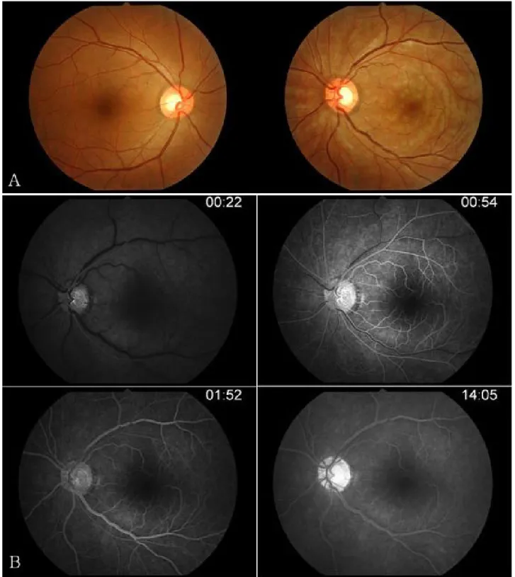
내원 당시 안과 검사상 나안 시력은 우안 1.0, 좌안 0.02였으며 교정되지 않았다. 안압은 우안 14, 좌안 15 mmHg 측정되었고 전방에 염증소견 관찰되지 않 았으며 전안부에 그 외 특이소견 없었다. 좌안에서 상 대적구심성동공장애가 관찰되었으며 안저검사상 좌안 의 후극부를 중심으로 백색의 다발성 및 미만성의 허혈 성 병변이 주로 혈관 주행을 따라 관찰되고 경도의 망 막정맥의 확장을 보이나 앵두반점이나 망막출혈은 관찰 되지 않았다(Fig. 1A). 좌안 유리체에 염증세포가 경 미하게 관찰되었고, 우안은 정상소견 보였다.
형광안저혈관조영술에서 좌안의 맥락막충만 지연, 동정맥통과시간 지연(1분 이상)이 관찰되었으나 명확
= 증례보고 =
Figure 1. (A) Fundus photograph shows whitish, multiple, diffuse ischemic lesions around the posterior pole and slightly engorged retinal veins in the left eye. (B) Fluorescein angiograph shows delayed choroidal filling and delayed arteriovenous transit time (more than 1 minute) in the left eye, but no apparently visible obstruction site.
한 망막동맥 폐쇄부위는 관찰되지 않았다(Fig. 1B).
광간섭단층촬영기(optical coherence tomography, OCT) 상에서 좌안 내측 망막에 reflectivity가 증가 된 소견이 관찰되었고 우안은 정상이었다.
감염이나 면역질환, 또는 악성종양과 관련된 망막혈 관염 내지는 불완전한 중심망막동맥폐쇄 의심 하에 혈 액검사 및 자기공명혈관조영술(magnetic resonance angiography), 목동맥초음파촬영술(carotid artery
Figure 2. Thirty days after steroid therapy. (A) Fundus photograph shows that the ischemic lesions disappeared and the engorged retinal veins diminished in the left eye. (B) Fluorescein angiograph shows normal choroidal fillling and normal arteriovenous transit time in the left eye.
ultrasonography)을 시행하였다.
말초혈액검사상 백혈구 27,400×103/ml (segmented neutrophil 27%, lymphocyte 5%, eosinophil 65%)로 호산구(eosinophil)가 증가된 소견 보였고,
적혈구침전속도(erythrocyte sedimentation rate, ESR) 29 mm/h, C-reactive protein (CRP) 3.17 mg/dl로 증가된 소견 보였다. perinuclear antineutrophil cytoplasmic antibody with
antimyeloperoxidase specificity (MPO-ANCA) 가 증가되어 있었다.
흉부 방사선검사에서 폐침윤 등 이상 소견은 보이지 않았으며, 타 병원에서 부비동염 수술시 시행한 조직 검사상 부비동 점막내 호산구 침윤 소견 관찰되었고, 환자가 팔, 다리가 저린 증상을 호소하여 시행한 신경 전도검사상 말초신경병증 소견을 나타냈다. 자기공명혈 관조영술 및 목동맥초음파촬영술에서 특이소견은 관찰 되지 않았다.
상기 소견으로 폐쇄성 망막혈관염을 동반한 척-스 트라우스 증후군으로 진단하였으며 내과에 의뢰하여 prednisolone 60 mg 및 hydroxyurea 1,000 mg 경구 복용을 시작하였고, 치료 시작 후 30일째 좌안 교 정시력 0.4로 호전되었다. 좌안 안저에서 초진시 보였 던 백색의 다발성 및 미만성의 허혈성 병변이 소실되었 으며 경도의 망막정맥의 확장도 감소하였다(Fig. 2A).
형광안저혈관조영술에서 좌안의 정상 맥락막충만 소견 보이며 정상 동정맥통과시간이 관찰되었다(Fig. 2B).
6개월이 지난 현재 prednisolone 7.5 mg 경구 복 용을 유지하는 중이며 좌안 교정시력은 0.4이고, 환자 는 여전히 팔, 다리가 저린 증상을 호소하고 있으나 그 외 전신적인 합병증이나 안과적 합병증은 관찰되지 않 고 있다.
고 찰
1951년 Churg and Strauss가 심한 천식, 열, 호산구증가증이 있으면서 전신적인 괴사성혈관염을 동반하는 환자 14명의 증례를 발표하면서 allergic granulomatosis and angiitis 또는 척-스트라우스 증후군(Churg-Strauss syndrome)이 보고되었다.8
1990년 the American College of Rheumato -logy에서는 척-스트라우스 증후군이라고 분류할 수 있는 다음과 같은 기준 6가지를 발표하였다: 천식, 호산 구증가증(말초혈액 검사에서 백혈구 수치 중 호산구의 비 율이 10% 이상), 단발신경병증(mononeuropathy) 또 는 다발신경병증(polyneuropathy), 흉부 방사선검사상 폐침윤, 부비동 이상(paranasal sinus abnormality), 조직 검사에서 혈관밖 호산구(extravascular eosinophil) 가 있는 소견. 이 6가지 중 4가지 이상 만족할 경우 척- 스트라우스 증후군이라고 분류하였다.2 비록 이러한 기준이 척-스트라우스 증후군을 진단하기 위해 고안된 것은 아니지만 현재는 진단 기준으로 널리 사용되고 있다.
Solans et al9이 척-스트라우스 증후군 32명 환 자에 대하여 의무 기록을 조사하여 임상 양상을 발표
한 바에 의하면 32명 중 24명(75%)에서 흉부 방사 선 소견상 이상 소견을 나타냈으며, 그 중 peripheral transient and patchy alveolar infiltrates가 가 장 흔한 소견이었다.
본 증례의 경우 위의 진단 기준 6가지 중 흉부 방사 선 검사상 폐침윤 소견을 제외한 다른 5가지를 만족하 여 진단하게 되었지만, 초기 검사 결과 흉부 방사선에 서 폐침윤 등 이상 소견을 보이지 않아 진단하는데 어 려움이 있었다.
본 증례에서와 같이 척-스트라우스 증후군 환자가 경 과 관찰 동안에 안과적 증상을 나타내는 경우는 드물다 고 알려져 있다.1,9,10 Solans et al9이 조사한 32명 환 자중 5명(15.6%)에서 안과적 증상을 나타냈는데, 2명 은 안와 거짓종양(orbital pseudotumor) 양상을 보 였고, 3명에서는 일과성흑암시 증상을 보였다. 그 중 중심망막동맥폐쇄를 보인 1명을 제외하곤 4명에서는 스테로이드 치료에 좋은 반응을 보였다고 한다. 본 증 례에서는 천식 또는 폐, 피부, 신경, 위장관과 관련된 증상들로 인해 내과에서 진단된 경우가 아니고, 좌안의 갑작스런 시력감소를 주소로 내원하여 안과에서 시행한 검사 결과로 인해 척-스트라우스 증후군이 진단된 증례 로 매우 드문 경우라고 할 수 있겠다.
척-스트라우스 증후군 진단에 있어 antineutrophil cytoplasmic antibodies (ANCA)의 유용성에 대하 여 보고된 바에 의하면 척-스트라우스 증후군 환자의 70%에서 ANCA를 가지고 있으며 대개 perinuclear ANCA with antimyeloperoxidase specificity (MPO-ANCA)인 것으로 알려져 있다.11
본 증례에서도 MPO-ANCA가 증가되어 있었다. 그 러나 중심망막동맥폐쇄를 동반한 척-스트라우스 증후 군 환자에서 MPO-ANCA 및 cytoplasmic ANCA 가 정상인 경우도 보고된 바가 있다.7
또한 Takanashi et al12은 척-스트라우스 증후군 에서 동반되는 안과적 증상을 두 군으로 나누었는데, 하나는 안와 거짓종양의 형태로 나타나는 것이고, 나머지 하나는 허혈성 혈관염의 형태로 나타나는 것이라고 하였 다. 허혈성 혈관염의 양상을 나타내는 경우에 ANCA 가 나타나는 경우가 많다고 하였는데 본 증례에서도 MPO-ANCA가 증가되어 있으면서 망막혈관염의 양 상을 나타내었다.
척-스트라우스 증후군의 예후 및 생존율은 대개 좋은 것으로 알려져 있다. 그리고 기타 다른 전신적인 혈관 염을 일으키는 질환들과 비교하여 낮은 사망률을 나타 낸다고 보고되어 있고, 스테로이드 단독 또는 면역억제 제와의 병합요법에 반응을 잘하는 것으로 알려져 있 다.1,9
본 증례에서도 스테로이드 치료에 반응하여 좌안 교 정시력이 0.02에서 0.4로 증가하였고, 안저 소견에서 초진시 보였던 허혈성 병변이 소실되었으며, 형광안저 혈관조영술에서도 정상 맥락막충만 소견 보이며 정상 동정맥통과시간 관찰되었다. 그러나 국외의 여러 증례 보고에서는 척-스트라우스 증후군에서 중심망막동맥폐 쇄가 동반된 경우에 스테로이드 단독 또는 면역억제제 의 병합요법에도 불구하고 시력 회복이 되지 않은 경우 가 많았다.3,4,7
본 증례에서처럼 안과 검사에서 폐쇄성 망막혈관염 소견을 보이는 경우에 전신 질환 동반 가능성을 염두에 두고 그에 대한 적절한 검사를 시행하는 것이 반드시 필요하다고 생각되며 폐쇄성 망막혈관염을 동반한 척- 스트라우스 증후군 1예를 경험하여 이를 보고하는 바 이다.
참고문헌
1) Guillevin L, Cohen P, Gayraud M, et al. Churg-Strauss syndrome. Clinical study and long-term follow-up of 96 patients. Medicine (Baltimore) 1999;78:26-37.
2) Masi AT, Hunder GG, Lie JT, et al. The American College of Rheumatology 1990 criteria for the classification of Churg-Strauss syndrome (allergic granulomatosis and angiitis).
Arthritis Rheum 1990;33:1094-100.
3) Hamann S, Johansen S. Combined central retinal artery and
vein occlusion in Churg-Strauss syndrome: case report. Acta Ophthalmol Scand 2006;84:703-6.
4) Udono T, Abe T, Sato H, Tamai M. Bilateral central retinal artery occlusion in Churg-Strauss syndrome. Am J Ophthalmol 2003;136:1181-3.
5) Partal A, Moshfeghi DM, Alcorn D. Churg-Strauss syndrome in a child: retina and optic nerve findings. Br J Ophthalmol 2004;88:971-2.
6) Yaman A, Ozbek Z, Saatci AO, et al. Topical steroids in the management of Churg-Strauss syndrome involving the conjunctiva. Cornea 2007;26:498-500.
7) Türkçüoğlu P, Isik A, Deniz N, et al. Central retinal artery occlusion in an ANCA negative Churg-Strauss syndrome patient. Int Ophthalmol 2007;27:369-71.
8) Churg J, Strauss L. Allergic granulomatosis, allergic angiitis and periarteritis nodosa. Am J Pathol 1951;27:277-301.
9) Solans R, Bosch A, Pérez-Bocanegra C, et al. Churg-Strauss syndrome: outcome and long-term follow-up of 32 patients.
Rheumatology 2001;40:763-71.
10) Chumbley LC, Harrison EG Jr, DeRemee RA. Allergic granulomatosis and angiitis (Churg-Strauss syndrome). Report and analysis of 30 cases. Mayo Clin Proc 1977;52:477-84.
11) Hoffman GS, Specks U. Antineutrophil cytoplasmic antibodies.
Arthritis Rheum 1998;41:1521-37.
12) Takanashi T, Uchida S, Arita M, et al. Orbital inflammatory pseudotumor and ischemic vasculitis in Churg-Strauss syndrome: report of two cases and review of the literature.
Ophthalmology 2001;108:1129-33.
=ABSTRACT=
A Case of Occlusive Retinal Vasculitis in Churg-Strauss Syndrome
Ji-Wook Yang, M.D., Young-Hoon Park, M.D.
Department of Ophthalmology and Visual Science, The Catholic University of Korea, Uijeongbu St. Mary’s Hospital, Gyeonggi-do, Korea
Purpose: To report a case of occlusive retinal vasculitis in Churg-Strauss syndrome.
Case summary: A-48-year-old man visited our clinic complaining of suddenly decreased visual acuity in the left eye. Two months previously he had an operation for sinusitis, and he had been taking medications for asthma. In the initial examination, his best corrected visual acuity was 0.02 in the left eye. Fundus examination showed whitish, multiple, diffuse ischemic lesions around the posterior pole and slightly engorged retinal veins in the left eye. Fluorescein angiography showed delayed choroidal filling and delayed arteriovenous transit time in the left eye, but no apparently visible obstruction site. The eosinophil count was elevated to 65% in the white blood cell differentiated count, and perinuclear antineutrophil cytoplasmic antibodies (ANCA) with antimyeloperoxidase specificity (MPO-ANCA) was increased, but a chest X-ray was normal. Eosinophil infiltrations in the mucosa of the paranasal sinus were found, and peripheral neuropathy was found in a nerve conduction study. Hence, we diagnosed the patient with Churg-Strauss syndrome accompanied by occlusive retinal vasculitis, and started steroid therapy. Thirty days later after steroid therapy, the best corrected visual acuity of the left eye was 0.4.
Conclusions: In patients with occlusive retinal vasculitis, we need to consider systemic diseases, such as Churg-Strauss syndrome.
J Korean Ophthalmol Soc 49(8):1335-1340, 2008
Key Words: Churg-Strauss syndrome, Occlusive retinal vasculitis
Address reprint requests to Young-Hoon Park, M.D.
Department of Ophthalmology, Uijeongbu St. Mary’s Hospital, The Catholic University of Korea
#65-1 Geumo-dong, Uijeongbu-city, Gyeonggi-do 480-130, Korea
Tel: 82-31-820-3110, Fax: 82-31-847-3418, E-mail: [email protected]