submit.radiology.or.kr J Korean Soc Radiol 2012;67(4):273-276
273 INTRODUCTION
Microglandular adenosis (MGA) of the breast is a rare be- nign proliferative disease of the breast (1). The condition arises due to infiltrative proliferation of small round glands lacking myoepithelial cell layers, which may mimic breast cancer. This subtype of adenosis has been more frequently associated with invasive carcinoma and in situ carcinoma than the other forms of adenosis in the breast. Several reports have described a spec- trum of the lesions, ranging from MGA to atypical MGA and cancer arising from MGA (2, 3). In the present work, we report the imaging and pathologic findings of a ductal carcinoma in situ arising in MGA.
CASE REPORT
A 57-year-old woman visited our hospital for the operation of
a palpable mass in the left breast. She detected the mass inciden- tally 1 month ago and underwent core biopsy concerning the mass in a local clinic. The histology report said that the section showed nodular growth consisted of lobules of mature adipo- cytes and hyperplastic lobules, suggestive of hamartoma. She wanted to remove the mass because of its large size. The mass was firm and ill-defined on palpation. Surgeon performed a sim- ple lumpectomy about the mass, based on the pathology report.
The mass was hardly separated from the surrounding tissue dur- ing the operation. The confirmed pathology was a ductal carci- noma in situ arising in MGA. We reviewed the imaging study.
Mammogram showed an ill-defined irregular isodense mass in the left breast (Fig. 1A). Some amount of fat was present within the lesion but no distinct margin or pseudocapsule was present. Suspicious microcalcifications were not noted in the mass. On a sonogram, a 4.5-cm irregular mass was demon- strated with indistinct margin in the left outer peripheral breast.
Case Report
pISSN 1738-2637
J Korean Soc Radiol 2012;67(4):273-276
Index terms Breast Neoplasm Microglandular Adenosis Ductal Carcinoma In Situ Mammography
Sonography
Received April 11, 2012; Accepted July 16, 2012 Corresponding author: Jin Kyung An, MD
Department of Radiology, Eulji Hospital, Eulji University School of Medicine, 14 Hangeulbiseok-gil,
Nowon-gu, Seoul 139-711, Korea.
Tel. 82-2-970-8290 Fax. 82-2-970-8346 E-mail: [email protected]
Copyrights © 2012 The Korean Society of Radiology
Microglandular adenosis of the breast is a benign proliferative lesion, and is a rare subtype of adenosis. The pathologic findings and clinical symptoms can mimic those of breast cancer. Microglandular adenosis has been frequently associated with invasive carcinoma and in situ carcinoma of the breast. Many reports have de- scribed a spectrum of the lesions, ranging from microglandular adenosis to cancer arising from microglandular adenosis. However, most of them have focused on the pathology, and there are a few cases that report imaging findings. In the present case, we report the imaging and pathologic findings of a ductal carcinoma in situ arising in microglandular adenosis. A 57-year-old woman detected a palpable mass in her left breast. Mammogram showed an ill-defined irregular isodense mass, and sonogram showed hyperechoic irregular mass with indistinct margin. The patient underwent breast conserving surgery and adjuvant radiotherapy.
Ductal Carcinoma In Situ of the Breast Arising in Microglandular Adenosis
1미세선 선증에서 발생한 유방의 관상피내암종1
Min Sun Jeong, MD
1, Jae Hee Kang, MD
2, Eun Kyung Kim, MD
3, Jeong Joo Woo, MD
1, Hyun Sook Kim, MD
1, Jin Kyung An, MD
1Departments of 1Radiology, 2Surgery, 3Pathology, Eulji Hospital, Eulji University School of Medicine, Seoul, Korea
Ductal Carcinoma In Situ of the Breast Arising in Microglandular Adenosis
submit.radiology.or.kr
J Korean Soc Radiol 2012;67(4):273-276
274
DISCUSSION
Microglandular adenosis (MGA) of the breast is an extremely rare proliferative benign condition. All of the reported cases of MGA are women subjects with the age range from 28 to 82 years, with the majority being in between 45-55 years old (4, 5).
The most common symptom of MGA is a palpable breast mass or breast thickening. Pathologically, the lesion consists of small round glandular structures with open lumens containing eosino- philic secretions, distributed in a hypocellular dense collagenous or fatty mammary stroma. A single layer of cuboidal epithelial cells lines the round glands without surrounding myoepithelial cell layer. Immunohistochemical stains demonstrate the glands of MGA, expressing S-100 protein and lacking myoepithelial lay- er with negative smooth muscle actin. Based on these clinical and histologic findings, MGA is likely to be confused with well- differentiated breast cancer, which presents as an aggressive his- tological feature. Progression to breast carcinoma has been re- ported in up to 27% of patients with MGA (4-6).
There have been several reports on imaging findings of MGA The echogenicity of the mass was predominantly hyperechoic
with some heterogeneously isoechoic areas (Fig. 1B, C). Abnor- mal lymph nodes were not noted in both axillae.
Consequently, the patient underwent breast conserving sur- gery of the left breast. The cut surface of the mass showed the adipose tissue with an irregular, whitish, firm area in the central portion. Microscopic findings showed round or solid glandular structures, infiltrating the fibrous septa and fatty mammary stro- ma. The small glands were lined by a single or multiple layers of atypical epithelial cells, as suggestive of atypical MGA. The large coalescent glands showed expansile growth with obliterat- ed lumens and marked cellular atypia, consistent with in situ carcinoma (Fig. 1D, E). Invasion was not present. The tumor cells were positive for cytokeratin 7, S-100 protein (Fig. 1F), but negative for estrogen receptor, progesterone receptor and Her2/
Neu. Most of the myoepithelial cells were negative for the smooth muscle actin on immunohistochemical staining (Fig.
1G). Permanent pathology confirmed a ductal carcinoma in situ arising in MGA. The patient was treated with adjuvant ra- diotherapy after the operation.
E A
F B
G C
D
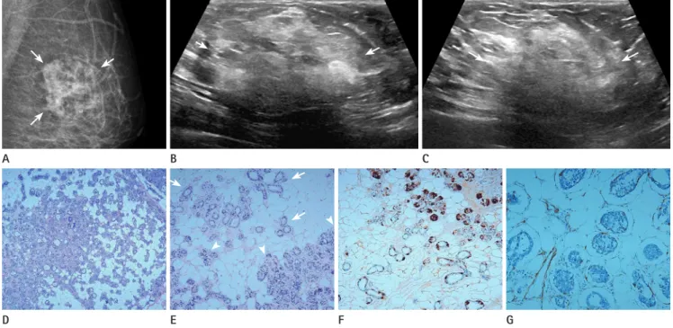
Fig. 1. A 57-year-old woman with a left palpable mass.
A. Mammogram shows an ill-defined irregular isodense mass on the left breast (arrows). Some amount of fat is present within the lesion.
B, C. Sonogram shows an irregular mass with indistinct margin (arrows). The echogenicity of the mass is predominantly hyperechoic with some heterogeneously isoechoic areas.
D. Microscopic finding shows solid type intraductal carcinoma extending to microglandular adenosis (H&E, × 100).
E. Transition from uncomplicated microglandular adenosis (arrows) to in situ carcinoma (arrowheads) is clearly noted (H&E, × 200).
F. The glands of microglandular adenosis and the tumor cells strongly express S-100 protein (× 200).
G. Most of myoepithelial cells are negative for smooth muscle actin on immunohistochemical staining (× 400).
Min Sun Jeong, et al
submit.radiology.or.kr J Korean Soc Radiol 2012;67(4):273-276
275
the imaging study in our case, we found the suspicious findings of an ill-defined margin and irregular shape, which we had missed on the initial evaluation. Our case was composed with intraductal carcinoma, infiltrating the fatty mammary stroma and fibrous septa. We presumed that the adipose tissue within the mass caused the sonographic hyperechogenicity.
In summary, MGA is a benign proliferative disease of the breast and has a spectrum from typical MGA to carcinoma aris- ing from MGA. Although additional study is necessary to set up the characteristic imaging findings of this tumor, they may tend to show suspicious finding in typical MGA, as well as in carcino- ma arising from MGA. Any suspicious finding should not be overlooked in an attempt to diagnose malignancy.
REFERENCES
1. Rosen PP. Adenosis and microglandular adenosis. In Rosen PP. Rosen’s Breast pathology. Philadelphia: Lippincott Wil- liams & Wilkins, 2001:152-161
2. Koenig C, Dadmanesh F, Bratthauer GL, Tavassoli FA. Car- cinoma arising in microglandular adenosis: an immuno- histochemical analysis of 20 intraepithelial and invasive neoplasms. Int J Surg Pathol 2000;8:303-315
3. Khalifeh IM, Albarracin C, Diaz LK, Symmans FW, Edgerton ME, Hwang RF, et al. Clinical, histopathologic, and immu- nohistochemical features of microglandular adenosis and transition into in situ and invasive carcinoma. Am J Surg Pathol 2008;32:544-552
4. Rosen PP. Microglandular adenosis. A benign lesion simu- lating invasive mammary carcinoma. Am J Surg Pathol 1983;7:137-144
5. Tavassoli FA, Norris HJ. Microglandular adenosis of the breast. A clinicopathologic study of 11 cases with ultra- structural observations. Am J Surg Pathol 1983;7:731-737 6. James BA, Cranor ML, Rosen PP. Carcinoma of the breast
arising in microglandular adenosis. Am J Clin Pathol 1993;
100:507-513
7. Kim DJ, Sun WY, Ryu DH, Park JW, Yun HY, Choi JW, et al.
Microglandular adenosis. J Breast Cancer 2011;14:72-75 8. Sabaté JM, Gómez A, Torrubia S, Matias-Guiu X, Alonso C,
Pericay C, et al. Microglandular adenosis of the breast in a BRCA1 mutation carrier: radiological features. Eur Radiol and carcinoma from MGA. Kim et al. (7) reported that MGA
showed ill-defined low echoic lesion on sonogram. Sabaté et al.
(8) reported the imaging findings of MGA in the BRCA1 mu- tation carrier. The mass showed parallel hypoechoic mass with relatively well-defined margins, irregular borders, discrete mi- crolobulations and angular margins. The mass was presented as hyperintense on T2-weighted image and showed homogeneous intermediate contrast enhancement with gradual delay en- hancement. Lee et al. (9) reported the imaging findings of inva- sive ductal carcinoma arising in atypical MGA. The mass was presented as hyperdense mass with indistinct margins and pleomorphic microcalcifications on a mammogram. It was in- compressible on sonogram with irregular shape and hypere- chogenicity.
In our case, fat component was demonstrated on both, the mammogram and sonogram. The majority of the mass has shown hyperechogenicity on a sonogram, which is an unusual finding to diagnose malignancy, because hyperechogenicity of the breast lesion is the most sensitive benign sonographic char- acteristics. Fat component and hyperechogenicity could be evocative of hamartoma. Choi and Ko (10) reported the inva- sive ductal carcinoma in a mammary hamartoma and summa- rized previously described 15 cases. Among the 15 cases, 12 carcinomas were located within the hamartoma and the re- maining three carcinomas involved both the hamartoma and adjacent normal breast tissue. The majority of the cases showed typical appearance of hamartoma on mammogram and/or so- nogram, and had suspicious findings within the hamartoma.
Typical hamartoma is a circumscribed fibrofatty mass with pseudocapsule, and is easily separated from the surrounding tissue from the operation. However, the mass, presented in our case, had no typical findings of hamartoma on both, imaging and operation. The only finding similar to hamartoma was the fat component. The mass had severe adhesion with surround- ing tissue on the operation, and there was not any pseudocap- sule on pathology. Therefore, hamartoma related lesion could be excluded on differentiation.
Linda et al. (11) reported that 0.4% of the malignant lesions were hyperechoic and all of them were palpable. They suggest- ed finding suspicious sonographic signs, because the hypere- choic malignant lesions were more likely to have noncircum- scribed margin and nonparallel orientation. On the review of
Ductal Carcinoma In Situ of the Breast Arising in Microglandular Adenosis
submit.radiology.or.kr
J Korean Soc Radiol 2012;67(4):273-276
276
hamartoma: case report and review of the literature. Korean J Radiol 2010;11:687-691
11. Linda A, Zuiani C, Lorenzon M, Furlan A, Girometti R, Londero V, et al. Hyperechoic lesions of the breast: not al- ways benign. AJR Am J Roentgenol 2011;196:1219-1224 2002;12:1479-1482
9. Lee YH, Dai YC, Lin IL, Tu CW. Young-aged woman with invasive ductal carcinoma arising in atypical microglandu- lar adenosis: a case report. Pathol Int 2010;60:685-689 10. Choi N, Ko ES. Invasive ductal carcinoma in a mammary
미세선 선증에서 발생한 유방의 관상피내암종1
정민선
1· 강재희
2· 김은경
3· 우정주
1· 김현숙
1· 안진경
1유방의 미세선 선증(microglandular adenosis)은 양성 증식성 병변으로 선증(adenosis)의 드문 아류형에 속한다. 미세선 선증의 병리 및 임상적 소견은 유방의 관상피내암 또는 침윤성 유방암과 유사한 소견을 보일 수 있으며 선증의 다른 아류 형보다 이들 유방암과의 관련성이 더 높은 것으로 알려져 있다. 미세선 선증, 비정형 미세선 선증, 미세선 선증에서 발생한 유방암으로 이어지는 일련의 스펙트럼이 보고되었으며, 대부분 병리적 소견에 대한 것으로 영상적 소견에 대해서는 드물게 보고되어 있다. 저자들은 본 증례에서 미세선 선증에서 발생한 유방의 관상피내암종에 대하여 보고하고자 한다. 57세 여 자 환자가 좌측 유방의 촉지 종괴를 주소로 내원하였다. 병변은 유방촬영상 동등밀도의 불규칙 형태를 보였고, 초음파상 경계가 불분명한 고에코의 불규칙 종괴로 나타났다. 환자는 유방보존수술과 보조방사선치료를 받았다.
을지대학교 을지병원 1영상의학과, 2외과, 3병리과