CONTENTS
Ⅰ. INTRODUCTION
Ⅱ. MATERIALS AND METHODS
Ⅲ. RESULTS
Ⅳ. DISCUSSION
Ⅴ. CONCLUSIONS REFERENCES KOREAN ABSTRACT
Ⅰ. INTRODUCTION
Almost immediately after the development of the ruby laser by Maiman in 1960, researchers postulated that it could apply to dental treatment. Stern and Sognnaes
30)in 1964 began looking at the possible uses of the ruby laser in dentistry. They were the first in a long list of investigators looking for a better way to treat dental patients with lasers. They began their laser studies on hard dental tissues by investigating the possible use of a ruby laser to reduce the subsurface demineralization.
29,31)Stern and Sognnaes found a reduction in permeability, to acid demineralization, of the exposed enamel. However, Adrian et al.
1)found that the ruby laser produced significant heat that caused damage to the pulp of the teeth.
During past decades, several in vitro studies with different laser devices were performed on dental hard
tissue applications. Results were not encouraging.
6,19,27,28,31)
Major problems have been thermal side effects, restricting the indications for lasers such as cw CO
2and cw and pulsed Nd:YAG laser to the vitrification of the residual dentinal caries.
16,18,28)Considering the ablation rate, the short pulsed CO
2laser seems to be suitable for hard tissue ablation.
However, problems such as heat generation and plasma formation persist
17,34)for an overview on these lasers for dental use.
Corresponding to the discouraging results of past in vitro studies, there have been few clinical investigations on the use of lasers for cavity preparation. In a retrospective clinical study, White
32)reported on restorations when a pulsed Nd:YAG laser was used for caries vitrification. Although he found the quality of restorations intact and all teeth remained vital, the sensitivity of the patients was not evaluated. Since these preliminary studies, research in this area has been sparse with but a few reports discussing the potential hard tissue uses. At present, the Food and Drug Administration has cleared laser soft tissue procedures with the CO
2, Nd:YAG, Ho:
YAG, Argon and Er:YAG lasers.
The Er:YAG laser at 2.94 ㎛
12,13)offers a new perspective for the effective removal of mineralized tooth tissues. Based on a thermally induced, preparations can be created by microexplosions without thermal damages to the adjacent hard and
Ablation Effects of Er:YAG Laser at Various Energy Levels, Different Repetition Rates and Irradiation Time on Enamel
Sae-Yong Lee, D.D.S.,M.S.D., Ki-Suk Kim, D.D.S.,M.S.D.,Ph.D.
Department of Oral Medicine, College of Dentistry, and Medical Laser Research Center
Dankook University
soft tissues.
7,9,13)Healthy enamel and dentin can be removed as well as carious tooth tissue. The laser-created defects contain rough walls without any signs of melting.
11)The ablation rate in dentin is very high in comparison to the pulsed CO
2laser at 9.6㎛ or the pulsed Nd:YAG laser.
5)The Er:YAG laser can be applied to primary carious lesions and secondary carious lesions under old restorations. It is also possible to remove cements or composites with an ablation efficiency comparable to that of healthy enamel and dentin.
10)On May 7,1998 the Food and Drug Administration cleared for marketing in the United States the first Er:YAG laser for use in preparing human dental cavities. Er:YAG laser was cleared for incisions, excisions, vaporization, ablation and hemostasis of soft and hard tissues in the mouth.
The purpose of this study was to investigate the ablation effect of Er:YAG laser on enamel at various irradiation parameters such as different energy level, repetition rates and irradiation times, using the first Er:YAG laser system developed recently in Korea.
Ⅱ. MATERIALS AND METHODS 1. Laser Apparatus
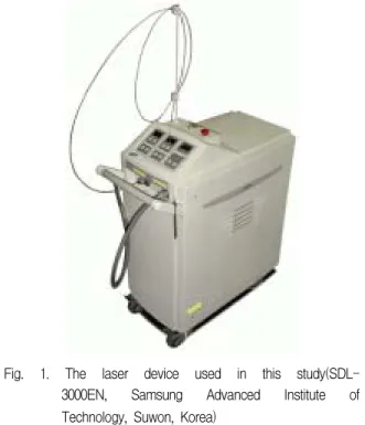
The laser device used in this study was the SDL-3000EN (Samsung Advanced Institute of Technology, Suwon, Korea). It has two different lasing medium, Nd:YAG and Er:YAG. Er:YAG laser device used in this study emits approximately 250 ㎲ pulsed laser and the wave length is 2.94 ㎛. The focal length was 9 mm and focal spot diameter was 300 ㎛.
The energy of laser apparatus used in this study is varied from 20 mJ up to 350 mJ per pulse and its peak output power is 3.5 W.
2. Samples
The tissue samples were derived from extracted adult molars. Immediately following extraction, the teeth were placed in ordinary tap water to which a small amount of bleach was added to disinfect the
samples. The time from extraction to experimentation varied considerably with the maximum being several weeks. Twenty one teeth were selected, and each of them was embedded in epoxy resin, and hardened.
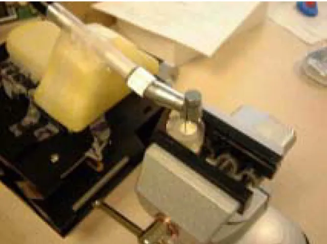
Enamel surfaces of all teeth were cut flat by buccal or lingual reduction with a slowly rotating diamond blade(Model 650, South Bay Technology Inc., USA) under running water(Fig. 2, 3). Each enamel surface of prepared teeth was divided into 6 partitions, to which Er:YAG laser would be applied. Therefore, 126 enamel samples were used in this study. Enamel samples were divided into 9 groups (40 mJ 2 Hz, 40 mJ 10 Hz, 40 mJ 20 Hz, 80 mJ 2 Hz, 80 mJ 10 Hz, 80 mJ 20 Hz, 120 mJ 2 Hz, 120 mJ 10 Hz, 120 mJ 20 Hz) in the first experiment to compare the ablation effects at various repetition rates under the identical total energy fluence at each energy level(8 J in 40 mJ groups, 16 J in 80 mJ groups, 24 mJ in 120 mJ groups). The other samples were divided into 12 groups (80 mJ 5 sec, 80 mJ 10 sec, 80 mJ 15 sec, 80 mJ 20 sec, 80 mJ 25 sec, 80 mJ 30 sec, 160 mJ 5 sec, 160 mJ 10 sec, 160 mJ 15 sec, 160 mJ 20 sec, 160 mJ 25 sec, 160 mJ 30 sec) in the second experiment to investigate the ablation effects with irradiation time.
Fig. 1. The laser device used in this study(SDL- 3000EN, Samsung Advanced Institute of Technology, Suwon, Korea)
Fig. 2. Teeth samples were cut flat by occlusal or buccal or lingual reduction with a slowly rotating diamond blade under running water.
3. Lasing
The laser was focused perpendicularly onto the surface of each enamel partitions using spherical lens with 9 mm focal length. Focal length was precisely reproduced using the guide pin attached on the side of handpiece. All teeth samples were clamped to a movable stage and were adjusted vertically and tilted so as to be in the focal point of the laser beam perpendicularly(Fig. 4, 5). And then, Er:YAG laser applied to each enamel partition.
4. Measuring
The equipment used for measuring the volume of ablation on enamel in this study was the NT-2000 (3-D Imaging Surface Structure Analyzer, WYKO Corp., USA)(Fig. 6, 7). It can measure volume of ablation utilizing vertical-scanning interferometry, and its resolution is 1∼3 nm.
5. Statistical Analysis
To determine the statistical significance for the difference of ablation rates between groups in two experiments, two-way ANOVA test and multiple comparison t-test was performed using StatView
Ⓡ4.0 program for Macintosh.
Fig. 3. Prepared enamel sample.
Fig. 4. Teeth samples were clamped to a movable stage and were adjusted vertically and tilted so as to be in the focal point of the laser beam perpendicularly.
Fig. 5. Focal length(9 mm) was precisely reproduced using the guide pin attached on the side of handpiece.
Fig. 6. Three-dimensional Imaging surface structure analyzing system (NT-2000).
Ⅲ. RESULTS
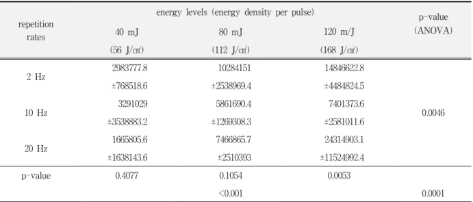
Table 1 shows the ablated volumes of the 2.94 ㎛ Er:YAG laser in enamel at energy levels of 40, 80, 120 mJ and at pulse repetition rates of 2, 10, 20 Hz. The ablation volumes of enamel in the groups of 40, 80 and 120 mJ (total energy; 8, 16 and 24 J) at 2, 10, and 20 Hz were 0.0017 to 0.0033, 0.001 to 0.0075 and 0.007 to 0.0243 mm3 respectively. From this table, it can be seen that energy fluence and repetition rates have
Table 1. Means and standard deviations of volumes(㎛3) ablated on enamel at 40, 80, 120 mJ of laser energy and at 2, 10, 20 Hz under the identical total energy and results of ANOVA test.
repetition rates
energy levels (energy density per pulse) p-value
(ANOVA)
40 mJ 80 mJ 120 m/J
(56 J/㎠) (112 J/㎠) (168 J/㎠)
2 Hz 2983777.8 10284151 14846622.8
0.0046
±768518.6 ±2538969.4 ±4484824.5
10 Hz 3291029 5861690.4 7401373.6
±3538883.2 ±1269308.3 ±2581011.6
20 Hz 1665805.6 7466865.7 24314903.1
±1638143.6 ±2510393 ±11524992.4
p-value 0.4077 0.1054 0.0053
<0.001 0.0001
Fig. 7. Tooth sample being measured volume of ablation by NT-2000.
strong influence on the variable ablation volume, as indicated the low p-values, <.0001 and .0046. The interaction of energy fluence and repetition rate also seems to have a strong influence.
Table 2, 3 and 4 show the differences of ablation
volumes of the Er:YAG laser in enamel at 2, 10 and
20 Hz measured after laser irradiation with different
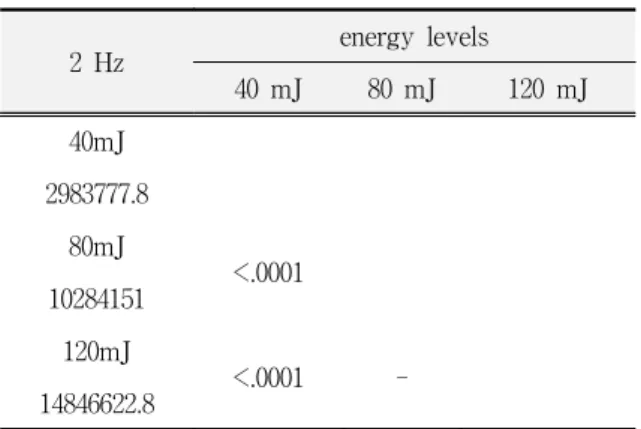
energy levels. These results mean that ablation rate
of high energy level is higher than that of low energy
level at an identical repetition rate. High ablation rate,
Table 2. Results of multiple comparison t-test for the ablation volumes(㎛3) of 40, 80 and 120 mJ of energy groups at 2 hertz of frequency.
2 Hz energy levels
40 mJ 80 mJ 120 mJ
40mJ 2983777.8
80mJ <.0001
10284151 120mJ
<.0001 -
14846622.8
Table 3. Results of multiple comparison t-test for the ablation volumes(㎛3) of 40, 80 and 120 mJ of energy groups at 10 hertz of frequency.
10 Hz energy levels
40 mJ 80 mJ 120 mJ
40mJ 3291029
80mJ -
5861690.4 120mJ
0.0444 -
7401373.6
Table 4. Results of multiple comparison t-test for the ablation volumes(㎛3) of 40, 80 and 120 mJ of energy groups at 20 hertz of frequency.
20 Hz energy levels
40 mJ 80 mJ 120 mJ
40mJ 1665805.6
80mJ 0.0008
7466865.7 120mJ
0.0008 0.0057
24314903.1
Table 5. Results of multiple comparison t-test for the ablation volumes(㎛3) of 2, 10 and 20 Hz groups at 40 mJ of energy level.
40 mJ repetition rates
2 Hz 10 Hz 20 Hz
2Hz 2983777.8
10Hz -
3291029 20Hz
- -
1665805.6
Table 6. Results of multiple comparison t-test for the ablation volumes(㎛3) of 2, 10 and 20 Hz groups at 80 mJ of energy level.
80 mJ repetition rates
2 Hz 10 Hz 20 Hz
2Hz 10284151
10Hz
0.0034 5861690.4
20Hz - -
7466865.7
Table 7. Results of multiple comparison t-test for the ablation volumes(㎛3) of 2, 10 and 120 Hz groups at 120 mJ of energy level.
120 mJ repetition rates
2 Hz 10 Hz 20 Hz
2Hz 14846622.8
10Hz 0.0055
7401373.6
20Hz - 0.0057
24314903.1
Table 9. Results of multiple comparison t-test for the ablation volumes(㎛3) of enamel after 5, 10, 15, 20, 25 and 30 sec of Er:YAG laser irradiations with 80 mJ of energy level.
80 mJ time(sec)
20 Hz 5 10 15 20 25 30
5 18570144.9
10 -
10839649.8
15 - -
10379801.6
20 - - -
7509271.7
25 - - - -
10392612.9
30 - - - - -
10165892.9
especially, can be seen at high energy level of 120 mJ and high repetition rate of 20 Hz.
Table 5, 6 and 7 shows that the differences of ablation volumes of the Er:YAG laser in enamel after laser irradiation at different repetition rates of 2, 10 and 20 Hz with an identical energy level. These
Table 10. Results of multiple comparison t-test for the ablation volumes(㎛3) of enamel after 5, 10, 15, 20, 25 and 30 sec of Er:YAG laser irradiations with 160 mJ of energy level.
160 mJ time(sec)
10 Hz 5 10 15 20 25 30
5 1839033.2
10 -
17501214.7
15 - -
23443698.5
20 - - -
21067544.7
25 - - - -
15236091.2
30 - - 0.025 0.047 -
15123761.7
results mean that high energy level of Er:YAG laser has significant increase of ablation rate in enamel, but low energy level does not have differences in ablation rates of Er:YAG laser between different repetition rates.
It can be seen in Table 8 that the ablation rate of
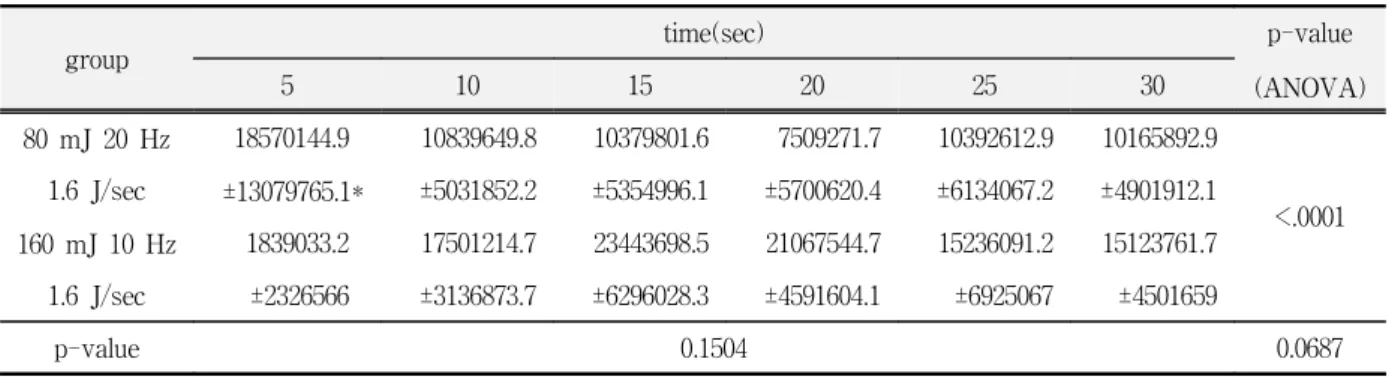
Table 8. Means and standard deviations of volumes(㎛3) ablated on enamel with time after 80 and 160 mJ ofEr:YAG laser irradiations and results of ANOVA test and unpaired t-tests.(㎛3)
group time(sec) p-value
5 10 15 20 25 30 (ANOVA)
80 mJ 20 Hz 18570144.9 10839649.8 10379801.6 7509271.7 10392612.9 10165892.9
<.0001 1.6 J/sec ±13079765.1* ±5031852.2 ±5354996.1 ±5700620.4 ±6134067.2 ±4901912.1
160 mJ 10 Hz 1839033.2 17501214.7 23443698.5 21067544.7 15236091.2 15123761.7
1.6 J/sec ±2326566 ±3136873.7 ±6296028.3 ±4591604.1 ±6925067 ±4501659
p-value 0.1504 0.0687
*=mean±standard deviation
Er:YAG laser at high energy level has higher than that at low energy laser, but there was no significant difference between irradiation times.
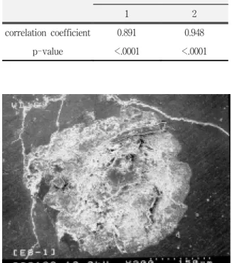
SEM photos and 3-D images of cut enamel surface for presentative samples can be seen in Fig. 8, 9 and 10. The Er:YAG lesions are partly marked by scaly or flaky rough areas without signs of serious thermal injuries. The arrangement of the individual hydroxyapatite crystal in the enamel unchanged. The pictures, rather, give the impression of a hard tooth substance having been ripped off by an explosion especially in the high energy level of 80 and 120 mJ.
After 200 pulses, the crater in the enamel is usually round or slightly oval with a sharp rim and a round, cone-shaped lesion without fissures or melted zoneson the inner wall surface can be observed. On
Table 11. Correlation coefficients showing correla- tion between ablation volumes(㎛3) and ablated surface roughness in experiment 1 and 2.
experiment
1 2
correlation coefficient 0.891 0.948
p-value <.0001 <.0001
Fig. 8. SEM and 3-D analyzer views of cut enamel surface after Er:YAG laser irradiation of 40 mJ and 20 Hz for 10 sec.
the 3-D images the lighter colored ring around the hole is solely due to the image of the conical hole. The area adjacent to the holes in enamel was slightly irregular without charring for all laser fluences ranging 8 to 24 mJ for 10 seconds.
Ⅳ. DISCUSSIONS
Stern et al.
29), and Scheinin et al
26). found a fissured enamel surface with cracks and pores greater than 1
㎛ in diameter by using a CO2 laser with 50 J/cm
2Fig. 9. SEM and 3-D analyzer views of cut enamel surface after Er:YAG laser irradiation of 80 mJ and 20 Hz for 10 sec.
Fig. 10. SEM and 3-D analyzer views of cut enamel surface after Er:YAG laser irradiation of 120 mJ and 20 Hz for 10 sec.
of output power. In a SEM overview, Kuroda and Fowler
15)investigated a crater in a human tooth that was treated with a focused beam of a CO
2laser at a radiant exposure of about 10,000 J/cm
2. He found that the crater and its surface had the smooth and rounded appearance of a solidified melt. This is the same results shown in a previous study.
8)Boehm et al.
2)stated in 1977 that the radial cracks are caused by expansion in response to heat and that the concentric fissures develop during the cooling process. They further considered a radiant exposure of 13-25 J/cm
2to be the critical threshold for enamel and dentin. At higher energy densities, cracking and fissuring are inevitable.
In contrast to the CO
2laser, the Er:YAG laser induced holes that were very different, because no-melt, no-fused zones and no cracks were seen.
This is in conformity with the investigation of hard tissue ablation with the Er:YAG laser carried out by Nuss et al.
25), Nelson et al.
24)and Keller and Hibst
8)and it is comparable because the enamel and dentin have the same inorganic matrix, the hydroxyapatite.
No increase of the damage zone around the crater by increasing radiant exposure was seen. The results of the Er:YAG laser application in this study also correspond to this tendency. Only minimal heating of the adjacent tissues can be seen and thus little or no damage to the dental hard substances can be found for the Er:YAG laser at a exposure of 114 to 2288 J/cm
2. There was not measured thermal effects in both Keller's study and present study even for a high radiant exposure of 100 J/cm
2and 2288 J/cm
2respectively. Comparing maximal dose of previous study, the exposure dose of this study was very high but there was few collateral damage to the dental tissue. Only little damage of the pulp can be expected as well. However, it is not clear whether the damage to the pulp is reversible and further investigations are indicated.
The Er:YAG (λ=2.94 ㎛) laser emits in the midinfrared near the IR peak of the water absorption curve and the OH
-absorption of hydroxyapatite.
These lasers represent the best near-term hope for a laser that can effectively remove dental hard
tissues. In brief, the radiation that these laser emits is strongly absorbed by water. The absorbed energy induces a rapid rise in temperature and pressure, and the heated material is explosively removed. Although the amount of water in dentin or enamel (20% and 10%, respectively) is relatively low, there appears to be enough absorption to initiate the ablation process.
Further, there is some evidence that carbonated hydroxyapatite, the mineral of dentin and enamel, also absorbs strongly in the midinfrared
20-23)because of the OH
-ions present in the structure.
The net result of this relatively high absorption is that the tissue is removed and little of the incident laser energy remains in the tissue to cause thermal damage. Hibst and Keller
7)measured the cutting rate for an Er:YAG laser. Using their 1.1-mm-diameter spot, a 50 J/cm
2radiant exposure, and a 10 Hz pulse repetition rate, one would expect to be able to remove a 2.7 mm x 1 mm x 4 mm volume of enamel in ∼ 30 seconds. The ablation volume of enamel surface was 0.007 mm
3being irradiated with 228.8 J/cm
2(160 mJ of output power, 1 Hz) in a pilot study. When irradiating at 10 Hz with 160 mJ of output power, the Er:YAG laser used in this study would be expected to take 154 seconds to remove a 2.7 mm x 1 mm x 4 mm volume of enamel. Nonetheless, it should not be thought the ablation rate of laser apparatus used in Hibst and Keller's study be higher than that of laser apparatus in this study, because the experimental designs were different.
It was thought that continuous irradiation should
increase the ablation volume of enamel proportionally
with irradiation time, the results of this study,
however, did not show proportional increase of the
ablation volume with irradiation time and there were
few differences in the ablation volumes among the
continuous irradiation groups for 5, 10, 15, 20, 25 and
30 second irradiation at 10 Hz with 160 mJ of output
power. Further study is required to confirm these
results, however, it is believed that major reason for
these results is as follows: In this study, the laser
focus was fixed in the holder and the focal length
became longer after lasing, consequently decreasing
a ablation rate by defocusing.
The detailed ablation mechanism of the Er:YAG laser is still unexplored. Some reflections therefore are described in Part I of Hibst's study,
7)In this context, the question arises as to whether the Er:YAG laser-induced cavities result only from a thermal evaporation process or from some additional processes.
Nelson et al.
24)stated in their study of bone ablation with the Er:YAG laser that some vapor produced by the laser energy will build up internal pressure until a microexplosion will take place with ejecting substrate in the form of microscopic particles. This can be one reason for the minimal damage of the surrounding tissue, because they found only a small thermal damage zone with little or no disruption of the remaining substrate. This is very similar to the pictures shown in this study on the ablation effect of enamel and dentin.
The conclusion must be that the major part of the incident energy is consumed in the ablative process and only a small fraction of the energy results in heating of the remaining tissue, so no damage occurs.
This is in conformity with previous observations of little heating of the crater surrounding tissues after Er:YAG laser treatment.
Several problems remain to be solved before the erbium lasers can be used safely and effectively for the treatment of dental hard tissues. The first and most important issue is the temperature rise within the tooth. The rise induced by a single erbium laser pulse is several hundred degrees at the site of absorption, but only a few hundred ㎛ from the ablation site, the temperature rise is but a few degrees. The temperature rise distant from the absorption site is, however, minimal only for a single pulse. It has been shown that multiple pulses induce a temperature increase that can be > 30℃ throughout the entire tooth.
3)Such a temperature rise likely does not damage the dentin or enamel immediately adjacent to the ablation site. Thus histologic examination of ablated teeth that were fixed immediately following laser irradiation fail to show changes induced by such minimal temperature rises.
Nonetheless, it is recognized that temperature rises of
5℃ or greater in the pulp for > ∼ 1 minute can necrose the pulp cavity.
36)The solution to the temperature increase is to cool the tooth surface with water.
4,30)Recent data indicate that the water flow rate can be relatively slow (e.g., 5 ml/min) to limit effectively the temperature rise to
< 3℃ even during prolonged ablation at 10 Hz.
4)Such water flow rates do inhibit the ablation but only minimally when compared with ablation in the absence of water flow. The ablation of tissue with an erbium laser through a layer of water seems contradictory when one considers that water strongly absorbs the incident laser radiation. However, it is clear that first segment of the incident laser pulse induces a cavity within the water layer through which the remainder of the pulse can propagate to the tissue surface. The challenge that remains is to incorporate the water spray mechanism into an ergonomic handpiece that can effectively deliver laser radiation.
The other major problem that erbium laser radiation can cause is cracking of the teeth.
3,14)Such cracking has been noted but mainly at higher radiant exposures.
It appears that these cracks are produced by shock waves that propagate into the teeth following ablation.
Such shock waves are an unavoidable aspect of the ablation process. Basically, to achieve ablation of a dental hard material, one needs to induce a pressure rise sufficient to explode the material away from the site of absorption. The challenge is to select a radiant exposure that achieves the desired ablation yet minimizes the unwanted side effect.
As can be seen, there has been much progress in the area of lasers in dentistry since the early report by Stern and Sognnaes.
30)These early researchers and those who followed wished to develop clinical uses for lasers with the aim of bringing the laser to the dental practitioner to imporve dental care. The progression has been slow, and some of the ambitions of the early researchers have yet to be realized.
To date, soft tissue applications have constituted
the primary area for the clinical use of lasers in
dentistry. Clinicians and patients alike have great
interest in the development of lasers for dental use.
In order to expand future applications in dentistry, developments must be based on understanding the effects of various wavelengths and parameters on laser/tissue interactions in the oral cavity. With this understanding, lasers can be developed to treat specific conditions or for specific purposes in the oral cavity. There are numerous other potential applications.
One of the most obvious applications of lasers is for the controlled removal of dental enamel, dentin, or bone. Using lasers to ablate hard dental tissue for bonding pretreatment, dental decay removal, and tooth preparation has received considerable attention by researchers.
35)This worthy goal can be achieved only after more information is available on tissue/laser interactions and technology can be developed to make use of this knowledge.
Replacement or supplementation of the dental drill is a real possibility for the future.
V. CONCLUSIONS
In this study, we have investigated the ablation effect of Er:YAG laser on enamel at various irradiation parameters such as different energy level, repetition rates and irradiation times, using the first Er:YAG laser system.
The results were as follows:
1. There was significant difference of ablation rate between energy levels.
2. The ablation was more effective at 20 Hz than 2 Hz or 10 Hz under the identical total energy using 120 mJ of output power, although there was no significant difference between various Hz using 40 mJ or 80 mJ of output power.
3. There was no increase of ablation rate with time when pulsed laser was irradiated to a enamel surface continuously.
In conclusion, it is believed that proper energy level and pulse repetition rate of Er:YAG laser should be used and accurate focal length should be maintained to ablate enamel effectively.
REFERENCES
1. Adrian, J.C., Bernier, J.L., Sprague, W.G.: Laser and the dental pulp. JADA., 83:113-117,1971.
2. Boehm, R., Rich, J., Webster, J., Janke, S.: Thermal stress effects and surface cracking associated with laser use on human teeth. J Biomech Eng., 99:189-194,1977.
3. Burkes, E.J., Hoke, J., Gomes, E., Wolbarsht, M.: Wet versus dry enamel ablation by Er:YAG laser. J Prosthetic Dentistry, 67:847-851,1992.
4. Cook, W.D.: Factors affecting the depth of cure of UV polymerized composites. J Dent Res., 1980; 59(5):800- 808.
5. Ertl, T., Muller, G.: Uberblick zu Lasertypen und deren Anwendungsprinzipien in der Zahnheilkunde. In: M ler G, Ertl T, eds. ngenwandte Laserzahnheilkunde, 1st ed.
Landsberg: Ecomed, 1995, III, pp 1-12.
6. Goldman, L., Hornby, P., Meyer, R., Goldman, B.: Impact of the laser on dental caries. Nature, 203:417,1964.
7. Hibst, R., Keller, U.: Experimental studies of the application of the Er:YAG laser on dental hard substances. I. Measurement of the ablation rate. Lasers Surg Med., 9:338-344,1989.
8. Hibst, R., Keller, U.: Experimental studies of the application of the Er:YAG laser on dental hard substances: II. Light microscopic and SEM investigations. Lasers in Surgery and Medicine, 9:345- 351,1989.
9. Hibst, R., Keller, U.: Heat effect of pulsed Er:YAG laser radiation. In: Joffe SN, At Atsumi K, eds. Laser surgery: Advanced characterization, therapeutics and systems. Los Angeles: Proceedings SPIE Vol 1200, 1990, pp 379-386.
10. Hibst, R., Keller, U.: Removal of dental filling materials by Er:YAG laser radiation. In: O rien SJ, Dederich DN, Wigdor H, Trent A, eds. aser in Orthopedic, Dental, and Veterinary Medicine. Los Angeles: Proceedings SPIE 1424, 1991, pp 120-126.
11. Keller, U., Hibst, R.: Ultrastructural changes of enamel and dentin following Er:YAG Laser radiation on teeth.
In: Joffe SN, Atsumi K, eds. Laser Surgery: Advanced Characterization, Therapeutics, and Systems II. Los Angeles: Proceedings SPIE 1200, 1990, pp 408-415.
12. Hibst, R., Keller, U., Steiner, R.: Experimental studies of the applications of the Er:YAG laser on dental hard substances. Lasers Surg Med., 8:145 (Abstr.),1988.
13. Hibst, R., Keller, U., Steiner, R.: The effect of pulsed Er:YAG laser radiation on dental tissues. Laser Med
Surg., 4:163-165,1988.
14. Koort, H.J., Frentzen, M.: Pulsed laser in dentistry- -Sense of Nonsense? SPIE Proceedings, 1424:87- 98,1991.
15. Kuroda, S., Fowler, B.O.: Compositional, structural-and phase changes in in vitro laser-irradiated human tooth enamel. Calcif Tissue Int., 36:361-369,1984.
16. Melcer, J., Chaumette, M.T., Melcer, F.: Dental pulp exposed to the CO2 laser beam. Lasers Surg Med., 7:347-352,1987.
17. Miserindino, L.J., Pick, R.M., eds.: "Lasers in Dentistry."
Chicago: Quintessence, 1995.
18. Myers, T.D., Myers, W.D.: The use of a laser for debridement of incipient caries. J Prosth Dent., 53:776-779,1985.
19. Neev, I., Liaw, L.L., Raney, D.V., Fujishige, I.T., Ho, P.T., Berns, M.W.: Selectivity and efficiency in the ablation of hard dental tissues with ArF pulsed excimer lasers. Lasers Surg Med., 11:499-510,1991.
20. Nelson, D.G.A., Jongebloed, W.L., Featherstone, J.D.B.:
Laser irradiation of human dental enamel. NZ Dent J., 82:74-77,1986.
21. Nelson, D.G.A., Shariati, M., Glena, R., Shields, C.P., Featherstone, J.D.B.: Effect of pulsed low energy infrared laser irradiation in artificial caries-like lesion formation. Caries Res., 20:289-299,1986.
22. Nelson, D.G.A., Wefel, J.S., Jongebloed, W.L., Featherstone, J.D.B.: Morphology, histology and crystallography of human dental enamel treated with pulsed low energy IR laser radiation. Caries Res., 21:411-426,1987.
23. Nelson, D.G.A., Williamson, B.E.: Low-temperature laser Raman spectroscopy of synthetic carbonated apatites and dental enamel. Aust J Chem., 3S:715-727,1982.
24. Nelson, J.S., Yow, L., Liaw, L.H., Macleay, L., Zavar, R.B., Orenstein, A., Wright, W.H., Andrews, J.J., Berns, M.W.: Ablation of bone and methacrylate by a prototype mid-infrared erbium: laser. Lasers Surg Med., 8:494-500,1988.
25. Nuss, R.C., Fabian, R.L., Sarkar, R., Puliafito, C.A.:
Infrared laser bone ablation. Lasers Surg Med., 8:381-391,1988.
26. Scheinin, A., Kantola, S.: Laser-induced effects on tooth structure. Crater production with a CO2 laser. Acta Odontol Scand., 27:173-181,1969.
27. Serebro, L., Segal, T., Nordenberg, D., Gorfil, C., Bar-Lev, M.: Examination of tooth pulp following laser beam irradiation. Lasers Surg., 7:236-239, Med 1987.
28. Shoij, S., Nakamura, M., Horiochi, H.: Histopathological changes in dental pulps irradiated by C02 laser: A preliminary report on laser pulpotomy. J Endod., 11:
379-384,1985.
29. Stern, R.H., Renger, H.L., Howell, F.V.: Laser effects on vital dental pulps. Br Dent J., 127:26-32,1969.
30. Stern, R.H., Sognnaes, R.F.: Laser beam effect on dental hard tissues. J Dent Res., 43 (Suppl.):873 (Abs 307),1964.
31. Stern, R.H., Vahl, J., Sognnaes, R.F.: Laser enamel:
Ultrastructural observations of pulsed carbon dioxide laser effects. J Dent Res., 51:455-460,1972.
32. White, J.M., Goodis, H.E., Setcos, J.C., Eakle, W.S., Hulscher, B.E., Rose, C.L.: Effects of pulsed Nd:YAG laser energy on human teeth. J Am Dent Assoc., 124:45-51,1993.
33. Wigdor, H.A., Visuri, S.R., Walsh, J.T.: Effect of water on dental material ablation of the Er:YAG laser. SPIE Proceedings 1994.
34. Wigdor, H.A., Walsh, J.T., Featherstone, J.D.B., Visuri, S.R., Fried, D., Waldvogel, J.L.: Lasers in dentistry.
Lasers Surg Med., 16:103-133,1995.
35. Wigdor, H.A., Walsh, J.T., Fried, D., Waldvogel, J.L.:
Lasers in dentistry. Lasers Surg Med., 16:103-133,1995.
36. Zach, L., Cohen, G.: Pulp response to externally applied heat. Oral Surg Oral Med Oral Surg., 19 (4):515-530,1965.
Corresponding Author : Ki-Suk Kim, Professor,
Department of Oral Medicine, School of Dentistry, Dankook University, San 7-1, Shinbudong, Cheonanm Choongnam 330-716 Korea