- CONTENTS -
Ⅰ. INTRODUCTION
Ⅱ. MATERIALS AND METHODS
Ⅲ. RESULTS
Ⅳ. DISCUSSION
Ⅴ. CONCLUSIONS REFERENCES KOREAN ABSTRACT
Ⅰ. INTRODUCTION
Occlusal appliances or splints are selectively used in the treatment of disorders of the temporomandi- bular joint (TMJ) and masticatory muscles. The purpose of occlusal appliance therapy is : to stabilize and improve the function of the TMJ ; to improve the function of the masticatory motor system and reduce abnormal muscle activity ; or to protect teeth from attrition and adverse traumatic load
1,2). Clark reviewed the design, theory, and effectiveness of occlusal appliance for specific symptoms and found a 70 ∼ 90% rate of clinical success
2). Clinical success has been reported in the treatment of TMJ and masticatory system using occlusal appliance
3-7). While the treatment effect is predictable, the explanation of the physiologic basis of the treatment response is less understood. Clark
described that five theories explained how the occlusal appliance actually work. These are : the occlusal disengagement theory ; the vertical dimension theory ; the maxillomandibular realignment theory ; the temporomandibular joint repositioning theory ; and the cognitive awareness theory
1,2).
Numerous testing modalities have been proposed for use in the diagnosis of patients presenting with temporomandibular disorders (TMD). In testing modalities, electromyography (EMG) represents a research tool for measuring muscle function and has been widely used since it was first introduced in dental research by Moyers in 1949. EMG has been used to study the functional mechanism of the occlusal appliance, as well as its therapeutic effects on the masticatory system
8).
Occlusal appliance is widely used in the treatment of TMD, but the mechanism of therapeutic effect is obscure and there have been few studies about the effect of occlusal appliance height on the TMJ and masticatory muscle
5,6). So, in order to evaluate the effect of changes in the height of the occlusal appliance on the masticatory muscles in various body postures, the EMG activities of masseter and anterior temporal muscles with various occlusal heights were observed in the uprighting, feeding and supine body postures in this study.
Effect of the Height of Occlusal Appliance on Masticatory Muscles in Various Body Postures
Kye-Ra Park
1, D.D.S., Bong-Jik Suh
1,2, D.D.S., M.S.D., Ph.D.
Department of Oral Medicine
1and Institute of Oral Bioscience
2,
School of Dentistry, Chonbuk National University
Ⅱ. MATERIALS AND METHODS 1. Subjects
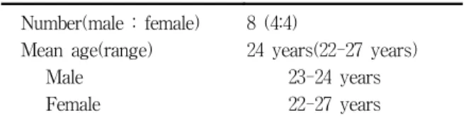
This study was performed on eight subjects, four women and four men, in the age range of 22 to 27 years with a mean of 24 years, dental students of School of Dentistry, Chonbuk National University (Table 1).
The following criteria were used for the selection of subjects.
(1) no signs and symptoms of TMD
(2) natural dentition, no missing tooth and no parafunctional oral habits
(3) absence of extensive restorations
(4) no history of orthodontic treatment and facial trauma
(5) absence of pathologic periodontal condition 2. Height of occlusal appliance
For each subject, a wafer was made of 2.0㎜
thickness transparent acrylic materials (Biocryl C
Ⓡ) by vacuum adapter (Biostar
Ⓡ). Transparent autopolymerized acrylic resin was added to the occlusal surface of the wafer to be made variable heights of occlusal appliance. The height of occlusal appliance was determined by the increase in the overbite of occlusion at the incisal surface of central incisor. Finally, 4 groups of the height, respectively, 0㎜ without occlusal appliance, 4.3(4.3
±0.2)㎜, 6.0(6.0±0.3)㎜ and 8.1(8.1±0.2)㎜ were selected.
Table 1. General characteristics of the variables of subjects
Number(male : female) Mean age(range) Male Female
8 (4:4)
24 years(22-27 years) 23-24 years 22-27 years
3. Body posture of subjects
The following three body postures were selected for this study.
(1) upright posture : sitting upright in a dental chair with the head supported and the Frankfort plane parallel to the floor
(2) feeding posture : sitting in a dental chair with head forward 30 degrees
(3) supine posture : supine in a dental chair with Frankfort plane vertical to the floor
4. Recordings of EMG activities
An eight-channel instruments, EM2 Bioelectric Processor
Ⓡ(Myotronics. U.S.A.) was used to record EMG activities. Prior to recording of EMG activities, all subjects were introduced to the EMG apparatus, and were carefully instructed about the test.
The bipolar surface electrodes were positioned on the muscular bellies parallel to muscular fibers of masseter and anterior temporal muscles.
Each subject was submitted to recordings of EMG activity, with and without occlusal appliance during maximum clenching for each body posture.
All subjects were instructed to clench as hard and rapidly as possible and to maintain it for 3 seconds. EMG recordings were made three times for each clenching. The mean value of the EMG activities of both masseter and anterior temporal muscles were used.
In order to minimize methodological error, the recordings of EMG muscle activities were made by the same examiner during all experimental procedures.
5. Statistical Analysis of Data
Statistical tests used ANOVA by the SPSSWIN
(version 8.0). Probability levels of p<0.05 were
considered statistically significant.
Ⅲ. RESULTS
1. EMG activities of masseter and anterior temporal muscle
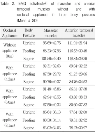
The EMG activities of masseter and anterior temporal muscles without and with occlusal appliance in three body postures were evaluated.
There were decreased EMG activities of masseter and anterior temporal muscles with occlusal appliance compared to without occlusal appliance and EMG activities tended to decrease, related to increasing heights (Table 2 and Figure 1, 2).
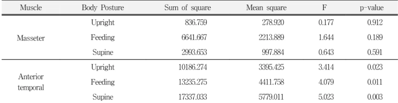
2. Comparison of EMG activities in three body postures
There were differences among the EMG activities of anterior temporal muscle in four groups of the height at three body postures (p<0.05) and no differences among the EMG activities of masseter muscle (Table 3).
Table 2. EMG activities(㎶) of masseter and anterior temporal muscles without and with occlusal appliance in three body postures (Mean ± SD)
Occlusal Appliance
Body Posture
Masseter muscles
Anterior temporal muscles Without
appliance (0㎜)
Upright 95.69±42.75 111.91±21.94 Feeding 88.23±37.96 116.53±30.48 Supine 101.56±42.40 118.84±29.36 With
appliance (4.3㎜)
Upright 92.31±32.63 89.04±32.22 Feeding 87.50±29.72 91.23±29.06 Supine 90.76±40.37 84.76±34.52 With
appliance (6.0㎜)
Upright 91.48±45.86 86.81±37.88 Feeding 62.94±43.55 83.88±38.33 Supine 87.50±40.32 80.60±37.82 With
appliance (8.1㎜)
Upright 85.64±36.13 77.64±32.00 Feeding 80.50±34.14 79.31±32.92 Supine 83.02±34.03 78.27±30.97
Fig 1. Mean EMG activity(㎶) of masseter muscle without and with occlusal appliance in three body postures
Fig 2. Mean EMG activity(㎶) of anterior temporal muscle without and with occlusal appliance in three body postures
3-1. Paired comparison of temporal EMG activity in upright body posture
In the uprighting body posture, only the EMG activity of anterior temporal muscle with 8.1㎜
height of occlusal appliance was significantly lower
than without occlusal appliance (Table 4).
Table 4. Paired comparison of temporal EMG activity (㎶) in the heights of occlusal appliance at uprighting body posture (Scheffe test)
Anterior temporal Muscle
0㎜ vs 4.3㎜ NS
0㎜ vs 6.0㎜ NS
0㎜ vs 8.1㎜ *
4.3㎜ vs 6.0㎜ NS
4.3㎜ vs 8.1㎜ NS
6.0㎜ vs 8.1㎜ NS
* p < 0.05
NS = non-significant
3-2. Paired comparison of temporal EMG activity in feeding body posture
In the feeding body posture, only the EMG activity of anterior temporal muscle with 8.1㎜
height of occlusal appliance was significantly lower than without occlusal appliance (Table 5).
3-3. Paired comparison of temporal EMG activity in supine body posture
In the supine body posture, the EMG activity of anterior temporal muscle during maximum clenching with 4.3, 6.0 and 8.1㎜ height of occlusal appliances was significantly lower than without
Table 5. Paired comparison of temporal EMG activity (㎶) in the heights of occlusal appliance at feeding body posture (Scheffe test)
Anterior temporal Muscle
0㎜ vs 4.3㎜ NS
0㎜ vs 6.0㎜ NS
0㎜ vs 8.1㎜ *
4.3㎜ vs 6.0㎜ NS
4.3㎜ vs 8.1㎜ NS
6.0㎜ vs 8.1㎜ NS
* p < 0.05
NS = non-significant
Table 6. Paired comparison of temporal EMG activity (㎶) in the heights of occlusal appliance at supine body posture (Scheffe test)
Anterior temporal Muscle
0㎜ vs 4.3㎜ *
0㎜ vs 6.0㎜ *
0㎜ vs 8.1㎜ *
4.3㎜ vs 6.0㎜ NS
4.3㎜ vs 8.1㎜ NS
6.0㎜ vs 8.1㎜ NS
* p < 0.05
NS = non-significant Table 3. Comparison of the EMG activities(㎶) in the heights of occlusal appliance (ANOVA)
Muscle Body Posture Sum of square Mean square F p-value
Masseter
Upright 836.759 278.920 0.177 0.912
Feeding 6641.667 2213.889 1.644 0.189
Supine 2993.653 997.884 0.643 0.591
Anterior temporal
Upright 10186.274 3395.425 3.414 0.023
Feeding 13235.275 4411.758 4.079 0.011
Supine 17337.033 5779.011 5.023 0.003
occlusal appliance and there were no differences among EMG activities of the anterior temporal muscle with 4.3, 6.0 and 8.1㎜ height of occlusal appliances (Table 6).
Ⅳ. DISCUSSION
The height of occlusal appliance is one of the factors which are considered in treatment of the TMD using occlusal appliance. And it may influence on masticatory muscular activities and therapeutic effects of treatment in TMD patients.
This study evaluated the effects of the height of the occlusal appliance on the myoelectrical activities of masseter and anterior temporal muscles in various body postures.
In this study, the EMG activities of anterior temporal muscle with occlusal appliances were significantly decreased compared to those without occlusal appliance. And there was no difference between the EMG activities of masseter muscle with and without occlusal appliance. The behavior of the EMG activities of masseter and anterior temporal muscles in healthy subjects with and without occlusal appliance in this study is in accordance with the findings of Lee et al
9). And Kawazoe et al
10)reported no difference between EMG potentials of masseter muscles with and without an occlusal appliance in normal subject but a significant decrease of EMG activities about 20%
with the splint in patients with myofascial pain-dysfunction syndrome during maximum voluntary clenching. Christensen reported that, in subjects without symptoms of mandibular dysfunction, the occlusal appliance tended to reduce the level of EMG activity in masseter muscle during maximal clenching
11).
However, Wood and Tobias reported that there was increase of the EMG activity of elevator muscles with the occlusal appliance in healthy subjects (masseter muscle 27%, anterior temporal muscle 15% increase
12).
In this study, the EMG activities of anterior temporal muscles during maximum clenching with
8.1㎜ height of occlusal appliance were significantly lower than without occlusal appliance in the upright and feeding body postures. And in the supine body posture, EMG activities of anterior temporal muscles with 4.3, 6.0 and 8.1㎜ heights of occlusal appliance were significantly lower than without occlusal appliance. There was decreasing tendency of the EMG activity of masseter muscle with occlusal appliance compared to without occlusal appliance, but there was statistically no difference among EMG activities of masseter muscle with and without occlusal appliance during maximum clenching in various body postures.
Previous works have reported the influence and clinical significance of the vertical height of occlusal appliances
5,6). Manns et al
5)treated sample of 75 TMJ pain patients with flat plane splints of one millimeter, four millimeters, and eight millimeters in thickness and the thinner splints took longer to reduce the pain symptoms. Also, Manns et al
6)studied that vertical dimension of least EMG activity was determined for each 60 patients, who were randomly divided into three groups according to the vertical dimension at which the occlusal appliance was adjusted(1.00, 4.25 and 8.25㎜). And the results showed a significant reduction of masster EMG activity in the mandibular postural position at the end of the 3-week treatment period for patient groups with 4.25 and 8.25㎜ occlusal appliances in comparison with the group with 1.00
㎜ occlusal appliance. But the studies about effect of the height of occlusal appliance during maximum clenching on the masticatory muscular activity in various body postures are lacking.
This study evaluated effect of the height of
occlusal appliance on EMG activities of masseter
and anterior temporal muscles with and without
occlusal appliances, but didn't consider effect of
body posture on EMG activities. However, body
posture may influence on masticatory muscular
activity. The influence of variation in body posture
on masticatory muscular activity has been studied
in healthy subject
13,14,15)and in patients with signs
and symptoms of mandibular dysfunction and
nocturnal bruxism
16,17). In healthy subjects, it has been demonstrated that during maximal voluntary clenching, masseter muscular activity decrease in the supine posture compared to the upright posture
15). In patient with temporomandibular dysfunction, it has also been demonstrated that masseter activity at rest did not change upon variation in body posture from seated upright to supine posture
16,17).
On the basis of the results of this study, it is suggested that the height of occlusal appliance has influence on EMG activities of masticatory muscle and further evaluation for the effect of the height of occlusal appliance on masticatory muscular activity would be necessary.
Ⅴ. CONCLUSIONS
The purpose of this study was to evaluate the effect of the height of occlusal appliance at various body postures on the masticatory muscle activities.
Eight healthy dental students without any signs and symptoms of temporomandibular disorders participated in this study. To record electro- myographic activity, EM2
Ⓡwas used. The occlusal appliance with the height of 4.3, 6.0 and 8.1㎜ was used and body postures were selected to upright, feeding, and supine.
The EMG activities of masseter and anterior temporal muscles in four heights of occlusal appliance during maximum clenching at three body postures were measured, analysed and evaluated.
The obtained results were as follows:
1. The EMG activities of anterior temporal muscle with occlusal appliance were significantly decreased compared to without occlusal appliances (p<0.05) and no difference between EMG activities of masseter muscle with and without occlusal appliance.
2. In the upright posture, the EMG activity of anterior temporal muscle with 8.1㎜ height of occlusal appliance during maximum clenching was significantly lower than without occlusal
appliance (p<0.05).
3. In the feeding posture, the EMG activity of anterior temporal muscle with 8.1㎜ height of occlusal appliance during maximum clenching was significantly lower than without occlusal appliance (p<0.05).
4. In the supine posture, the EMG activity of anterior temporal muscle with occlusal appliances of 4.3, 6.0 and 8.1㎜ height during maximum clenching was significantly lower than without occlusal appliance (p<0.05).
REFERENCES
1. Clark, G.T. : A critical evaluation of orthopedic interocclusal appliane therapy:design, theory, and overall effectiveness, J. Am. Dent. Assoc.
108:359-364, 1984.
2. Mohl, N.D., Zarb, G.A., Carlsson, G.E. and Rugh, J.D.
: A textbook of occlusion. 1st ed., Quintessence Publishing Co., Inc., Chicago, 1988.
3. Greene, C. and Laskin, D. : Splint therapy for the myofascial pain and dysfunction syndrome : A comparative study. J. Am. Dent. Assoc. 84: 624, 1972.
4. Carraro, J.J., and Caffesse, R.G. : Effect of occlusal splints on TMJ symptomatology, J. Prosthet. Dent., 40; 563-566, 1978.
5. Manns, A., Miralles, R., Santander, H., and Valdivia, J. : Influence of the vertical dimension in the treatment of myofascial pain-dysfunction syndrome.
J. Prosthet Dent 50: 700-709, 1983.
6. Manns, A., Miralles, R., and Cumsille, F. : Influence of vertical dimension on masseter muscle electromyo- graphic activity in patients with mandibular dysfunction. J. Prosthet. Dent., 53: 243-247, 1985.
7. Sheikholeslam, Holmgren, and Riise : Clinical and EMG study of long term effects of an occlusal splint on the temporal and masseter muscles in patients with functional disorders and nocturnal bruxism. J.
Oral rehabil., 13: 137, 1986.
8. Dahlström, L. : Electromyographic studies of craniomandibular disorders : a review of the literature. J. Oral Rehabil., 16: 1:20, 1989.
9. Lee, E.H., Suh, B.J., and Oh, H.M. : An electromyographic Study on Changes of Mandibular Position, Korean Journal of Oral Medicine, 24(1):
49-58, 1999.
10. Kwazoe, Y., Kotani, H., Hamada, T., and Yamada, S.
: Effect of occlusal splints on the electromyographic activities of masseter muscles during maximum clenching in patients with myofascial pain- dysfunction syndrome. J. Prosthet. Dent., 43: 578-580, 1980.
11. Christensen, L.V. : Effects of an occlusal splint on integrated electromyography of masseter muscle in experimental tooth clenching in man. J. of Oral Rehabil., 7:281, 1980.
12. Wood, W.W., and Tobias, D.L. : EMG response to alteration of tooth contacts on occlusal splint during maximal clenching, J. Prosthet. Dent., 51: 394-396, 1984.
13. Lund D., Nishiyama T., Moller E. : Postural activity in muscles of mastication with the subjects upright, inclined and supine, Scand. J. Dent. Res., 78: 417-424, 1970.
14. Hairston L., Blanton L : An electromygraphic study of mandibular position in response to change in body position. J. Prosthet. Dent., 49:271-275, 1983.
15. Mirralles, R., Bull, R., Lolas, F., and Manns, A. : Functional dissociation between two elevator mandibular muscles of different body position. J.
Gnathol., 6: 97-105, 1987.
16. Moller, E., Sheikholeslam, A., and Lous, I. : Deliberate relaxation of the temporal and masster muscles in subjects with functional disorders on the chewing apparatus, Scand. J. Dent. Res., 79: 478-483, 1971.
17. Holmgren, K., Sheikholeslam, A., Riise, C. : An electromyographic study of the immediate effects of an occlusal splint on the postural activity of the anterior temporal and masster muscles in different body positions with and without visual input. J. Oral Rehail., 12: 483-490, 1985.