CONTENTS
Ⅰ. INTRODUCTION
Ⅱ. MATERIALS AND METHODS
Ⅲ. RESULTS
Ⅳ. DISCUSSION
Ⅴ. CONCLUSIONS REFERENCES KOREAN ABSTRACT
Ⅰ. INTRODUCTION
Temporomandibular disorders are a heterogenous set of clinical conditions, characterized by pain and dysfunction of the masticatory system. Pain in the masticatory muscles, in the temporomandibular joint, and in associated hard and soft tissues, limitation in jaw function, and sounds in the TMJ are the common signs and symptoms of Temporomandibular disorders.
1,2,3)Imaging is the only method of obtaining visual information on the status of the joint tissues short of arthroscopy or open joint surgery. Its primary purpose is to provide information to assist the diagnosis and treatment planning process.
Radiography has long been the primary means for diagnosing organic diseases of the TMJ.
However, it has been difficult to determine which
radiographic signs are characteristic of individual diseases of the joint. Among the classical radiographic signs of joint disease decreased joint space has been found to be correlated with crepitus, a clinical sign of structural damage to the joint.
4,5)Reduction of the joint space, subcortical sclerosis and flattening of the lateral part of the condyle have been found to be intercorrelated and frequent among patients with crepitus, pain and joint dysfunction.
6)Despite TMJ imaging s long history of research and clinical application, the quality of information gleaned from imaging is often less than desired.
The small size of the TMJ, the widely varying fossa and condylar morphology and the surrounding dense osseous structures make clear and undi- storted imaging of the joint hard tissue technically difficult. To overcome these obstacles, multiple conventional radiographic technique have been introduced over the years.
In the past decade, impressive technological advances (computed tomography and magnetic resonance imaging) have come into widespread use making possible the characterization of both hard and soft tissue in almost any desired plane.
7,8)Conventional TMJ radiography has an established role in the detection of structural bone changes and sagittal tomography has been shown
Study on the Conventional Tomographic Findings for the Patients with Temporomandibular Disorders
Seong-Jong Seo, D.D.S.,M.S.D., Ki-Suk Kim, D.D.S.,M.S.D.,Ph.D.,
Department of Oral Medicine, School of Dentistry, and Medical Laser Research Center,
Dankook University
to yield the most information.
9)Chritstiansen et al.
10)assessed the frequency and severity of the classical radiographic signs of joint disease: sclerosis, decreased joint space, osteo- phytosis, cysts or pseudocysts and coritcal/
subcortical rarefaction. They reported these classical signs were correlated with the additional radiographic findings of lateral joint reciprocal reshaping, changes in condylar angulation and in the shape of the condyle, articular eminence, and glenoid fossa. They found that condylar transverse size, condylar angulation, condylar shape, size and shape of the glenoid fossa, and position of the coronoid process tip changed in the diseased joints.
In that study, those variables were found to be interrelated and significantly correlated to the radiographic severity of disease.
In the study of Tanimoto et al., Autopsy specimens were examined both radiographically and macrosopically to compare direct computed tomography with conventional tomography for their diagnostic yield of the structural bone changes in the temporomandibular joint. They concluded that conventional tomography is superior to computed tomography in the diagnosis of single structural bone changes but comparable for comprehensive diagnosis of TMJ disease.
11)The purpose of this study was to depict, by means of conventional tomography, the bone changes that take place in a temporomandibular
Fig. 1. Scanora used in tomographic imaging of TMJ
joint of patient with temporomandibular disorder and to correlate these changes to different variables such as condylar angulation, condylar type, condylar position and bone change type.
Ⅱ. MATERIALS AND METHODS 1. Subjects
A series of 256 patients, referred to the Department of Oral Medicine and Orofacial pain and TMJ disorder clinic, Dental Hospital, Dankook University, between July and December 1999 was examined with conventional tomography. From this total, a subsample of 73 which showed symptoms and signs of a unilateral internal derangement in joints was selected for this study.
2. Tomographic equipment
Tomographic imaging was performed using a
multidirectional tomograph (SCANORA, Orion
Corporation Soredex, Helsinki, Finland). Scanora is
a multifunction x-ray unit designed for radiographic
examination of dento-maxillo-facial regions. The
multifunction feature means that the imaging
elements include arrangements for examinations
utilizing both narrow scanning beam and
multidirectional tomography principles. Although the
imaging procedure are computer controlled, and
tomographic imaging is included, SCANORA is not a computed tomography device. Components of SCANORA include an imaging element, patient chair, x-ray generator.
Corrected tomographs were taken of the right and left TMJs in the sagittal plane as part of routine TMJ examination. A submentovertex projection was used to correct for orientation of the condylar heads with respect to the midline and to calculate the depth of cut. All cuts were 4 mm thick and collimated to include only the TMJ area. Four cuts were taken in maximum intercuspation and one cut was taken at maximum opening for each TMJ. The average exposure factors for the Scanora unit were 72 kV, 3.5 mA, 82 sec (range 57-85 kV).
All radiographs were viewed by the same observer under standardized conditions and masked to eliminate extraneous light. The observer was not blinded with respect to patient name or identification of right and left sides but history regarding TMJ symptoms was not given until after radiographic observations were made.
3. Scoring of Bone change
Tomograms showing bone change in the frontal and 4 sagittal views of joint were counted and the number was used as a score of bone change for each subject.
4. Bone change type
Radiographic observations of TMJs were recorded according to definitions described in previous report.
11)All tomograms were assessed for the following features:
Concavity: a hollowed-out area on the bony surface of the joint with a well-defined cortical outline.
Cyst: a well-defined, localized area of bone destruction beneath an intact cortical outline of the joint surface.
Erosion: a localized area of decreased density of the joint surface and adjacent subcortical bone.
Flattening: a flat bony contour deviating from the convex form.
Osteophyte: a marginal bony outgrowth.
Sclerosis: a localized area of increased density of the cortical bony joint surface extending into the subcortical bone.
5. Condylar angulation
Submentovertex(SMV) image was used to determine the horizontal condylar angle. Right and left condylar angle to axis of both external meatus was measured on SMV image. According to the measured condylar angle, the transcranial projections for taking lateral and frontal views were performed in both intercuspal and maximum opening position.
6. Condylar shape
Condylar shape were assessed from frontal view of condyle. Condylar shape was divided into round, angled, convex and flat types.
12)7. Joint space and condylar position
To estimate the joint space, medial 2nd tomographic image among 4 sagittal cuts of joint was used and every image was traced onto acetate overlays with a 0.3 mm diameter lead pencil. The horizontal reference plane defined by the superior glenoid fossa tangent, parallel to the superior border of each tomogram, was assumed parallel to Frankfort Horizontal. The reference planes for joint space were drafted onto the tracing papers. Linear
Round Angled Convex Flat Fig. 2. Four type of condylar shape
Fig. 3. The reference plane and three spaces of TMJ
joint spaces were defined as posterior, superior, and anterior in the joint(Fig. 2). Measurements were made manually to the nearest 1/50 mm by Vernier caliper.
Condylar position was determined from the ratio of posterior space to anterior space. Center position was defined when the ratio was in the range of the mean ratio±standard deviations. Posterior position was defined when the posterior ratio was below the mean ratio-standard deviation and anterior position
Table 1. Condylar angles(CA) and differences of condylar angles between both condyles according to sex
Sex Means of
CA SD Means of
difference of CA SD n
F 17.72 6.34 4.8 4 44
M 20.9 7.84 5.4 3.7 29
P-value .0078 .3552
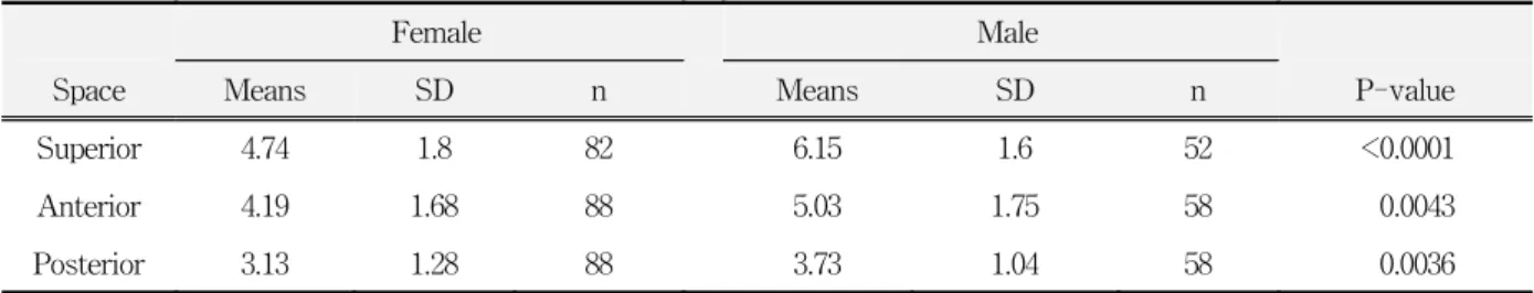
Table 2. Comparison of joint spaces between male & female patients
Female Male
Space Means SD n Means SD n P-value
Superior 4.74 1.8 82 6.15 1.6 52 <0.0001
Anterior 4.19 1.68 88 5.03 1.75 58 0.0043
Posterior 3.13 1.28 88 3.73 1.04 58 0.0036
when the posterior ratio was above the mean ratio+standard deviation.
8. Statistic analysis
The relationship between the bony changes of the factors such as condylar angulation, condylar shape and condylar position was tested by contingency table analyses. The differences of condylar angle and joint space between female and male and between both joints were compared using unpaired and paired t-tests respectively. The differences among condylar shape or condylar position were tested by Kruscal-Wallis test and multiple comparison test.
Ⅲ. RESULTS
Table 1 shows that the long axis of the condylar head is angled at approximately 18 and 21°for female and male patients respectively in horizontal plane and there is significant difference between them (p=0.0078). There, however, is no significance in the difference of both condylar angles between female and male patients.
The measurements of joint spaces can be seen in table 2. There are significant differences in the joint spaces between female and male patients and also significant differences among anterior, posterior and superior joint spaces.
There does not seem to be a relationship between
sex and condylar position as seen in table 3,
although there are much more posterior condylar
positions than others.
Table 3. Observed frequencies of condylar positions for sex
Sex Center Anterior Posterior Totals
F 20 27 41 88
M 15 18 25 58
Totals 35 45 66 146
Table 4. Observed frequencies of condylar types for sex and results of x²-tests
Condylar types P=.0068
Sex A C F R Totals
F 16 49 9 14 88
M 5 32 16* 3 56
Totals 21 81 25 17 144
A:angled C:convex F:flat R:round
* 95% significant
Table 5. Observed frequencies of bone change types for sex and result of x²-tests
Bone change type
Sex C E F N P Totals
F 5 9 4 65 5 88
M 3 3 1 48 1 56
Totals 8 12 5 113 6 144
C:concave E:erosion F:flat P:osteophyte N:no change
It can be seen there is significant relationship between sex and condylar type (p=0.0068) indicating that flat condyle seems to be prominent in male patients in table 4. There is, however, no relationship between sex and bone change type (table 5).
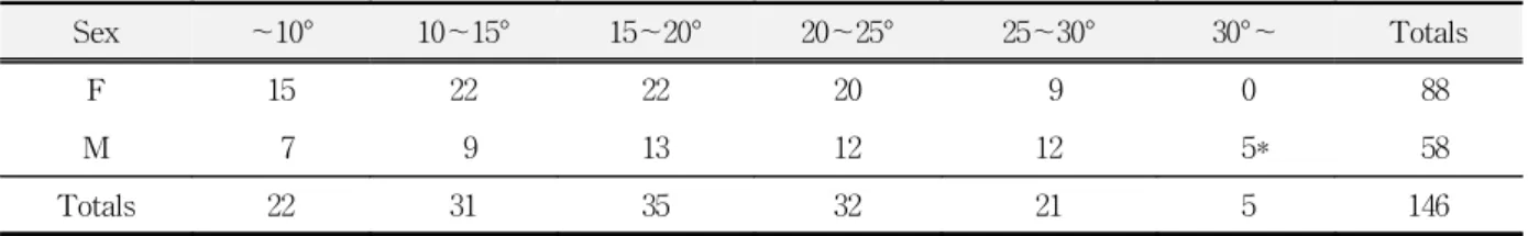
Table 6 and 9 shows that there is significant difference between condylar angles of male and female patients(p=0.0208) and above 30°of condyles are more in male than female. The bony change has a significant relationship between sex and condylar
Table 6. Means and standard deviation of condylar angles measured according to sex and results of unpaired t-test
Sex n Mean SD
F 88 17.72 6.34
M 58 20.9 7.84
P-value .0208
Table 7. Means and standard deviation of condylar angles measured according to condylar position and results of multiple com- parison tests
Position n Mean SD P-value
C 35 19.37 7
A 45 16.96 7.36 .0201
P 66 20.15 6.83
C:center position A:anterior position P:posterior position
Table 8. Means and standard deviations of condylar angles measured according to condylar type and results of multiple comparison test
Condylar Type n Means SD P-value
A 21 18.33 6.07
.0218
F 25 18.2 7.41
C 81 19.85 7.02
R 17 15.47 51.27
A:angled F:flat C:convex R:round
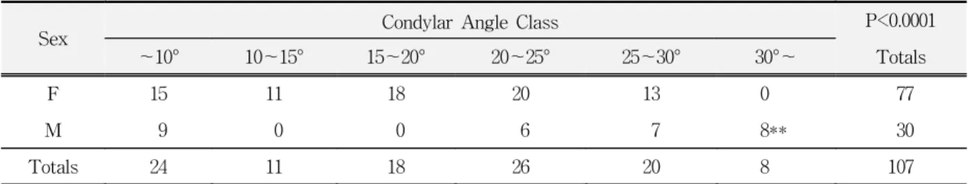
angle (p<0.0001) and increases in the condyle of male patient with large angle(above 30 degrees) of condyle(p<0.01)(table 13).
Table 7 shows that the angles of posterior
positioned condyles are larger than those of anterior
positioned condyles(p=0.0201) and table 8 shows that
the angles of convex condyles are larger than those
of round condyles(p=0.0218), although there are no
differences compared with angled or flat condyles.
Table 10. Means and standard deviations of joint spaces according to angle and results of multiple comparison test
Anterior Superior Posterior
Mean SD Mean SD Mean SD
0∼10° 4.26 2.01 5.26 2.21 3.48 1.46
10∼15° 4.49 1.57 5.06 1.67 3.32 1.12
15∼20° 3.93 2.15 5.23 1.84 3.37 1.38
20∼25° 4.68 3.2 5.46 2.09 3.36 1.17
25∼30° 5.46 3.17 5.04 1.2 3.19 0.9
30°∼ 5.07 5.2 6.95 1.3 3.9 1.5
P-value .044 - -
Table 11. Observed frequencies of bone changes for condylar angle class and condylar position and results of x²-test
Condylar position P<0.0001 Angle class Center Anterior Posterior Totals
0∼10° 9 10 5 24
10∼15° 0 7* 4 11
15∼20° 7 4 7 18
20∼25° 8 7 11 26
25∼30° 0 0 20** 20
30°∼ 4 0 4 8
Totals 28 28 51 107
* 95% significant ** 99% significant
Table 10 presents the joint spaces according to angle. Anterior joint space has differences among angle classes (p=0.044), but superior or posterior joint spaces has no difference among them.
Bony change has a significant relationship between condylar angle and position (p<0.0001) and bony changes increase in anterior positioned condyle within 10 to 15°of condylar angle and in posterior positioned condyle within 25 to 30°of condylar angle (table 11).
Table 12 shows bony changes has a significant relationship between condylar type and angle (p<0.0001). Bony changes increase in the angled type of condyle with 20-25°or above 30°of angle (p<0.01) and in the flat type of condyle with 25-30°
of angle(p<0.05).
Table 12. Observed frequencies of bone changes for condylar angle class and condylar type and results of x²-test
Angle class Condylar type P<0.0001
A C F R Totals
0∼10° 0 14 5 5 24
10∼15° 0 10 0 1 11
15∼20° 8** 8 2 0 18
20∼25° 4 13 9 0 26
25∼30° 0 7 9* 4 20
30°∼ 4** 4 0 0 8
Totals 16 56 25 10 107
* 95% significant ** 99% significant A: angled F: flat C: convex R: round Table 9. Observed frequencies of condylar angle class for sex and results of x²-test
Sex ∼10° 10∼15° 15∼20° 20∼25° 25∼30° 30°∼ Totals
F 15 22 22 20 9 0 88
M 7 9 13 12 12 5* 58
Totals 22 31 35 32 21 5 146
* 95% significant
Table 14 represents that the bony change has a significant relationship between condylar angle and age (p<0.0001). The bony change increases in above 40 years of patients with 10∼15°of condylar angle (p<0.01) and in 4th decade of patients with above 30°of condylar angle (p<0.05).
Table 14 and 15 represent that the bony change has a significant relationship between condylar angle and age (p<0.0001) and between condylar and sex (p=0.0008) respectively. The bony change increases in above 40 years of patients with 10- 1 5°of condylar angle (p<0.01) and in 4th decade of patients with above 30°of condylar angle (p<0.05).
The bony change increases in the TMJ of male patients in the range of 3 to 6°of differences between both condylar angles.
Table 14. Observed frequencies of bone changes for condylar angle class and age class and results of x²-test
Angle class Age class(year) P<0.0001 10∼19 20∼29 30∼39 40∼ Totals
0∼10° 0 19 5 0 24
10∼15° 4 1 0 6** 11
15∼20° 8 4 2 4 18
20∼25° 9 13 1 3 26
25∼30° 8 0 5 7 20
30°∼ 4 0 4* 0 8
Totals 33 37 17 20 107
* 95% significant ** 99% significant
The bony changes have a significant relationship between the difference of both condylar angles and age(p<0.0001, table 16), condylar type(p<0.0001, table 17), condylar position (p<0.0004, table 18), or bone change type (p<0.0001, table 19) respectively.
The bony change increases with age and difference of angle within 9°of difference, although it is not
Table 15. Observed frequencies of bone changes for sex and differences of condylar angles between both joints and results of x²-test
Sex Difference of Angles P<0.0008 0∼3° 3∼6° 6∼9° 9∼12° 12°∼ Totals
F 23 14 26 4 10 77
M 4 17** 9 0 0 30
Totals 27 31 35 4 10 107
** 99% significant
Table 16. Observed frequencies of bone changes for age class and difference of condylar angle between both joints and results of x²-test
Age Difference of both condylar angles P<0.0001 0∼3° 3∼6° 6∼9° 9∼12° 12°∼ Totals
10∼19 4 8 21** 0 0 33
20∼29 1 18** 8 0 10** 37
30∼39 11** 5 1 0 0 17
40∼ 11** 0 5 4** 0 20
Totals 27 31 35 4 10 107
** 99% significant Table 13. Observed frequencies of bone changes for sex and condylar angle class and results of x²-test
Sex Condylar Angle Class P<0.0001
∼10° 10∼15° 15∼20° 20∼25° 25∼30° 30°∼ Totals
F 15 11 18 20 13 0 77
M 9 0 0 6 7 8** 30
Totals 24 11 18 26 20 8 107
Table 17. Observed frequencies of bone changes for condylar type and difference of condylar angles between both joints and results of x²-test
Difference of both condylar angles
Type P<0.0001
A C F R Totals
0∼3° 0 19 7 1 27
3∼6° 4 27** 0 0 31
6∼9° 8 10 13 4 35
9∼12° 4** 0 0 0 4
12°∼ 0 0 5 5** 10
Totals 16 56 25 10 107
* 95% significant ** 99% significant A: angled F: flat C: convex R: round
Table 18. Observed frequencies of bone changes for condylar position and condylar angle difference class and results of x²-test
Difference of both condylar angles
Condylar position P=0.0004
C A P Totals
0∼3° 7 8 12 27
3∼6° 12 10 9 31
6∼9° 5 5 25** 35
9∼12° 4** 0 0 4
12°∼ 0 5 5 10
Totals 28 28 51 107
C: center A: anterior P: posterior ** 99% significant
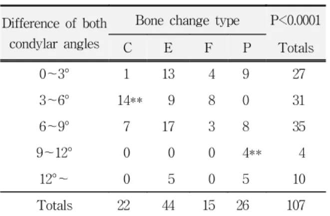
consistent with the condyles having above 9°of difference (table 16). Table 17 shows the bony change increases in order of convex, angled and round type of condyle with the difference of both condylar angles. Table 18 shows the bony change increases in the posterior positioned condyle with 6 to 9°of difference between both condylar angles.
Table 19. Observed frequencies of bone changes for condylar angle difference class and bone change type and results of x²-test
Difference of both condylar angles
Bone change type P<0.0001
C E F P Totals
0∼3° 1 13 4 9 27
3∼6° 14** 9 8 0 31
6∼9° 7 17 3 8 35
9∼12° 0 0 0 4** 4
12°∼ 0 5 0 5 10
Totals 22 44 15 26 107
C:concave E:erosion F:flat P:osteophyte ** 99% significant
Ⅳ. DISCUSSION
Tomography is body section radiogrphy.
Tomography has become the standard for comprehensive evaluation of the bony components of the TMJ, because it allows visualization of the temporal and condylar component. In addition, it allows the best evaluation of condyle position.
13)Interpretation of a technically correct tomogram is straightforward because its projection can be viewed in standard anatomical planes. Despite its many advantages, full capability tomographic equipment is expensive to use and is not used extensively in dental clinic.
Therefore, corrected tomographic equipment (SCANORA multifunction x-ray unit), which is less expensive spiral tomographic system, was used in this study.
Both tomograhic and plain projections have
distortion effects if the angles of the x-ray beam
are not related to the horizontal axis of the condyle
and mandibular fossa. Hence a cephalostat is
required such as head support and chin rest used in
this study. The following radiological principles
should be kept in mind. Projections should be taken
in two or more planes. Axial correction should be
made of the condylar axis by the use of preliminary
submentovertex view followed by orientation with a cephalostat. Solberg
14)suggested that sagittal views should be taken in the medial, central and lateral parts of the joint to represent maxillomandibular positions of clinical relevance. In this study 4 sagittal views were taken with a 4 mm of focal thickness from medial pole of the joint to investigate extensive bone change of the condylar head. The frontal view is most valuable in demonstrating condylar remodelling and other changes.
15)Often the changes seen in frontal view are not well identified in sagittal projections. It is reasonable, therefore, to propose that at least the following tomographic views be recommended to examine the TMJ: closed sagittal views (medial, central or 2 central, lateral cuts), maximally open sagittal view (central cut only), frontal plane view with the jaw open, and panoramic survey of the jaw region.
14)This study was carried out to determine whether there is a relationship between the bony change of condylar head and one of predisposing factors such as condylar position, condylar type, condylar angulation and bony change type. It is believed that the use of MRI for this purpose is justified. Although MRI has a good resolving power for soft tissues, it was thought conventional tomography might be enough to assess the bony changes.
The bony contour of the upper component of the temporomandibular joint is thought to be an important factor in the etiology of internal derangement.
16-23)Kurita et al.
26)found that a flattened articular eminence was more common in disc displacement without reduction than in disc displacement with reduction. It has been suggested that flattening occurs with progressive internal derangement.
21-25)It has also been reported that the greatest difference in the steepness of the eminence is seen between the joints with bony changes and those without.
21)It is well known that osteo- arthritic changes are more frequent in disc displacement without reduction than disc displacement with reduction.
Those results suggested that flattening of the
articular eminence may occur in disc displacement without reduction as a result of remodelling or degenerative changes secondary to internal derangement. This hypothesis was supported by Kurita's finding that disc displacement without reduction was more frequent in older individuals where degenerative changes are more likely to develop.
26)In this study on the bony change of the condylar head, the results also show that the bony changes in the condylar head are more frequent in disc displacement without reduction (33%) than in disc displacement with reduction (10%). It can be, therefore, thought that anatomy of the condylar head as well as the articular eminence may predispose to disc displacement and eventually bring to degenerative change.
Joint anatomy varies remarkably between individuals and often between sides in the same individual, complicating interpretation and achievement of consistent, quality imaging. Yale
12)identified four basic condylar morphologies, which were used to classify the condyles in this study. He classified basic morphology of 3008 condyles in dry mandibles finding 3% round, 11.6% angled, 58.3%
convex, 25.2% flat and 1.2% other. The result of this study shows 11.8% round, 14.6% angled, 56.3%
convex, and 17.4% flat. The differences in flat and round condyles between Yale's and this studies are thought to be due to the variability of naturally occurring condylar shapes described previously.
When evaluating the clinical presentation of a TMJ disorders, one should be aware of possible disturbing influences of the contralateral joint. In the past study the overall difference in predictability of unilateral versus bilateral cases was small.
27)In the present study, only unilateral cases were selected to investigate the difference between symptom and symptom-free sides of TMJ.
There were, however, few differences between them and all joints were examined simultaneously in this study to investigate the effects on the bone change by various factors.
Stengenga et al. reported that three major
categories of symptom and signs might be
distinguished in patients with osteoarthrosis and internal derangement, i.e., changes in joint mechanics, pain and tenderness, and radiographi- cally detectable degenerative changes.
However, pain-related symptoms or radiographic information did not appear to be useful discriminating symptom categories, as opposed to variables related to joint mechanics. They suggested that diagnostic criteria should be primarily based on the latter variables.
In their study, the distinction between reducing and permanent disk displacement categories was used as the standard. However, the results from the analyses performed suggest that this distinction still leaves considerable variability within these groups. Besides the characteristic signs of the classical internal derangement groups, less common symptoms and signs were seen within these groups. They reported, on the other hand, that radiographically visible degenerative changes were present in about half of the number of patients with reducing disk displacement.
This observation indicated that bony changes may precede permanent disk displacement, which supports previous findings.
28-30)In the present study, the results also support those findings although radiographically visible bony changes were present in 10% of the number of patients with reducing disk displacement and 33% of the number of patients with permanent disk displacement, which were lower than the results of the previous study.
31)In Liedberg's study,
32)the condylar position was assessed as posterior, central, or anterior, whereas Blair et al.
33)and Kopp and Rockler
34)had two more scores, namely inferior and superior. In this study the inferior and superior positions also were used to combine with anterior, posterior or center positions, but many positions are likely to decrease the significant difference between the joint spaces in various condylar positions. Therefore, this study assessed the condylar position as posterior, central, or anterior.
The standard sagittal technique involves aligning
the tomographic layer perpendicular to the average horizontal and vertical condylar angulation measurements. Fifteen or 20 degree is the most often used horizontal angulation, and the usual vertical angulation is zero degree.
35)In this study, the results show that the average condylar angles of male and female subjects are 20.9°and 17.7°
and are in the range of standard criteria, although the angle of male is significantly larger than that of female. Angulation of the condylar long axis in both horizontal and vertical planes is extremely variable.
12)If the central X-ray beam is not aligned paralleled with the long axis of the condyle in both the horizontal and vertical planes, an oblique projection occurs in which the medial condylar pole may be projected outside of the true condylar profile image. The broad range of variation in horizontal and vertical angulation often produces projection errors and artifacts, particularly if they are not considered and accommodated in the projection technique. Impressions obtained from imaging must be tempered with an appreciation for the multiple potential sources of distortion.
This study showed differences in the angle of the mandibular condyle in joints with different bony changes. A clear pattern was found with larger condylar angle in joints with more advanced pathologic bony changes related to temporoman- dibular joint disorders. These observations support speculations in earlier studies.
36-42)In this study, the differences of angles between both condyles were investigated and the bony changes according to the difference of angles were compared.
The results suggest that larger difference of angles between both condyles or higher angle of the condyle increases the bony change in the condyle as well as the condyle with larger angle.
This means that the difference of angles between both condyles should be also considered as a important factor as much as condylar angle.
In the previous study, Westesson et al.
36)reported
that they didn't suggested a causal relationship
exist between internal derangement and a high
condylar angle clearly, although a statistically
significant association was documented. They suggested two possible explanations:
(1) joints with a higher condylar angle may have a greater tendency for disk displacement and degenerative joint disease, and (2) remodeling changes of the bone that occur in association with internal derangement and degenerative joint disease may result in an increased condyle angel.
Mechanically, the first theory that joints with a high condylar angle have a greater tendency for internal derangement to develop makes sense.
In a joint with a high condylar angle, there seems to be a possibility for more stretching in the lateral attachment of the disk to the condyle during anterior translation of the condyle than there is in a joint with a low condylar angle. Then, the condyle will move straight forward on opening and there will be some stretching in the lateral attachment of the condylar angle, the more pronounced this stretching will be. The lateral attachment does not have the same elasticity as the posterior disk attachment, and if it is stretched beyond its capacity, it could result in permanent elongation that will subsequently lead to displacement of the disk and internal derangement.
The first theory is supported by the results of this study The second theory, however, condyle angle might be increased in association with degenerative joint disease is not supported by this results.
Although it is not included in the results of this study, the statistic result showed that bony changes increase, but condylar angle decrease in the joint with the disk displacement without reduction compared with disk displacement with reduction against the previous study.
36)Longitudinal imaging studies will be necessary to answer the questions raised by this investigation.
The results of the past study support that clinical and conventional radiographic information is sufficient to predict the stage of osteoarthrosis and internal derangement regarding disk displacement in many cases. In personal opinion, routine use of invasive or expensive procedures like arthrography and MRI si not realistic. These techniques should
be preserved for difficult or confusing diagnostic cases and for research purposes.
14)In this study, however, there are significant correlation in bone changes among various factors seen in tomographs such as condylar angulation, condylar type, condylar position and bone change type. These results, therefore, suggested that some tomographs should be included to predict the predisposing factors relative to anatomy of TMJ.
V. CONCLUSIONS
In conclusion, these results support the hypothesis that the condylar shape, condylar angulation and condylar position are related to the development of bony change in the joint. Bony changes of condylar head are more likely to be found in the joints of male patients with posterior positioned and above 250 of high condylar angulation and especially in the flat or angle type of condyle when the age of patients is in the 2nd or 4th decade respectively, and when the difference of both condylar angles is in the range of 9 to 12 degrees.
REFERENCES
1. Okeson JP (ed). Orofacial Pain : Guidelines for Assessment, Diagnosis, and Management. Chicago:
Quintessence, 1996.
2. Chung, S.C., Lee, S.W., Kim, Y.K.: Clinical symptoms and patterns of mandibular movement in the patients with TMJ dysfunction. J Korean Academy of Oral Medicine, 10 : 5-16, 1985.
3. Choi, J.K.: A clinical study on the MPDS patients. J Korean Academy of Oral Medicine, 7 : 47-58, 1982.
4. Kopp S, Rockler G: Realtion between clinical and radiographic findings in patients with mandibular pain or dysfunction. Acta Radiol Diagn, 20 : 465-477, 1979.
5. Park, B.I., Han, K.S.: The relation between clinical sign and radiolographic findings in temporomandi- bular disorders. J Korean Academy of Oral Medicine, 14 : 57-66, 1989.
6. Kopp S, Rockler B: Relationship between radiographic signs in the temporomandibular joint and hand joints.
Acta Odontol Scand, 37 : 169-175, 1979.
7. Ryu, S.S., Kee, W.C., Choi, J.K.: A relationship between the joint effusion and the presence of pain and disc displacemnet in the temporomandibular joint.
Korean J of Oral Medicine, 25 : 63-71, 2000.
8. Kwon, J.H., Kee, W.C., Choi, J.K.: Configuration of temporomandibular joint articular disc in magnetic resonance images and its relationship to treatment response of anterior disc displacement without reduction. Korean J of Oral Medicine, 25 : 73-85, 2000.
9. Omnell K-A, Petersson A: Raidograhpy of the temporomandibular joint utilizing oblique lateral transcranial projections. Comparison of inforamtion obtained with standardized technique and individu- alized technique. Odontol Revy, 27 : 77-92, 1976.
10. Christiansen EL, Thompson JR, Kopp SFO, Hasso AN, Hinshaw DB Jr.: Radiographic signs of tempo- romandibular joint disease: An investigation utilizing x-ray computed tomography. Dentomaxillofac Radiol, 14 : 83-92, 1985.
11. Tanimoto K, Petersson A, Rohlin M, Hansson LG, Johansen CC.: Comparison of computed with conventional tomography in the evaluation of tempo- romandibular joint disease: a study of autopsy specimens. Dentomaxillofac Radiol, 19 : 21-27, 1990.
12. Yale SH, Allison BD, Hauptifuehrer JD: An epidemilolgic assessment of mandibular condyle morphlolgy. Oral Surg Oral Med Oral Pathol, 21 : 169-177, 1966.
13. Blaschke DD: Temporomandibular joint. In Goaz P, White S(ed). Oral radiology: principle and interpretation. pp508-601. St Louis, 1982, CV Mosby.
14. William KS: Temporomandibular disorders: functional and radiological considerations. Br Dent J, 160 : 195-200, 1986.
15. Mahan PA, Gibbs CJ, Mauderli A: Superior and inferior lateral pterygoid activity(absract). J Dent Res, 61 : 272, 1982.
16. Bell WE: Clinical management of temporomandibular disorders. Chicago: Year Book Medical Publishers, 1982.
17. Atkinson WB, Bates RE: The effects of the angle of the articular eminence on anterior disk displacement.
J Prosthet Dent, 49 : 554-555, 1983.
18. Hall MB, Gibbs CC, Sclar AG: Association between the prominence of the articular eminence and displaced TMJ disks. J Craniomandib Pract, 3 : 237-239, 1985.
19. Sato S, Kawamura H, Motegi K, Takahashi K:
Morphology of the mandibular fossa and the articular
eminence in temporomandibular joints with anterior disk displacement. Int J Oral Maxillofac Surg, 25 : 236-238, 1996.
20. Pullinger AG, Bibb CA, Ding X, Baldioceda F:
Contour mapping of the TMJ temporal component and the relationship to articular soft tissue thickness and disk displacement. Oral Surg Oral Med Oral Pathol, 76 : 636-646, 1993.
21. Ren YF, Isberg A, Westesson PL: Steepness of the articular eminence in the temporomandibular joint.
Tomographic comparison between asymptomatic volunteers with normal disk position and patients with disk displacement. Oral Surg Oral Med Oral Pathol Oral Radiol Endod, 80 : 258-266, 1995.
22. Panmekiate S, Petersson A, Akerman S: Angulation and prominence of the posterior slope of the eminence of the temporomandibular joint in relation to disc position. Dentomaxillofac Radiol, 20 : 205-208, 1991.
23. Galante G, Paesani D, Tallents RH, Hatala MA, Katzberg RW, Murphy W: Angle of the articular eminence in patients with temporomandibular joint dysfunction and asymptomatic volunteers. Oral Surg Oral Med Oral Pathol Oral Radiol Endod, 80 : 242-249, 1995.
24. Moffett BC, Johnson LC, McCabe JB, Askew HC:
Articular remodeling in the adult temporomandibular joint. Am J Anat, 115 : 119-142, 1964.
25. Toyama M, Kurita K, Westesson PL, Sakuma S, Ariji E, Rivera R: Decreased disk-eminence ratio is associated with advanced stages of temporoman- dibular joint internal derangement. Dentomaxillofac Radiol, 28 : 301-304, 1999.
26. Kurita H, Ohtsuka A, Kobayashi H, Kurashina K: Is the morphology of the articular eminence of the temporomandibular joint a predisposing factor for disc displacement? Dentomaxillofac Radiol, 29 : 159-162, 2000.
27. Roberts C, Katzberg TW, Tallents RH, et al.: The clinical predictability of internal derangements the temporomandibular joint. Oral Surg Oral Med Oral Pathol, 71 : 412-414, 1991.
28. Boering G: Temporomandibular Joint Arthrosis. An Analysis of 400 Cases. Leiden: Stafleu, 1966.
29. Schiffman E, Anderson G, Fricton J, et al.: Diagnostic criteria for intraarticular TM disorders. Community Dent Oral Epidemiol, 17 : 252-257, 1989.
30. Pullinger A, Seligma D: TMJ osteoarthritis: A differentiation of diagnostic subgroups by symptom history and demographics. J Craniomandib Pract, 4 :
251-256, 1987.
31. Boudewijn S, Lambert GM, Bart van der Kuijl, Geert B. Classification of temporomandibular joint osteoar- throsis and internal derangement. Part I: Diagnostic significance of clinical and radiographic symptoms and signs. J Craniomandib Pract, 10 : 96-106, 1992.
32. Lieberg J, Rohlin M, Westesson P-L. Observer performance inassessment of condylar position in temporomandibular joint radiograms. Acta Odontol Scand, 43 : 53-58, 1985.
33. Blair GS, Chalmers IM, Leggat TG, Watson Buchanan W: Circular tomography of the temporomandibular joint. Oral Surg, 35 : 416-427, 1973.
34. Kopp S, Rockler B: Variation in interpretation of radiographs of temporomandibular and hand joints.
Dentomaxillofac Radiol, 7 : 95-102, 1978.
35. Zarb GA, Carlsson GE, Sessle BJ, Mohl ND:
Temporomandibular joint and masticatory muscle disorders. Copenhagen: Munksgaard. 445-447, 1995.
36. Per-Lennart Westesson, Joseph AB, Ross HT, Mark PH. Increased horizontal angle of the mandibular condyle in abnormal temporomandibular joints : A magnetic resonance imaging study. Oral Surg Oral Med Oral Pathol, 72 : 359-363, 1991.
37. Huls A, Schulte W, Voigt K: Neue Aspekte der Myoarhropathien durch die Computertomographie.
Dtsch Zahnarztl Z, 36 : 776-778, 1981.
38. Huls A, Schulte W, Voigt K, Ehrlich-Treuenstatt V:
Computed tomography of the temporomandibular joint: new diagnostic possibilities and initial clinical results. Electromedica, 51 : 14-19, 1983.
39. Huls A, Walter E, Schulte W: Konventionelle Rontgendiagnostik und Computertomographie der Kiefergelenke bei Myoarthropathien. Radiologie, 24 : 310-318, 1984.
40. Huls A, Walter E, Schulte W, Freesmayer WB:
Computertomographische Stadieneinteilung des dysfunktionellen Gelenkkopfumbaus. Dtsch Zahnarztl Z, 40 : 37-51, 1985.
41. Huls A, Walter E, Schulte W: Zur Darstellung des Discus articularis im Computertomogramm. Dtsch Zahnartzl Z, 40 : 326-333, 1985.
42. Westesson P-L, Liedberg J: Horizontal condylar angle in relation to internal derangement of the temporo- mandibular joint. Oral Surg Oral Med Oral Pathol, 64 : 391-394, 1987.
Corresponding Author : Ki-Suk Kim, Professor,
Department of Oral Medicine, School of Dentistry, Dankook University, San 7-1, Shinbudong, Cheonan, Choongnam 330-716, Korea