CONTENTS
Ⅰ. INTRODUCTION
Ⅱ. MATERIALS AND METHODS
Ⅲ. RESULTS
Ⅳ. DISCUSSION
Ⅴ. CONCLUSION REFERENCES KOREAN ABSTRACT EXPLANATION OF FIGURES
Ⅰ. INTRODUCTION
It is believed that stress is related with a variety of disease.
1,2)But the belief was based on somewhat metaphysical philosophy and was lacking firm scientifical evidence. In those recent years, numerous studies have focused on the mechanism how stress is related with the physiological and pathological changes of various organ. And now we are gaining ample evidence of the relation between the stress and the body at concrete science level
3).
Pert suggested that the three systems - the neural, immune and endocrine system - must be seen as forming a single 'psychosomatic network'
4). It can be said that we have found the language with which the mind and the body talk to each other. And now we hope to find the language between the stress and the disease. Which pathway is involved in the onset and
the progress of stress-induced disease?
There are many stress-related symptoms and diseases in the orofacial tissue
5). Recently, quite a few studies have suggested that stress is strongly associated with orofacial diseases. We have been particulary interested in the clinical observations that it's not uncommon to see that the patients under stressfull conditions show signs and symptoms of xerostomia and decreased function of salivary glands. In the former studies, Hong and Park prooved that stress could actually cause the cell death of submandibular glands of rats
6). But, nevertheless, the mechanism of it is still unclear. If we can obtain the insight into the pathway and the mechanism, It can hold great promise for the prevention and the treatment of xerostomia and decreased salivary function related with stress.
In the present study, we examined the expression of clusterin in the salivary glands as stress marker under adrenalectomized condition by immunohis- tochemistry, in order to inquire the relationship between glucocorticoid and salivary gland.
Ⅱ. MATERIALS AND METHODS Experimental animals and tissue preparation
40 Sprague-Dawley, 7-week-old rats(165-209g/
bw) were purchased from Dae-Han Experimental
An Effect of Endocrinological Changes related to the Stress on the Submandibular Gland in Rats
Bong-Youl Chang, D.M.D., M.S.D., Ph.D., Sung-Hoon Kim, D.M.D., M.S.D., Ph.D., Yang-Hyun Chun, D.M.D., M.S.D., Ph.D., Jung-Pyo Hong, D.M.D., M.S.D., Ph.D.
Department of Oral Diagnosis & Oral Medicine, College of Dentistry, Kyung Hee University
Animal Research Center, Seoul, Korea. They were maintained at 20-23℃ and fed ad libitum on a normal laboratory diet. The rats were divided into 2 groups:
ADX group and DEX group. The rats of ADX group were adrenalectomized and not given any other experimental conditions. and the rats of DEX group were adrenalectomized and injected dexamethasone (1.5*10
-4mg/g I.V./day) through all the periods of experiment. All the experimental animals were sacrificed at the day of 0, 1, 3, 5 and 7 after the experiment and the submandibular glands were excised immediately. The tissues were fixed and processed for immunohistochemistry. After fixation in Bouin solution overnight, the tissues were embedded in paraffin resin for immunohistoche- mistry. Serial paraffin sections (4∼6㎛) were cut, placed on poly-L-lysine coated slides, and stored at -70℃ until use.
Preparation of clusterin antibody
Prior to raising antibody against clusterin, a synthetic peptide corresponding to the sequence of 144-158 amino acids (NH
2-GDRIDSLMENDRQQS- COOH;1865.9 atomic mass units) from the porcine clusterin α-subunit was prepared by Fmoc peptide synthesis procedure and purified by repeated HPLC(High Pressure Liquid Chromatography). The α-peptide (2.2mg) was conjugated to 2mg of cathionized BSA using SuperCarrier EDC system(Pierce Rockford, IL, USA). A New Zealand white rabbit was injected at multiple sites subcutaneously with the conjugated peptide-carrier in complete Freund's adjuvant. Starting one week after the first injection, the rabbit was boosted weekly for 2 weeks with the same conjugate in incomplete Freund's adjuvant. Ten days after the last third injection, the rabbit was bled and antiserum was collected.
Immunohistochemistry
Immunohistochemical analysis was performed on the paraffin section by ABC (avidin-biotin-
peroxidase complex) method as described previously using the anti-clusterin α-peptide antiserum
7). Briefly, the sections were deparaffinized in xylene, hydrated, washed in phosphate buffered saline (PBS), and incubated twice in methanol containing 0.5% H
2O
2for 5 min each at room temperature. After rinsing with PBS, slides were incubated in PBS containing 0.1% Triton X-100 for 10 min, and then immersed in normal goat serum diluted with rabbit IgG anti-clusterin peptide for 24 hr at 4℃. After washing with PBS, biotinylated goat anti-rabbit immunoglobulin was applied to the slides for 1 hr at room temperature. The slides were then washed in PBS and incubated with avidin-biotin-peroxidase complex (ABC kit ; Vector Lab., Burlingame, CA, USA) for 1 hr. Thereafter, immunohistochemical reactions were detected by color development using diaminobenzidine solution (100mM Tris, pH 7.4, 0.01% H
2O
2, 0.05% diaminobenzidine hydrochloride).
Ⅲ. RESULTS
To localize the expression of clusterin protein in the submandibular glands, the tissues of each group were fixed and processed for immunohistochemistry using the specific antibody against a clusterin α -subunit peptide.
Through all the perioids of experiment, There was no significant histological change in both group.
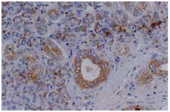
In the ADX group(adrenalectomized and not given any other experimental conditions), clusterin was observed in both ductal cells and acinic cells throughout the entire period of experiment with dark brown color.
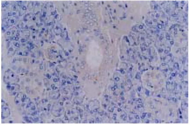
But, in the DEX group(adrenalectomized and injected dexamethasone through all the periods of experiment), there were somewhat localized expressions of clusterin in few acinic cells, and, in general, there was no significant expression in both ductal cells and acinic cells.
Ⅳ. DISCUSSION
The new paradigm of mind and body is emerging.
The mind and the body are no longer viewed as discrete entities in the mechanistic view, but as two closely related manifestations of a single process of life. So, it is of no surprise to assume that the illness of the mind can cause the illness of the body. It has long been believed that stress is related with a variety of disease
1,2). But the belief was based on somewhat metaphysical philosophy and was lacking firm scientifical evidence. In those recent years, numerous studies have focused on the mechanism how stress is related with the physiological and pathological changes of various organ. And now we are gaining ample evidence of the relation between the stress and the body at concrete science level
6). Pert et al. introduced the concept of 'psychosomatic network'
4). They identified a group of peptides, as the molecular messengers that facilitate the conversation between the nervous system and the immune system. They have found that these messengers interconnect three distinct systems - the nervous, immune, and endocrine system - into a single network. In the traditional view these three systems are separate and serve different functions, but, the recent peptide research has shown that these conceptual separations are merely historical artifacts that can no longer be maintained. Pert suggested that the three systems must be seen as forming a single 'psychosomatic network'. It can be said that we have found the language with which the mind and body talk to each other. And now we hope to find the language between the stress and the disease. Which pathway is involved in the onset and the progress of stress-induced disease?
Stress is the nonspecific response of the body to any demand
8)and can also be defined as an interactional process between the individual and the environment
3).
It is clear that stress can lead down the physiological function even at the cellular and molecular level, so stress can be the potential cause of disease. The evidence that stress may cause disease is established for a large number of factors and diseases
3).
Especially, there are many stress-related symptoms and diseases in the orofacial tissues
5), and that is the reflection of the fact that the orofacial tissue is emotionally charged or highly reactive to psychologic influences, representing directly or symbolically our major instincts and desiresfood, sex, hostility and so on.
Recently, quite a few studies have suggested that stress is strongly associated with orofacial diseases.
Chun and Hong
9)indicated that stress causes various forms of diseases in the region including orofacial psychosomatic diseases in which emotional stress appears to play a major role (lichen planus, aphthous stomatitis), orofacial diseases in which psychologic factors appear to play a role (erythema multiforme, benign mucous membrane pemphigoid, geographic tongue), orofacial infections where emotional stress is a significant predisposing factor (recurrent herpes labialis, acute necrotizing ulcerative gingivitis), orofacial lesions induced by neurotic habits inflicting trauma (biting of oral tissues, physical trauma with foreign objects, leukoplakia due to smoking, bruxism and clenching), neurotic orofacial symptoms (xerostomia, halitosis, burning mouth syndrome, altered or loss of taste perception, pain or discomfort with no tissue change), and orofacial pain induced by emotional stress (temporomandibular disorders, muscle tension headache, atypical odontalgia).
As doctors of dentistry, We have been particulary interested in the clinical observations that it's not uncommon to see that the patients under stressfull conditions show signs and symptoms of xerostomia and decreased function of salivary glands.
Xerostomia is the subjective feeling of dry mouth
which may or may not be caused by salivary gland
hypofuction. Saliva is an important body fluid which
plays several vital roles. In addition to moistening
and protecting the oral tissue, it acts as an aqueous
solvent necessary for taste and aids oral function as
well as the digestion of food. Also, saliva has an
antibacterial action which inhibits or prevents the
onset of dental caries and inflammation
10).
Xerostomia is rarely seen in isolation, but with time,
the changes of quantity and quality of saliva can be
devastating to oral health and may severely affect the general well-being and lifestyle of the patient. In deed, the oral tissues become susceptible to infection, and the ability for oral function may be disturbed. In addition, a number of pathologic conditions can develope, such as halitosis, burning mouth syndrome, gustatory changes and other oral inflammatory diseases
12). In the former studies, Hong and Park prooved that stress could actually cause the cell death of submandibular glands of rats
6). But, nevertheless, the mechanism of it is still unclear. If we can obtain the insight into the pathway and the mechanism, it can hold great promise for the prevention and the treatment of xerostomia and decreased salivary function related with stress.
The salivary glands, the major salivary glands including the parotid, the submandibular and the sublingual glands, and the several minor glands, produce and secrete saliva. Saliva is formed by units of secretory cells of which there are 3 types, serous, mucous and seromucous, and is modified by cells of the ductal system as it passes toward the oral cavity
14). The composition of the final saliva, as well as the primary saliva, is quite possibly unique for each salivary gland and is determined by the nature of the secretory endpiece cells and ductal elements of that gland
10).
Therefore, when salivary glands are affected by a variety of stimuli, the cells of the salivary glands may adapt to them by eliciting various cellular responses, such as structural and biochemical transformations. But if the stimulus is lying outside an acceptable range of normality, it may cause such changes at the clinical and subclinical level as alteration in the production, flow rate, the response to stimuli, the components and the immune function of saliva.
Johnson et al. showed that glucocorticoid can have an effect on the composition of saliva. Parotid saliva collected from adrenalectomized rats exhibited a two-fold greater proportion of proline-rich proteins and reductions in other major secretory proteins.
Treatment of adrenalectomized rats with dexamethasone largely prevented the changes in
salivary protein composition. This shows that glucocorticoid secreted in response to the stress can make an effect on the composition of saliva. So, stress can actually cause the changes of salivary protein composition, and the dysfunctional changes of saliva
15).
Stress triggers a variety of changes in the body including GAS(general adaptation syndrome) through multiple pathways
16). One of the most important pathways involved in stress is HPA axis(Hypothalamo-pituitary adrenal axis)
17). When stress activates the HPA axis, glucocorticoids are massively secreted by adrenal cortex.
In the former study of Park and Hong, restraint stress induced apoptosis of submandibular gland of rats
6). But the question, 'Which pathway is involved in the Stress-induced apoptosis of submandibular gland?" was not answered. To solve this problem, we performed adrenalectomy to block the HPA axis pathway. and then we observed and evaluate the effect of glucocorticoid on the change of the submandibular glands of rats.
The rats of DEX group were injected dexame- thasone everyday to compensate the deficiency of glucocorticoid. Dexamethasone is glucocorticoid agonist about 30 times as potent as cortisol, while showing almost zero mineralocorticoid activity, which is a good character for selective re-activation of glucocorticoid activity.
The rats of ADX group were given no other condition after adrenalectomy to observe the effect of glucocorticoid deficiency on submandibular gland.
Clusterin is an intriguing, ubiquitous, and highly conserved glycoprotein. It is secreted as a heterodimer of 70∼80kDa glycoprotein, comprising α and β subunits
18,19). It has potential amphipathic helical domains that allow this protein to bind to hydrophilic molecules, as well as potential heparin-binding domains responsible for interaction with the cell membranes and extracellular matrices
20).
Although the function of clusterin remains unclear,
there has been a speculation that it may play a role
in cell aggregation, lipid transport, sperm maturation,
regulation of the complement cascade, membrane recycling, and apoptosis
21).
A notable property of clusterin is its massive induction during apoptosis
24,25,26,27)in castration- mediated prostatic involution
28), in several models of kidney injury
29,30), and in neuronal development
31). On the basis of its elevated expression in apoptotic tissues, it was originally proposed that the protein might be causally involved in apoptosis
28). In contrast to earlier notion, however, it has been recently suggested that clusterin expression is not enhanced but rather is down-regulated in the cells undergoing apoptosis
32), and that its expression in the apoptotic tissue is restricted to the vital neighboring cells
32,33). Sensibar et al.
34)also provided evidence that clusterin plays a role in the protection of TNF-induced cell death. Therefore, it is likely that clusterin overexpression is a reactive antiapoptotic and cytoprotective response to environmental changes rather than a causative factor in cell death. The cytoprotective mechanism of clusterin has been proposed based on the findings that it inhibits complement-mediated lysis, binds to surface active toxic hydrophobic compounds and neutralize at or near the cell membranes by formation of soluble complex, and preserves the integrity of the barrier as a potent cell aggregation and adhesion molecule
35,36,37). Since clusterin is associated with various forms of diseases in response to different stress, it should be considered as a nonspecific sensitive cellular response to various tissue insults and a marker of pathological injury. So, Hong et al.
proposed that clusterin expressed as a part of physiologic stress response can be used as a 'Stress Marker'
6).
In the present study, we examined the expression of clusterin in the salivary glands as stress marker under adrenalectomized condition by immunohisto- chemistry, in order to inquire the relationship between glucocorticoid and salivary gland.
Adrenalectomy and subsequent deficiency of glucocorticoids lead to opposite result depending on the target organs. Thymus and dentate gyrus of hippocampus are those two examples which are
firmly established as standard model of apoptotic and anti-apoptotic action of glucocorticoids. the exact mechanism why the same endocrinological factor elicite opposite result has to be elucidated yet.
It is well known that glucocorticoids cause the death of thymocytes in vivo and in vitro. and the process has been clearly shown to be apoptosis, as characterized by cell shrinkage, membrane alterations, nuclear collapse, and chromatin fragmentation into oligonucleosomes
38).
Aldosterone, another type of steroid is known to have thymolytic action, but the thymolytic action of aldosterone was prooved to be mediated by type II glucocorticoid receptor, not by its specific receptor
39). The thymolytic action of dexamethasone could be inhibited by cycloheximide, suggesting that this cell death program requires a fully operating protein synthesis machinery and perhaps the induction of new proteins
40).
Restraint stress causes thymic involution. And the endogenous glucocorticoids was shown to be involved in restraint-induced thymic involution and cell apoptosis. Exposure of mice to restraint stress led to involution of the thymus and apoptosis of thymocytes. Tarcic et al. investigated the role of endogenous glucocorticoids in restraint stress- induced changes in the thymus by three experimental approaches: surgical adrenalectomy, chemical adrenalectomy, and blocking of GC receptors by a specific type II receptor antagonist. In all of these three conditions, the effects of restraint on the thymus were wholly or partially abrogated. This indicate that glucocorticoids are involved in the restraint-induced effects on the thymus
41).
Intestinal intraepithelial lymphocytes were also
susceptible to the exogenous and endogenous
glucocorticoids. The mice treated with dexame-
thasone, and the mice subjected to the water
immersion stress both showed apoptosis of intestinal
intraepithelial lymphocytes, but different sensitivity
to steroid-induced apoptosis may exist among the
subsets in relation to their Bcl-2 expression
42).
Fraker et al. showed that thymic atrophy and
lymphopenia associated with malnutrition might
involve the glucocorticoids. Those mice maintained on a zinc deficient diet showed thymic atrophy, but adrenalectomy prevented those atrophic effect
43). Insuline administration to rat caused hypoglycemia and subsequent apoptosis of thymocytes. this effect was inhibited by glucose or RU486, a glucocorticoid receptor antagonist. This suggest that insuline- induced hypoglycemia causes thymocyte apoptosis by promoting glucocorticoid secretion from the adrenal gland
44).
Pharmacological doses of corticosterone reduce bone formation by increasing osteoblast apoptosis;
they reduce growth cartilage width, probably by inhibiting chondrocyte proliferation and increasing the apoptosis of terminal hypertrophic chon- drocytes
45).
Sloviter et al. showed that adrenalectomy of adult male rats resulted in a nearly complete and selective loss of hippocampal granule cells. Corticosterone replacement prevented both the adrenalectomy- induced granule cell loss and the attenuated physiological response. Thus, the adrenal glands play a role in maintaining the structural integrity of the normal adult brain
46).
Gerth et. al prooved that adrenalectomy-induced loss of neuronal death in hippocampus is mediated by direct glucocorticoid, not by corticotropin releasing hormone increased by adrenalectomy- induced loss of negative feedback
47).
Gould et al. showed that type 2 adrenal steroid receptor activation protected granule cells from degeneration in rat pups
48).
Hu et al. observed that the neuronal apoptosis of hippocampus began immediately after adrenale- ctomy, and once the apoptois began, corticosterone could not reverse the process. And healthy cells were protected after corticosterone replacement
49). In another study of Sloviter, it was also shown that dexamethasone does not restore normal hippocampal structure once granule cell loss has occurred
50). Besides hippocampus, glucocorticoids were shown to protect cells of other organs from apoptosis.
Withdrawal of glucocorticoid from defined media triggers apoptosis in human mammary epithelial
cells, despite the presence of epidermal growth factor and insulin. The apoptosis was also independent of CD 95/ FAs receptor activation, or phosphatidyl- inositol 3-kinase activity, thus establishing the existence of a novel epithelial cell survival pathway mediated by glucocorticoids
51).
It was also shown that Dexamethasone inhibited involution and programmed cell death in all the mammary glands
52).
Kimura et al. showed that endogenous glucocor- ticoids are an important factor for pancreatic acinar cell survival and endogenous glucocorticoids may protect acinar cells by decreasing their sensitivity to the induction of cell death during acute pancreatitis
53).
As a conclusion, it is unwise to assume that all results in any one system are generally applicable.
It was shown that depending on the target organ, and the states of differentitation, there are varying sensitivity to glucocorticoids and different mechanisms glucocorticoids act. So the apoptotic or anti-apoptotic effects of glucocorticoids are influenced by a complex network of interactive signaling systems. Before we fully understand the action of these steroids concerning the apoptosis, it will be necessary to understand how these networks mesh.
In our result, adrenalectomy on the rats resulted in the expression of clusterin in the submandibular glands. On the contrary, the adrenalectomized rats given dexamethasone showed little expression of clusterin. On the basis that clusterin is expressed for the protection of cells in response to the stress, we suggest that the glucocorticoids are needed for the protection of salivary gland cells.
V. CONCLUSION
In the present study, we examined the expression
of clusterin as stress marker in the salivary glands
under adrenalectomized condition by using the
technique of immunohistochemistry, in order to find
the relationship between glucocorticoid and salivary
gland.
40 Sprague-Dawley, 7-week-old rats(165-209g/
bw) were used for the experiment, and divided into 2 groups : ADX group and DEX group. The rats of ADX group were adrenalectomized and not given any other experimental conditions. And the rats of DEX group were adrenalectomized and injected dexamethasone(1.5*10
-4mg/g I.V./day) through all the periods of experiment. All the experimental animals were sacrificed at the day of 0, 1, 3, 5 and 7 after the experiment and the submandibular glands were excised immediately. The tissues were fixed and processed for immunohistochemistry and examined the expression of the clusterin. The results were as follows.
1. Through all the perioids of experiment, There were no significant histological changes in both group.
2. In the immunohistochemistry, ADX group showed expression of clusterin through the period of experiment. On the basis that clusterin appears to protect the cells in the physical response to the stress, This implies that the effect of adrenalectomy on the submandibular glands was similar to the one under the stress condition.
3. DEX group, that were given dexamethasone after adrenalectomy, show little or no expression of clustein. This implies that dexamethasone antagonized the effect of adrenalectomy on the submandibular glands.
The overall results suggest that glucocorticoid, which is secreted in the adrenal cortex has an important role in protecting the salivary gland.
REFERENCES
1. Weiss, J.M. : Psychological factors in stress and diseases. Sci Am, 226:104-13, 1972.
2. Cohen, S. and Wills, T.A. : Stress, social support, and the buffering hypothesis. Psycho Bulletin, 98:310-57, 1985.
3. Selye, H. : Selye's guide to stress research. Vol. I, Van Nostrand Reinhold Ltd., Canada, 1980.
4. Pert, C., Michael, R., Richard, W. and Miles, H. :
Neuropeptide and Their Receptors : A Psychosomatic Network. J Immuno, 135(2):820-26, 1985.
5. Shkla, G. and McCarthy, P.L. : The oral manife- stations of systemic disease. Boston and London, Butterworths, 1976.
6. Park, H.K., Chun, Y.H., Lee, J.Y., Cho, H.G. and Hong, J.P. : Expression of Clusterin in the Salivary Gland under Restraint Stress. Kor J Stress Res, 6(2):33-44, 1998.
7. Hsu, S.M., Raine, L. and Fanger, H. : Use of avidin-biotin-peroxidase complex (ABC) in immunoperoxidase techniques : a comparison between ABC and unlabeled antibody (PAP) procedures. J Histochem Cytochem, 29:577-80, 1980.
8. Kutash, I.L. and Schlesinger, L.B. : Handbook of stress and anxiety. Jossey-Bass Inc, California, 1980.
9. Chun, Y.H. and Hong, J.P. : Stress and orofacial diseases. Kor J Stress Res, 3(1):57-72, 1995.
10. Dobrosielski-Vergona, K. : Biology of the salivary glands. CRC Press, Inc, Boca Raton, 1993.
11. Rankow, R.M. and Polayes, I.M. : Diseases of the salivary glands. W.B. Saunders Co, Philadelphia, 1980.
12. Field, E.A., Longman, L.P., Bucknall, R., Kaye, S.B., Higham, S.M. and Edgar, W.M. : The establishment of a xerostomia clinic : a prospective study. Br J Oral Maxillofac Surg, 35(2):96-103, 1997.
13. Sreebny, L.M. : Recognition and treatment of salivary induced conditions. Int Den J, 39:197-204, 1989.
14. Lynch, M.A., Brightman, V.J. and Greenber, M.S. : Burket's oral medicine. J.B. 9th Ed, Lippincott Co, philadelphia, 1994.
15. Johnson, D.A., Etzel, K.R., Alvares, O.F. and Cortez, J.E. : Regulation of parotid salivary proteins by glucocorticoids. J Dent Res, 66(10):1563-8, 1987.
16. Selye, H. : A syndrome produced by diverse noxious agents. Nature 138:32, 1936.
17. Axelrod, J. and Reisine, T.D. : Stress hormone : their interaction and regulation. Science 224:452-459, 1984.
18. de Silva, H.V., Harmony, J.A.K., Stuart, W.D., Gil, C.M. and Robbins, J. : Apoprotein J-structure and tissue distribution. Biochem, 29:5380-9, 1990.
19. de Silva, H.V., Stuart, W.D. and Park, Y.B. : Purification and characterization of apolipoprotein J. J Biol Chem, 265:14292-7, 1990.
20. de Silva, H.V., Stuart, W.D. and Duvic, C.R. : A 70kDa apolipoprotein designated apoJ is a marker for subclasses of human plasma high density lipoproteins. J Biol Chem, 265, 13240-7, 1990.
21. Fritz, J.B. and Murphy, B. : Insight into a mul- tifunctional protein. TEM, 4(2);41-5, 1993.
22. Purello, M., Betuzzi, S., DiPietro, C. and et al. : The gene for SP-40,40, human homologue of rat sulfated glycoprotein 2, rat clusterin, and rat testosterone- repressed prostatic messenger 2, maps to chromosome 8. Genomics, 10:151-6, 1991.
23. Jenne, D.E. and Tschopp, J. : Molecular structure and functional characterization of a human complement cytolysis inhibitor found in blood and seminal plasma:
identity to sulfated glycoprotein 2, a constituent of rat testis fluid. Proc Natl Acad Sci USA, 86:7123-7, 1989.
24. Michel, D., Chatelanin, G., North, S. and Brun, G. : Stress-induced transcription of the clusterin/apoJ gene. Biochem J, 328:45-50, 1997.
25. Jordan-Starck, T.C., Witte, D.P., Aronow, B.J. and Harmony, J.A.K. : Apolipoprotein J : a membrane policeman? Curr Opin Lipidol, 3:75-85, 1992.
26. McGeer, P.L., Kawamata, T. and Walker, D.G. : Distribution of clusterin in Alzheimer brain tissue.
Brain Res, 579:337-41, 1992.
27. Laskins, J., Bennett, S.A.L., Chen, J.H., Arnold, J.M., Morrissey, C., Wong, P., O'Sullivan, J. and Tenniswood, M. : Clusterin biogenesis is altered during apoptosis in the regressing rat ventral prostate. J Biol Chem, 273:27887-95, 1998.
28. Leger, J.G., Montnetit, M.L. and Tenniswood, M.P. : Characterization and cloning of androgen-repressed mRNAs from rat ventral prostate. Biochem Biophys Res Commun, 147:196-203, 1987.
29. Silkensen, J.R., Skubitz, K.M. and Skubitz, A.P. : Clusterin promotes the aggregation and adhesion of renal porcine epithelial cells. J Clin Invest, 96(6):2646-53, 1995.
30. Min, B.H., Jeong, W.Y., Kang, S.W., Crabo, B.G., Foster, D.N., Chun, B.G., Bendayan, M. and Park, I.S.
: Transient expression of clusterin (sulphated glycoprotein-2) during development of rat pancreas. J Endocrinol, 158:43-52, 1998.
31. O'Bryan, M.K., Cheema, S.S., Bartlett, P.F., Murphy, B.F., and Pearse : Clusterin levels increase during neuronal development. J Neurobiol, 24(4):421-32, 1993.
32. Koch-Brandt, C. and Morgans, C. : Clusterin : a role in cell survival in the face of apoptosis? Prog Mol Subcell Biol, 16:130-49, 1996.
33. Nishida, K., Kawasaki, S., Adachi, W. and Kinoshita, S. : Apolipoprotein J expression in human ocular surface epithelium. Ophthalmol Vis Sci, 37:2285-92,
1996.
34. Sensibar, J.A., Sutkowski, D.M., Raffo, A., Buttyan, R., Griswold, M.D., Sylvester, S.R., Kozlowski, J.M.
and Lee, C. : Prevention of cell death induced by tumor necrosis factor alpha in LNCaP cells by overexpression of sulfated glycoprotein-2 (clusterin).
Cancer Res, 55(11):2431-7, 1995.
35. Aronow, B.J., Lumd, S.D. and Brown, T.L. : Apoli- poprotein J expression at fluid-tissue interfaces:
potential role in barrier cytoprotection. Proc Natl Acad Sci USA, 90:725-9. 1993.
36. Swertfeger, D.K., Witte, D.P., Stuart, W.D., Rockman, H.A. and Harmony, J.A.K. : Apolipoprotein J/clusterin induction in myocarditis. Am J Pathol, 148(6):1971-83, 1996.
37. Ahuja, H.S., Tenniswood, M.T., Lockshin, R. and Zakeri, Z. : Expression of clusterin in cell differentiation and cell death. Biochem Cell Biol, 72:523-30, 1994.
38. Cohen, J.J. : Glucocorticoid-induced apoptosis in the thymus. Semin Immunol, 4(6):363-9, 1992.
39. Ben, R.K., Schimchowitsch, S., Stoeckel, M.E., Felix, J.M. and Sakly, M. : Implication of type II glucocorticoid receptors in aldosterone induced apoptosis of rat thymocytes. Arch Physiol Biochem, 105(2):216-24, 1997.
40. Nieto, M.A., Gonzalez, A., Gambon, F., Diaz-Espada, F. and Lopez-Rivas, A. : Apoptosis in human thymocytes after treatment with glucocorticoids. Clin Exp Immunol, 88(2):341-4, 1992.
41. Tarcic, N., Ovadia, H., Weiss, D.W. and Weidenfeld, J. : Restraint stress-induced thymic involution and cell apoptosis are dependent on Endogenous glucocorticoids. J Neuroimmunol, 82(1):40-6, 1998.
42. Murosaki, S., Inagaki-Ohara, K., Kusaka, H., Ikeda, H.
and Yoshikai, Y. : Apoptosis of intestinal intraepithelial lymphocytes induced by exogenous and endogenous glucocorticoids. Microbiol Immunol, 41(2):139-48, 1997.
43. Fraker, P.J., Osati-Ashtiani, F., Wagner, M.A. and King, L.E. : Possible roles for glucocorticoids and apoptosis in the suppression of lymphopoiesis during zinc deficiency: a review. J Am Coll Nutr, 14(1):11-7, 1995.
44. Morishita, S., Sato, E.F., Takahashi, K., Manabe, M.
and Inoue, M. : Insulin-induced hypoglycemia elicits thymocyte apoptosis in the rat. Diabetes Res Clin Pract, 40(1):1-7, 1998.
45. Silvestrini, G., Ballanti, P., Patacchioli, F.R., Mocetti,
P., Di Grezia, R., Wedard, B.M., Angelucci, L. and Bonucci, E. : Evaluation of apoptosis and the glucocorticoid receptor in the cartilage growth plate and metaphyseal bone cells of rats after high-dose treatment with corticosterone. Bone, 26(1):33-42, 2000.
46. Sloviter, R.S., Valiquette, G., Abrams, G.M., Ronk, E.C., Sollas, A.L., Paul, L.A. and Neubort, S. : Selective loss of hippocampal granule cells in the mature rat brain after adrenalectomy. Science, 243(4890):535-8, 1989.
47. Gerth, A., Hatalski, C.G., Avishai-Eliner, S. and Baram, T.Z. : Corticotropin releasing hormone antagonist does not prevent adrenalectomy-induced apoptosis in the dentate gyrus of the rat hippocampus. Stress, 2(3):159-69, 1998.
48. Gould, E., Tanapat, P. and McEwen, B.S. : Activation of the type 2 adrenal steroid receptor can rescue granule cells from death during development. Brain Res Dev Brain Res, 101(1-2):265-8, 1997.
49. Hu, Z., Yuri, K., Ozawa, H., Lu, H. and Kawata, M.
: The in vivo time course for elimination of adrenalectomy-induced apoptotic profiles from the granule cell layer of the rat hippocampus. J Neurosci, 17(11):3981-9, 1997.
50. Sloviter, R.S., Sollas, A.L. and Neubort, S. : Hippocampal dentate granule cell degeneration after adrenalectomy in the rat is not reversed by dexamethasone. Brain Res, 682(1-2):227-30, 1995.
51. Moran, T.J., Gray S., Mikosz, C.A. and Conzen, S.D.
: The glucocorticoid receptor mediates a survival signal in human mammary epithelial cells. Cancer Res, 60(4):867-72, 2000.
52. Feng, Z., Marti, A., Jehn, B., Altermatt, H.J., Chicaiza, G. and Jaggi, R. : Glucocorticoid and progesterone inhibit involution and programmed cell death in the mouse mammary gland. J Cell Biol, 131(4):1095-103, 1995.
53. Kimura, K., Shimosegawa, T., Sasano, H., Abe, R., Satoh, A., Masamune, A., Koizumi, M., Nagura, H.
and Toyota, T. : Endogenous glucocorticoids decrease the acinar cell sensitivity to apoptosis during cerulein pancreatitis in rats. Gastroenterology, 114(2):372-81, 1998.
국문초록
스트레스와 관련된 내분비계 변화가
백서 악하선 조직에 미치는 영향에 관한 실험적 연구
경희대학교 치과대학 구강내과학 교실 장봉열․김성훈․전양현․홍정표
현대에는 날로 복잡해지는 생활양식의 변화에 따라 다양한 스트레스에 노출되고 그로 인한 면역계, 신경계 또 는 각종 장기의 기능이상이 점차로 증가되고 있다.
최근 치과에 내원하는 환자들 중 구강건조증을 호소하는 환자들의 다수는 스트레스에 노출되어 있고, 임상적으 로도 방사선 타액선 기능 검사에서 타액선, 특히 악하선의 기능이 현저하게 저하되어 있는 소견을 관찰할 수 있 다.
더욱이 이는 타액선 조영술에서 이미 보고된 바 있는 면역성 질환인 Sjögren 증후군과는 다른 양상으로 관찰되 고 있어, 이에 본인은 백서에 스트레스와 관련이 깊은 내분비적 환경 변화를 유도함으로써 이와같은 변화가 악하 선 조직의 병리적 변화와 어떠한 연관성을 갖는 지를 관찰하고자 본 실험을 시행하였다.
생후 7주된 Sprague-Dawley계 웅성 백서(165-209g/bw) 40마리를 2 개의 실험군(부신 제거군 : ADX 군, 부신 제거 후 dexamethasone 투여군 : DEX 군)으로 나누어 실험하였다. ADX 군은 외과적으로 부신을 제거한 후 다른 조건을 부여하지 않았고, DEX 군은 외과적으로 부신을 제거한 후 매일 dexamethasone(1.5*10
-4mg/g I.V./day) 을 투여하였으며, 이들을 각각 실험 후 즉시, 1일, 3일, 5일, 7일에 희생시켰다. 그 후에는 즉시 악하선을 적출하여 면역조직화학법으로 Clusterin의 발현 정도 및 부위를 관찰하였다.
그 결과는 다음과 같았다.
1. 광학현미경 하에서, 양 군 모두 유의할만한 조직학적 변화는 관찰되지 않았다.
2. ADX 군에서는 실험기간 전반에 걸쳐 도관세포와 선포세포에서 clusterin이 발현되었다.
3. DEX 군에서는 소수의 선세포에서 국소적으로 clusterin이 관찰되었으나, 전반적으로 도관세포, 선포세포에서 공히 clusterin이 관찰되지 않았다.
부신을 제거한 군에서는 실험기간 전반에 걸쳐 clusterin이 발현되었는데, 이는 clusterin이 스트레스의 생리적 반응의 결과로서 세포보호작용을 한다는 사실에 기초하여 볼 때, 부신을 제거하였을 때도 스트레스를 받았을 때 와 같은 영향이 백서의 악하선에 가해졌을 것으로 생각된다.
반면, 부신제거후 glucocorticoid agonist인 dexamethasone을 투여하였을 때, clusterin이 감소 내지 관찰되지
않았던 것은 부신제거에 의해 악하선에 가해졌던 영향을 dexamethasone이 길항하여 나타난 결과로 볼 수 있어,
스트레스에 의해 부신으로부터 분비되는 glucocorticoid가 타액선을 보호하는데 중요한 작용을 하는 것을 시사한
다.
Explanation of Figures
Fig. 1 : Immunolocalization of clusterin protein in the submandibular gland of rat of DEX group after the adrenalectomy and the injection of dexamethasone at day 0 of the experiment.
Fig. 2 : Immunolocalization of clusterin protein in the submandibular gland of rat of DEX group at day 1 of the experiment.
Fig. 3 : Immunolocalization of clusterin protein in the submandibular gland of rat of DEX group at day 3 of the experiment.
Fig. 4 : Immunolocalization of clusterin protein in the submandibular gland of rat of DEX group at day 7 of the experiment.
Fig. 5 : Immunolocalization of clusterin protein in the submandibular gland of rat of ADX group after the adrenalectomy at day 0 of the experiment.
Fig. 6 : Immunolocalization of clusterin protein in the submandibular gland of rat of ADX group at day 1 of the experiment.
Fig. 7 : Immunolocalization of clusterin protein in the submandibular gland of rat of ADX group at day 3 of the experiment.
Fig. 8 : Immunolocalization of clusterin protein in the submandibular gland of rat of ADX group at day 5 of the experiment.
Fig. 9 : Immunolocalization of clusterin protein in the submandibular gland of rat of ADX group at day 7 of the experiment.
Fig. 1. Fig. 2.
Fig. 3. Fig. 4.
Fig. 5. Fig. 6.
Fig. 7. Fig. 8.
Fig. 9.