Ablation Rate and Intrapulpal Temperature by Addition of Water Spray During Er:YAG Laser Irradiation
Jung-Moon Kim, D.D.S., Mee-Eun Kim, D.D.S.,M.S.D.,Ph.D., Ki-Suk Kim, D.D.S.,M.S.D.,Ph.D.
Department of Oral Medicine, College of Dentistry and Medical Laser Research Center, Dankook University
Er:YAG laser has been considered a promising alternative to dental drill and many researches indicate that adjustment to variable parameters, including water flow rate, pulse energy and pulse repetition rate, can be made to improve ablation ability and efficiency of the laser. Of these parameters, addition of water spray during irradiation has been thought to ablate dental hard tissue more rapidly and safely.
The purpose of this study was to investigate tooth ablation amount by Er:YAG laser irradiation as related to varied water flow rates added and, ultimately to find the most effective water flow rate for ablation. In addition, the temperature change of pulp chamber during irradiation was also monitored on the irradiated and opposite pulpal walls, respectively.
An Er:YAG laser with contact mode was employed. Extracted human molars were split into two pieces for ablation experiment. Pulse energies of 200 and 300 mJ with a pulse repetition rate of 20 Hz and 5 water flow rates (1.6, 3.0, 5.0, 7.0, and 10.0 ml/min) were applied. Each irradiation was performed for 3 seconds. According to these parameters, experimental groups were divided into 10 subgroups which consisted of 5 specimens. For temperature experiment, another 5 tooth-specimens were prepared in the manner that pulp chamber was open through access cavity preparation and two temperature-measuring probes were placed respectively on the irradiated and the opposite walls of pulp chamber.
From the experiment on ablation amount related to different water flow rates, it was shown that the least water flow rate of 1.6 ml/min ablated more than any other water flow rates (p<0.000). When the irradiation for 3 seconds, combined with the pulse repetition time of 20Hz and the water flow rate of 1.6 ml/min was done to tooth specimen, the temperature rise was not noticeable both on the irradiated and the opposite pulpal walls (less than 3℃) and there was no significant difference in temperature rise between the two pulse energies, 200 and 300 mJ.
From the results of this study, it is suggested that tooth ablation with Er:YAG laser can be done effectively and safely at a energy between 200 and 300 mJ/pulse and a pulse repetition rate of 20 Hz when the lasing is conjugated with the water flow rate of 1.6ml/min.
Key words : Er:YAG laser, Ablation efficiency, Water flow rate, Temperature
Corresponding author : Prof. Mee-Eun Kim
Dept. of Oral Medicine, College of Dentistry, Dankook University Sinbu-dong san 7-1, Chunan, Choongnam 330-716 Korea
Email : [email protected]
received: 2004-12-02 accepted: 2005-04-26
* This study was funded by Dankook Medical Laser Research Center.(R12-2001-050-07001-0 (2004))
Ⅰ. INTRODUCTION
Er:YAG laser, a promising alternative to dental drill, emits the 2.94 μm radiation which is strongly absorbed by water with the same absorption wavelength
1-3). Because both enamel and dentin contain a small amount of water in their substances, Er:YAG laser application leads to volumetric expansion and micro-explosions that result in hard tissue ablation
4). Er:YAG laser ablation markedly decreases unpleasant sound and vibration
5), while producing a good quality of cutting surface comparable to that produced by a dental drill
6,7). To obtain more efficient and safer tooth ablation with Er:YAG laser, a number of studies has been performed on irradiation variables. Several researchers
8-12)investigated the effects of Er:YAG laser on dental hard tissue when combined with a fine water mist and concluded that simultaneous application of laser beam and water spray produced little thermal damage on dental pulp and better ablation efficiency.
However, if water flow rate was too high, vaporization of water occurred without ablation and if it was too low, ablation was insufficient and heating of the tooth became significant
9). In other words, determination of appropriate water flow rate is of importance and it can be influenced by variable parameters such as water content of the target tissue and irradiation parameters including pulse energy, pulse repetition rates and so on. The results of our previous study13) indicated a water flow rate of 1.69 ml/min for enamel and dentin ablation at a pulse energy of 250 ml/min and for dentin ablation at 400 mJ and a water flow rate of 6.75 ml/min for enamel ablation at 400 mJ, regardless of pulse repetition rate of 5, 10, 20 Hz.
Effective and safe ablation requires the appropriate water flow rate which corresponds properly to irradiation conditions. The purposes of this study were to investigate tooth ablation rate by Er:YAG laser as related to varied water flow rates added during irradiation and to monitor change of intrapulpal temperature during irradiation.
Ⅱ. MATERIALS AND METHODS 1. Ablation rate
An Er:YAG laser, emitting light at a wavelength of 2.94μm, (SDL-3300E, B&B Systems Co., Korea) was used and it comprised a contact-type hadnpiece with the quartz tip. Its maximum average power was 6 W.
For preparation of tooth specimens, extracted human healthy molar teeth were cleaned and sliced into two pieces in bucco-lingual direction using a disk on a low-speed handpiece.
To determine the most effective water flow rate for ablation, five rates of water spray were applied over the enamel surfaces during irradiation; 1.6, 3.0, 5.0, 7.0 and 10.0 ml/min. With each water condition, two pulse energies of 200 and 300 mJ were also examined and pulse repetition time was set at 20 Hz.
The specimens were randomly divided into 10 groups according to water flow rate and pulse energy. Each experimental group consisted of five tooth specimens. Quantitative measurements by a scale were performed to compare ablation efficiency.
Before irradiation, specimens were weighed after the application of water spray for 9 seconds and drying with air syringe for 40 seconds to minimize potential measurement errors resulting from absorption of water that might have occurred during the lasing of the specimen.
Irradiation was performed in contact mode with a tangential position of the quartz tip with 800 μm in diameter and water spray was directed at the ablation site. The irradiation was done three times for three seconds.
After irradiation, the specimen was dried using an
air syringe for 40 seconds and weighed again. The
amount of ablation was determined to be the weight
differences before and after lasing divided by three
to account for the fact that each specimen was
irradiated three times for three seconds.
2. Temperature change
After cleaning extracted healthy human molars, access cavity was made from occlusal surface of each tooth and pulp remnants were eliminated. Each tooth specimen was embedded into resin block (auto-polymerized), pulp cavity was filled with saline and occlusal surface was covered with resin capping on which two holes were made to place the temperature-measuring probes (Pen Recorder LR8100, Yokogawa Co., Japan) on the irradiated and opposite walls in pulp cavity (Fig. 1). To prevent water sprayed during lasing from permeating pulp cavity and possibly affecting intrapulpal tempe- rature, the gap between hole and probe was reinforced by using plaster adhesives.
Temperature was monitored and recorded during the whole irradiation procedures and irradiation was performed during 3 seconds with two pulse energies of 200 and 300 mJ. Other irradiation variables were set at a pulse repetition rate of 20 Hz and a water flow rate of 1.6 ml/min. Each experimental group consisted of 5 specimens.
3. Statistical analysis
The differences of ablation rate and intrapulpal termperature were analyzed using two-way ANOVA and multiple comparison t-tests.
Fig. 1. A schematic of tooth specimen to monitor change of temperature in dental pulp by using temperature-measuring probes
Ⅲ. RESULTS 1. Ablation rate
Five water flow rates indicating 1.6, 3.0, 5.0, 7.0 and 10.0 ml/min were compared to find the most effective water flow rate for ablation of dental hard tissue with two pulse energies of 200 and 300 mJ and a pulse repetition rate of 20Hz. There were significant differences between energy groups (p=0.009) as well as among water groups(p=0.000) but there was no interaction between the two parameters, energy and water flow rate(p=0.808).
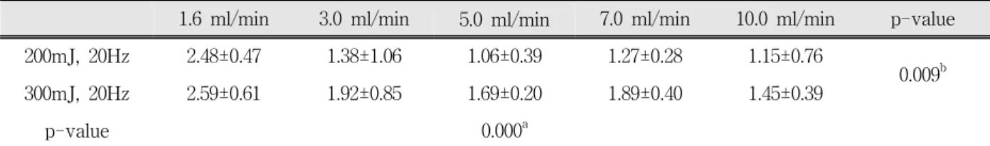
Table 1 shows the ablation weight of dental hard tissue related to pulse energy and water flow rate.
The higher energy of 300 mJ/pulse ablated more than 200 mJ/pulse(p=0.009), regardless of water flow rate. When comparing the ablation efficiency related to water flow rate, the least water flow rate of 1.6 ml/min produced the most ablation of dental enamel and it exhibited high significant difference in comparison to the other rates. There were no significant differences between the other water flow rates (Fig. 2 & Table 2).
Fig. 2. The ablation weight of enamel related to pulse energy and water flow rate. There were significant differences between a water flow rate of 1.6 ml/min and other rates, whereas there existed no significant differences between the other water flow rates except 1.6 ml/min. Pulse repetition rate was 20 Hz and each application of laser beam was performed for 3 seconds.
1.6 ml/min 3.0 ml/min 5.0 ml/min 7.0 ml/min 10.0 ml/min p-value
200mJ, 20Hz 2.48±0.47 1.38±1.06 1.06±0.39 1.27±0.28 1.15±0.76
0.009b
300mJ, 20Hz 2.59±0.61 1.92±0.85 1.69±0.20 1.89±0.40 1.45±0.39
p-value 0.000a
a stands for statistical significance between the two energies of 200 and 300mJ and b for statistical significance among the five water flow rates. Each application of laser beam was performed for 3 seconds.
Table 1. Mean and standard deviations of the ablation weights (mg) related to pulse energy and water flow rate.
Water flow rate 1.6 ml/min 3.0 ml/min 5.0 ml/min 7.0 ml/min 10.0 ml/min
1.6 ml/min 0.001 0.000 0.001 0.000
3.0 ml/min 0.286 0.785 0.177
5.0 ml/min 0.425 0.770
7.0 ml/min 0.278
10.0 ml/min
Numerical value indicates p-value of t-tests between the water flow rate groups.
Table 2. The results of multiple comparison t-test for the differences of the ablation weights between the water flow rate groups.
2. Temperature Experiment
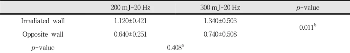
Table 3 shows the temperature rise in dental pulp related to pulse energy and the results of ANOVA test. There existed no significant difference of temperature rise in dental pulp between energies of 200 and 300 mJ/pulse(p=0.408), and there was significant difference between the irradiated and opposite walls of dental pulp of the specimens (p=0.011), when irradiation for 3 seconds was performed with conjunction of water flow of 1.6ml/min. All the condition experimented in this study exhibited a bit higher temperature rise induced by irradiation on the irradiated walls compared to the opposite walls, but all of them were still in safe temperature range for pulp protection.
Fig. 3. The temperature change on the irradiated and opposite pulpal walls during Er:YAG laser ablation. Pulse repetition rate was 20 Hz and each laser beam was applied for 3 seconds in conjunction with water flow rate of 1.6 ml/min. There was significant difference of the temperature change between the irradiated and opposite walls (p=0.011) while there existed no significant differences between 200 and 300 mJ/pulse (p=0.408).
200 mJ-20 Hz 300 mJ-20 Hz p-value
Irradiated wall 1.120±0.421 1.340±0.503
0.011b
Opposite wall 0.640±0.251 0.740±0.508
p-value 0.408a
a stands for statistical insignificance between 200 and 300 mJ and b for statistical significance between the irradiated and opposite pulpal walls. During 3-second irradiation, water flow with a rate of 1.6 ml/min was added on tooth surface.
Table 3. Temperature change (△T(℃)) in the dental pulp induced by Er:YAG laser irradiation.
Ⅳ. DISCUSSION
Dentin and enamel have high absorption peaks in the infrared region at 2.9 ㎛.
14)The laser beam delivered by Er:YAG laser works by utilizing the water in dental hard tissue which absorbs the radiant energy of the laser, and is heated to boiling, producing water vapor. The expansion of water as it becomes vaporized builds up pressure within the irradiated site until a micro-explosion occurs and a small portion of tissue is ablated
9).
The risk of thermal damage to the tooth by the Er:YAG laser is very low. Most of the energy is released in the ablation effect, whereas only a slight amount of the energy from the laser dissipates as heat in the superficial layers of the tooth
2,15). The following laser pulse removes the heated material.
This explains the safety of Er:YAG laser ablation with the minimal thermal side effect. However, once the available water in dental hard tissue has been vaporized and a small amount of ablation has occurred, no additional water is available for absorbing the energy
16). Without the addition of a water spray, the tooth surface becomes dried out and overheated, consequently leading to thermal damage to pulp and inhibition of ablation efficiency.
The addition of water during irradiation not only enhances ablation ability, but offers thermal protection to the pulp
9,17). However, if the rate of water flow is too high, it may result in a film of water that is too thick at the ablation site, requiring a greater amount of energy to be consumed for its removal, thereby decreasing ablation rate
18). Too much water also compromises vision of dentists
during the laser ablation
19).
Thus, it is of importance to determine the appropriate water flow rate at a given irradiation condition. In the previous study
13)of ours mentioned earlier, the effect of water on dental hard tissue ablation with Er:YAG laser was investigated as it relates to energy (250 and 400 mJ/pulse) and pulse repetition rate (5, 10, 20 Hz). When comparing three rates of 1.69, 6.75 and 13.50 ml/min, the results suggested, to obtain the most effective ablation, a water flow rate of 1.69 ml/min for enamel and dentin ablation at a pulse energy of 250 ml/min and for dentin ablation at 400 mJ and a water flow rate of 6.75 ml/min for enamel ablation at 400 mJ, regardless of pulse repetition rate of 5, 10, 20 Hz.
Because the study had shown the least water flow rate of 1.69 ml/min to be the most effective, the authors wanted the water flow rate lower than 1.69 ml/min to be selected for this study and 1.6 ml/min was the lowest one within allowable limit of the Er:YAG laser system employed.
Five rates of water flow exhibiting 1.6, 3.0, 5.0, 7.0 and 10.0 ml/min were compared here, combined with a pulse repetition rate of 20 Hz with pulse energies of 200 and 300 mJ and it was the least rate of 1.6 ml/min that produced the most effective ablation at both 200 and 300 mJ. When regarding the pulse energies used in the two studies, it is thought to be that they showed similar findings.
Although the water flow rate of 1.6 ml/min was
relatively low compared to other studies, there
existed no significant temperature rise in pulp
chamber during irradiation of 3 seconds. It is
generally accepted that the pulpal temperature rise
of less than 5℃ during irradiation is safe for the pulp
20). To obtain more accurate information on pulpal temperature change, the irradiated and opposite pulpal walls of each specimen were respectively monitored by temperature-measuring probes in this study. The temperature rise on the irradiated wall was higher than one on the opposite wall, but it was still in safety range for survival of pulp.
When regarding another study
21)of ours on irradiation time that recommended the irradiation time of less and 3 seconds per application in fixed position, the water flow rate of 1.6 ml/min added enables safer and more efficient ablation with Er:YAG laser.
The tooth specimen prepared for detection of temperature rise in this study is different from a vital tooth condition in oral cavity of a human body, suggesting that the temperature change during irradiation would also be different from our findings.
Considering there are lots of capillaries for blood circulation in dental pulp, it is unlikely that the temperature rise in vital pulp tissue due to irradiation would be higher than in our specimens.
In conclusion, it is suggested from the results of this study that tooth ablation with Er:YAG laser can be done effectively and safely at a energy between 200 and 300 mJ/pulse and a pulse repetition rate of 20 Hz when the irradiation is combined with a water flow rate of 1.6 ml/min.
REFERENCES
1. Hibst R, Keller U. Experimental studies of the application of the Er:YAG laser on dental hard substances: I. Measurement of the ablation rate.
Lasers Surg Med 1989;9:338-344.
2. Hibst R, Keller U. Experimental studies of the application of the Er:YAG laser on dental hard substances: II. Light microscopic and SEM investigations. Lasers Surg Med 1989;9:345-351.
3. Paghdiwala AF, Vaidyanathan TK, Paghdiwala MF.
Evaluation of Er:YAG laser radiation of hard dental tissues : analysis of temperature changes, depth of cuts and structural defects. Scanning Microsc 1993;7:983-997.
4. Jayawardena JA, Kato J, Moriya K, et al. Pulpal response to exposure with Er:YAG laser. Oral Surg Oral Med Oral Pathol Oral Radiol Endod 2001;91:
222-229.
5. Dostálová T, Jelínková H, Kučerová H et al.
Noncontact Er:YAG laser ablation: Clinical evaluation. Lasers Surg Med 1998;16:273-282.
6. Patel BCM, Rickwood KR. Morphological changes induced by short pulse hydrogen fluoride laser radiation on dental hard tissue and restorative materials. Laser Surg Med 1997;21:1-6.
7. Pelagalli J, Gimbel CB, Hansen RT, et al. Investi- gational study of the use of Er:YAG laser versus dental drill for caries removal and cavity preparation- phase I. J Clin Laser Med Surg 1998;15: 109-115.
8. Hoke J, Burkes E, Gomes E, Wolbarsht M. Er:YAG (2.94 ㎛) laser effect on dental tissues. J Laser Applic 1990;2:61-65.
9. Burkes EJ, Hoke JA, Gomes ED, Wolbarsht ML. Wet versus dry enamel ablation of Er:YAG laser. J Prosthet Dent 1992;67:847-851.
10. Armengol V, Jean A, and Marion D. Temperature rise during Er:YAG laser and Nd:YAP laser ablation of dentin. J Endod 2000;26:138-141.
11. Hossain M, Nakamura Y, and Yamada Y et al.
Ablation depths and morphological changes in human enamel and dentin after Er:YAG laser irradiation with or without water mist. J Clin Laser Med Surg 1999;17:105-109.
12. Visuri SR, Walsh JT, and Wigdor HA. Erbium laser ablation of dental hard tissue: Effect of water cooling.
Lasers Surg Med 1996; 18: 294-300.
13. Kim ME, Jeoung DJ, Kim KS. Effects of water flow on dental hard tissue ablation using Er:YAG laser. J Clin Laser Med Surg 2003;21:139-144.
14. Nagasawa A. Researches and development of lasers in dental and oral surgery. In, K. Atsumi (Ed). New Frontiers in Laser Medicine and Surgery.
Amsterdam, 1983, Excerpta Medica, pp.233-241.
15. Keller U, Hibst R. Effects of Er:YAG laser in caries treatment: a clinical pilot study. Lasers Surg Med 1997;20:32-38.
16. Wolbarsht M. Laser surgery; CO2 or HF. IEEE J Quantum Electronics QE-20 1984;12:1427-1432.
17. Cozean C, Arcoria CJ, Pelagalli J, et al. Dentistry for the 1st century? Erbium:YAG laser for teeth. J Am Dent Assoc 1997;128:1080-1087.
18. Hibst R, Keller U. Effects of water spray and repetition rate on temperature elevation during Er:
YAG laser ablation of dentin. SPIE 1996;2623:
139-144.
19. Glockner K, Rumpler J, Ebeleseder K, et al. Intra- pulpal temperature during preparation with the Er:YAG laser compared to the conventional burr: an in vitro study. J Clin Laser Med Surg 1998;16:
153-157.
국문요약
Er:YAG laser를 이용한 치아삭제시 물분사량이 삭제율과 치수내 온도변화에 미치는 영향
단국대학교 치과대학 구강내과학 교실, 단국대학교 의학레이저 연구소 김정문․김미은․김기석
기존의 치과용 핸드피스를 대체할 수 있는 효과적인 치아 삭제 방법인 Er:YAG 레이저를 이용하여 보다 효과 적으로 치아를 삭제하기 위해 여러 가지 변수들, 즉 펄스에너지, 조사반복율 및 레이저 조사 동안의 물분사량에 대한 다양한 연구가 진행되어 왔다. 특히 이 중에서도 물분사량은 삭제 효율이 높이면서 치수를 보호할 수 있는 중요한 요소로 여겨지고 있다. 레이저 조사 동안 분사되는 물의 양이 적으면 치아에 균열이나 탄화를 유발하고 치수손상을 야기할 수 있는 위험이 있는 반면, 물의 양이 지나치게 많으면 삭제효율이 저하되고 레이저 시술 동안 치과의사의 시야확보를 방해할 수 있으므로, 조사조건에 따른 가장 적절한 물 분사량을 결정하는 것이 아주 중요하다.
본 연구에서는 특정조사조건에서 치아법랑질 삭제에 가장 효과적인 물분사량을 결정하고, 그 물분사량을 적용 하였을 때 치수내에서 발생하는 온도변화를 측정하여 안전성 여부를 함께 평가하고자 하였다. 발거된 건전치아 를 표본으로 하여, 20 Hz의 조사반복율, 200 mJ 및 300 mJ의 펄스에너지의 조사조건에서 1.6, 3.0, 5.0, 7.0, 10.0 ml/min의 서로 다른 물분사량을 적용하여 치아삭제 효과를 평가하였다. 이때 레이저 조사시간은 3초로 고정하였 다. 삭제효율은 조사 전후의 치아무게를 측정하여 그 차이로 결정하였으며 온도측정을 위해서는 별도의 치아를 준비하여 온도측정장치를 조사측과 반대측의 치수벽에 위치하여 레이저조사 동안의 온도변화를 추적하였다.
실험결과, 200 mJ과 300 mJ 모두에서 1.6 ml/min의 가장 적은 물분사량이 치아삭제효율이 가장 좋았다. 또한 이 조건에서의 온도변화를 측정한 경우에도 치수손상을 일으키지 않을 정도의 미미한 온도상승만을 보여주었다.
ANOVA 분석의 결과 조사부위(조사측과 비조사 반대측)에 따른 유의한 차이가 나타났으나(p<0.05), 펄스에너지 에 따라 각각 비교하였을 때는 유의한 차이를 보여주지 않았다.
그러므로 본 실험의 결과에 따르면, 1.6 ml/min의 비교적 적은 양의 물을 레이저 조사시에 함께 분사해 준다면 200~300 mJ의 펄스에너지, 20 Hz의 조사반복율, 3초의 레이저조사시간이라는 조건에서는 치수손상을 일으키지 않는 안전한 범위에서 가장 효과적으로 치아를 삭제할 수 있을 것으로 생각된다.
주제어 : Er:YAG laser, 삭제 효율, 물 분사량, 온도
20. Selzer S, Bender I. Biologic considerations in dental procedures. 3rd Ed. The dental pulp. Philadelphia, 1990, Lippincott, pp.200-201.
21. Kim KS, Kim ME, Shin EJ. Irradiation time and ablation rate of enamel in contact and non-contact irradiation with Er:YAG laser. J Clin Laser Med Surg Accepted 21 November 2003.