CONTENTS
Ⅰ. INTRODUCTION
Ⅱ. MATERIALS AND METHODS
Ⅲ. RESULTS
Ⅳ. DISCUSSION
Ⅴ. CONCLUSION REFERENCES KOREAN ABSTRACT EXPLANATION OF FIGURES
Ⅰ. INTRODUCTION
In the course of daily life, there is a continuous ongoing interaction between the individual and environment.
1)Stress, one type of the interaction, is the nonspecific response of the body to any demand.
2)Human body can adapt to stress acceptable for each case but not excessive one, which can produce alterations in some organ and tissue. Recently, dramatic changes in the pace of life apparently seem to have contributed to increase the occurrence rate of stress-related symptoms and diseases. Stress can have an effect on human body which is apt to maintain the homeostasis and modulate three main systems such as autonomic nervous, hormonal and immune response systems.
2)So the term 'psychoneuroimmunology' has developed in order to elucidate the relationship between psychosocial stress and diseases. There are many stress-related symptoms and diseases in the orofacial tissue such as lichen planus, aphthous stomatitis, geographic tongue, recurrent herpes labialis, xerostomia, halitosis, burning mouth syndrome, tension-type headache, atypical odontalgia and temporomandibular disorders.
3)Apoptosis is a fundamental biochemical cell death pathway for normal tissue homeostasis, cellular differentiation, and development within a multicellular organism
4).
A family of interleukin 1β-converting enzyme (ICE) cysteine proteases
5)(now called caspase) are clearly activated in apoptosis and appear to be required for certain aspects of apoptosis. Among them, caspase-3 (CPP32/Yama/apopain)- like protease is responsible for the cleavage of some substrates at the onset of apoptosis
6). It has recently been shown that caspase-3, in particular, is a key player in the DNA fragmentation process and other morphological changes associated with apoptosis
7). With the developing understanding of mechanisms regulating apoptosis, it is becoming increasingly clear that a number of cytotoxic stimuli, including chemotherapeutic agents, operate
Caspase-3 Expression in the Submandibular Gland of Rats under Restraint Stress
Woon-Bong Chung, D.M.D., M.S.D., Ph.D., Sung-Hee Jung, D.M.D., Yang-Hyun Chun, D.M.D., M.S.D., Ph.D., Jin-Yong Lee
*, D.M.D., M.S.D., Ph.D.,
Jung-Pyo Hong, D.M.D., M.S.D., Ph.D.
Department of Oral Diagnosis & Oral Medicine, Dept. of Oral Microbiology
*,
College of Dentistry, Kyung Hee University
through similar mechanisms. Indeed, some of the insight into mechanisms regulating apoptosis has come from the examination of chemotherapy- induced death
8). It now appears that many inducers of cell death ultimately converge on the activation of caspase-3(-like) proteases, which then appear to launch the terminal and execution stages of apoptosis.
Among the stress-related orofacial diseases, although previously investigations have been addressed to dry mouth, halitosis and burning mouth syndrome, the pathologic mechanism of the salivary glands induced by stress remains vague.
There is a reported evidence that repeated stress, even of temporary duration, is able to influence directly or indirectly the morphofunctional state of the salivary glands
9), suggesting a functional linkage. Although apoptosis of salivary gland cells has been demonstrated in several pathological conditions, the definite role of apoptosis in the postnatal development of the salivary glands is unknown yet
10). It has been also shown that apoptosis of the submandibular gland may occur under the restraint stress
11). A linkage between stress and apoptosis has been well demonstrated in postmenopaused women in whom a significant alteration in the salivary compositions is attributed to symptomatic activation in response to psychologic stress
12).
In order to have a better understanding of the pathologic mechanism in submandibular gland disease, emphasizing the significant role of stress in the disease, the present study was performed to investigate the expression of caspase-3 with respect to apoptosis in cells of the submandibular gland under psychological stress by restraint.
Ⅱ. MATERIALS AND METHODS Experimental animals and tissue preparation
Thirty-six Sprague-Dawley rats(8-week-old) were purchased from Dae-Han Experimental Animal Research Center, Seoul, Korea. They were
maintained at 20-23℃ and fed ad libitum on a normal laboratory diet. The rats were divided into 2 groups: restraint stress (32 rats) and control groups (4 rats). The rats of restraint stress group were placed in the plastic cages during the experiment. The stress groups were then sacrificed at 30min, 1hr, 3hr, 6hr, 24hr, and 3, 5, 7day of the experiment and the submandibular glands were collected immediately. The submandibular glands were frozen in dry ice, embedded in O.C.T.
compound (Polyfreeze
TM, cat, #19636, Polysciences, Inc.) in cryomolds, and stored at -70℃ until use.
Preparation of Frozen sections
Serial frozen sections were cut (4-8㎛), placed on positive-charged and RNA-free microscope slides, and stored at -70℃ until use. Frozen sections were allowed to come to room temperature (30 minutes) and fixed in cold acetone for 10 minutes, kept refrigerated. The specimens were rinsed in three changes of 0.01M phosphate buffered saline (PBS) solution.
Immunofluorescence
Anti-caspase-3 (H-277 ; Santa Cruz Biotechno- logy, U.S.A.) used for immunofluorescen-ce is a rabbit polyclonal antibody raised against a recombinant protein corresponding to amino acids 1-277 representing the full-length precursor form of caspase-3 of human origin. The anti-caspase-3 is reactive with the p11 and p20 subunits and the precursor of caspase-3 (also designated CPP32) of mouse, rat and human origin.
Immunofluorescence analysis was performed on
the frozen section by the method as described
previously. After the fixation, the sections were
washed in PBS three times and incubated with the
rabbit anti-caspase-3 in humidity at room
temperature that was diluted 1:50 with concentrated
-reagent diluents (PBS with carrier protein and
0.09% sodium azide). After 2hr, the specimen were
rinsed with PBS solution and then were incubated
with fluorescein anti-rabbit IgG (H+L) (10mM HEPES, 0.15M NaCl, pH7.5, 0,08% sodium azide, 1.5mg/ml active conjugate, VECTOR laboratories) that was diluted with concentrated-reagent diluents for 20min in dark humidity at room temperature adjusting to the final concentration of 5μg/ml. The slides were then washed in PBS and mounted in VECTASHIELD
®Mounting Medium for retaining fluorescence. Thereafter, immunofluorescence reactions were examined under the fluorescent microscope and photographed.
Ⅲ. RESULTS
To localize the expression of caspase-3 in the submandibular glands, the glands from each group were fixed and processed for immunofluorescence using the specific antibody against caspase-3.
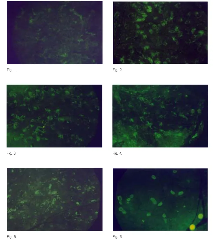
In the normal control group, caspase-3 immuno- reactivity was not detected at any time during the experiment (Fig. 1).
In the restraint stress group, immunoreaction of caspase-3 appeared to be strong 30min after the application of the stress (Fig. 2, 7), and then gradually decreased (Fig. 3-11).
The expression of caspase-3 was found in both acini and ductal cells, but the caspase-3 in the acini cells disappeared earlier than in ductal cells (Fig.
2-11).
Ⅳ. DISCUSSION
We are exposed to various stresses in every moment of daily life. Black
13)addressed that no disease is exempt from psychological influences, and, as a corollary, all diseases have psychological repercussions.
It has been suggested that psychological stress could play a key role in triggering suppression of the immune system and alter the susceptibility of animal and man to infectious agents, influencing the onset, course and outcome of certain infectious pathologies.
14)Recently, quite a few studies have shown that stresses are closely associated with
diseases, especially with orofacial diseases.
Under stressful circumstances, there appears to be an increase in circulating levels of hormones produced within the hypothalamic-pituitary-adrenal axis (HPA).
15)This stimulation prepares the subject to be able to meet the demands of the stress situation. Such things as increase in heart rate, blood pressure, and respiration and a redirection of oxygen and nutrients to organs that require additional energy to function with stress are required to survive under stress conditions.
1)Any response to stress cannot occur without centrally mediated mechanism, that can selectively modulate the response to different incoming signals.
Stress stimulates parvocelluar neurosecretory
neurons of the paraventricular nucleus to release
corticotrophin releasing factor (CRF) into the
hypothalamo-pituitary portal circulation, thereby
stimulating adrenocorticotrophic hormone (ACTH)
release from the pituitary, which then acts on the
adrenal cortex to stimulate the secretion of
corticosteroids.
16)In response to acute stress, there
is a rapid increase in levels of both CRH mRNA
and arginine vasopressin (AVP) mRNA.
17)AVP is
a weaker ACTH secretagogue than CRF releasing
from parvocellular neurons and also acts
synergistically with CRH to regulate ACTH
secretion.
16)On the other hand, repeated or chronic
stress may result in a desensitization of the HPA
axis to the homotypic stressor whereby the
response to subsequent stresses is reduced and
may even be abolished.
18)But exposure of
chronically stressed rats to a different (heterotypic)
stressor induces greater and more rapid increases
in plasma ACTH and corticosterone.
19)Glucocorticoid itself controls the rate of protein
synthesis and influences on carbohydrate, protein,
fat metabolism, and inflammation, physiologically.
20)Weinstein et al.
21)suggested that glucocorticoid
excess, the third most common cause of
osteoporosis, may affect the birth or death rate of
bone cells, thus exhibits osteoblast or osteocyte
apoptosis. Lane
22)provided also the evidence that
thymocytes, immature T cells, can be induced to
undergo apoptosis at an even more rapid rate in vivo and in vitro by treatment with glucocorticoids.
Restraint stress induces thymus involution and the reduction in thymus weight seems to be, at least in part, a consequence of glucocorticoid-induced apoptosis.
23)Hong et al.
24)reported previously that increased expression of clusterin mRNA was observed in the submandibular glands of rats under streptozotocin-induced diabetes mellitus (DM) as well as stress conditions. In DM-stress group, clusterin mRNA was prominently expressed in the submandibular glands at 5 day of the experiment.
25)On the contrary, in rats under the DM-stress condition but received insulin, clusterin mRNA was not observed in the submandibular glands.
26)Therefore, the expression of clusterin, which is known as cell protective protein, indicated that stress could induce apoptosis in the submandibular gland.
Glucose may cause cell injury directly or by deranging electrolyte homeostasis.
27)Baumgartner- Parzer et al.
28)reported that glucose toxicity in human umbilical vein endothelial cells results in cell death especially at high concentrations and high ambient glucose (30 mmol/l) induces apoptosis in cultured human umbilical vein endothelial cells after both short-term and long-term exposure to glucose.
Apoptosis, or programmed cell death, is essential for the development and homeostasis of multicellular organisms.
29)It is an active form of cellular suicide encoded by an endogenous program that can be triggered by either internal or external causes. The morphological alterations of programmed cell death include cellular shrinkage, membrane blebbing, and chromatin condensation.
30)Derangements of apoptosis contribute to the pathogenesis of several human diseases including cancer, acquired immunodeficiency syndrome, and neurodegenerative disorders.
31,32,33)In contrast to necrosis, apoptosis does not provoke the release of cell content into the surrounding tissue of inflammatory reactions.
34)Despite its biological importance, the molecular
mechanism behind apoptosis remains to be defined.
Recently, systematic genetic analysis of Caenor- habditis elegans has identified three genes (ced-3, ced-4, and ced-9) that are important in the regulation of nematode cell death. The proteins encoded by ced-3 and ced-4 are required for all somatic cell deaths that occur during the development of Caenorhabditis elegans.
35)Mutations of ced-3 and ced-4 abolish the apoptotic capability of cells that normally die during development.
36)By contrast, ced-9, which encodes a protein that is homologous to the mammalian gene bcl-2, functions to suppress somatic cell death in the nematode.
37)The function of ced-9 can be partially substituted by ectopic expression of bcl-2.
38)These results suggest that components of the apoptotic machinery are conserved throughout evolution and that mammalian counterparts of ced-3 and ced-4 may play an important role in the mammalian cell death pathway. To date, no homologues of ced-4 have been identified. By contrast, numerous relatives of ced-3 have been characterized, comprising a new gene family of cysteine proteases.
The first mammalian homologue of ced-3
identified was interleukin-1β-converting enzyme
(ICE),
5)an cysteine protease in volved in the
processing and activation of pro-interleukin-1β to
an active cytokine.
39,40)Overexpression of ICE or
Ced-3 in Rat-1 cells induced apoptosis, suggesting
that ICE may act as the functional mammalian
homologues of Ced-3.
41)However, later studies
refute this possibility, since ICE-deficient mice
develop normally and express no overt defects in
apoptosis, except possibly in the Fas pathway.
42,43)Furthermore, apoptotic extracts prepared from
chicken DU249 cells failed to cleave the primary
substrate of ICE, pro-interleukin-1β. Instead, an
ICE-like activity in these extracts, termed prICE,
cleaved the nuclear repair enzyme poly (ADP-
ribose) polymerase (PARP) into characteristic
fragments,
44)thereby abolishing their critical
homeostatic functions. Purified ICE failed to cleave
PARP,
6,44)suggesting that prICE was distinct from
ICE. Therfore, Hangjun Daun et al. also found that ICE-like enzyme, rather than ICE itself, plays a role in the mammalian cell death pathway.
Five members of the ICE/Ced-3 family have been recently identified and include Nedd-2/ICH1,
45,46)Yama/CPP32/Apopain,
6,47,48)TX/ICH2/ICE rel-II,
49,50,51)
ICE rel-III,
49,52)and Mch2. All family members share sequence homology with ICE/Ced-3 and contain an active site QACRG pentapeptide in which the cysteine residues is catalytic. Overexpression of these proteins in a variety of cells causes apoptosis.
Among the ICE/Ced-3 family members thus far cloned, evidence is growing that caspase-`3 may act as a distal effector of the apoptotic machinery.
Caspase-3 has been shown to cleave the death substrate PARP, in addition to being inhibitable by the cowpox serpin, CrmA.
6)Activated caspase-3 (or apopain), comprised of p17 and p12 subunits, was purified from cell extracts using a tetrapeptide aldehyde inhibitor corresponding to amino acids at the PARP cleavage site.
48)Depletion of caspase-3 from these extracts abrogated their apoptotic potential in vitro.
48)This apoptotic activity could be restored by adding back purified caspase-3 to the depleted extracts.
48)Though the evidence is compelling that caspase-3 serves as a functional mammalian homologue of Ced-3, one cannot exclude a role for other ICE/Ced-3 family members in the cell death pathway. For instance, caspase-3 may be the distal effector of a proteolytic cascade that is comprised of (or activated by) other related family members. A precedent for this is the activation of caspase-3 by purified ICE or granzyme B in vitro.
6,53)Alternatively, there may exist a redundant cell death pathway in which involved ICE/Ced-3 family members play a role.
Porter and Janicke
54)studied the specific requirements of caspase-3 in apoptosis. They suggested that caspase-3 is essential for normal brain development and is important or essential in other apoptotic scenarios in a remarkable tissue-, cell type- or death stimulus-specific manner.
Caspase-3 is also required for some typical hallmarks of apoptosis, and is indispensible for
apoptotic chromatin condensation and DNA fragmentaion in all cell types examined. Thus, caspase-3 is essential for certain processes associated with the dismantling of the cell and the formation of apoptotic bodies, but it may also function before or at the stage when commitment to loss of cell viability is made.
Essmann et al.
55)also suggested that the differential regulation of the homologue GDP dissociation inhibitors Rho-GDI 1 and D4-GDI during drug-induced apoptosis by proteolysis mediated by caspase-3 but not by caspase-1.
Owing to their crucial role as modulators of Rho GTPases, that might in turn have a significant impact on the mechanisms that induce the cytoskelotal and morphological changes in apoptotic cells.
Marissen et al
56)reported that the cleavage of eukaryotic translation initiation factor 4G (elF4G) by caspase 3 was described as a possible event contributing to translation inhibition that could regulate translation during apoptosis. Hartmann et al.
57)reported that caspase-3 act as a vulnerability factor and final effector in apoptotic death of dopaminergic neurons in Parkinson's disease.
Recently a few studies have suggested that
stress is strongly associated with orofacial
diseases. Chun and Hong
58)indicated that stress
causes various forms of diseases in the region
including orofacial psychosomatic diseases in which
emotional stress appears to play a major role
(lichen planus, aphthous stomatitis), orofacial
diseases in which psychologic factors appear to
play a role (erythema multiforme, benign mucous
membrane pemphigoid, geographic tongue),
orofacial infections where emotional stress is a
significant predisposing factor (recurrent herpes
labialis, acute necrotizing ulcerative gingivitis),
orofacial lesions induced by neurotic habits
inflicting trauma (biting of oral tissues, physical
trauma with foreign objects, leukoplakia due to
smoking, bruxism and clenching), neurotic orofacial
symptoms (xerostomia, halitosis, burning mouth
syndrome, altered or loss of taste perception, pain
or discomfort with no tissue change), and orofacial pain induced by emotional stress (temporomandi- bular disorders, muscle tension headache, atypical odontalgia).
Xerostomia is the subjective feeling of dry mouth which may or may not be caused by salivary gland hypofuction. Saliva is an important body fluid which plays several vital roles. In addition to moistening and protecting the oral tissue, it acts as an aqueous solvent necessary for taste and aids oral function as well as the digestion of food. Also, saliva has an antibacterial action which inhibits or prevents the onset of dental caries and inflammation.
59,60)Xerostomia is rarely seen in isolation, but with time, the changes of quantity and quality of saliva can be devastating to oral health and may severely affect the general well-being and lifestyle of the patient. In deed, the oral tissues become susceptible to infection, and the ability for oral function may be disturbed. In addition, a number of pathologic conditions can develope, such as halitosis, burning mouth syndrome, gustatory changes and other oral inflammatory diseases.
61,62)Emotional stress induces a variety of responses in the body. In most instances alarm-like reactions and depression and anxiety inhibit, while pleasurable, relaxed sensations promote to increasing salivation.
59,63)Xerostomia can be caused by various factors that can affect salivation, including the use of xerogenic drugs, aging, radiation therapy, systemic or local diseases such as Sjögren syndrome and diabetes, dehydration, and psychogenic factors such as emotional stress.
61,64)Such xerostomia can induce many complications, and so can be regarded as a very important condition in clinic. In spite of studies on the relationship between stress and diseases, the mechanism of the stress-related orofacial diseases remains vague. In the present study, to provide it, stress response of the salivary gland of rats was determined by examining the expression of caspase-3, known as a key enzyme in apoptosis.
During the period of cellular changes, especially in
apoptosis, caspase-3 can be expressed prior to morphological changes.
Although apoptosis of salivary gland cells has been demonstrated in several pathological condi- tions, the role of apoptosis in the postnatal development of the salivary glands is largely unknown. Hecht et al.
10)studied the development of the rat submandibular gland (SMG) during its transition from the perinatal stage to the mature adult stage. They suggested that the loss of Type I cells and reduction of SMG immunoreactivity during development of the intercalated ducts of the adult rat SMG is due, at least in part, to apoptosis.
Scott et al.
65)also suggested that pathological atrophy is largely similar to physiological atrophy, providing a mechanism for acinar cell survival under adverse conditions, with the possibility of eventual recovery. Their results confirmed that totally obstructed glands undergo a rapid, progressive severe atrophy amounting to absolute losses of over 85% of acinar tissue by two weeks.
It has been reported that morphology and function of the salivary glands are vulnerable to stress: the inhibition of salivary gland function is due to central influences from higher centers acting on the salivary centers and thereby suppressing reflex activity
66); the submandibular gland is more sensitive to cold than the sublingual gland
67); repeated stress, even of temporary duration, is able to influence directly or indirectly the morphofunctional state of the submandibular gland
9); acute emotional painful stress enhances free-radical lipid oxidation in the salivary gland tissues and reduces the activities of antioxidant enzymes in it
68). In addition, the submandibular gland is reportedly more age-related, in the pattern of fluid secretion with an intense gustatory stimulus than parotid gland
69).
Chung et al.
70)reported that clusterin (cell
protective protein in stress condition) was
expressed in the salivary gland of the rat in the
early period of the application of cold stress but
disappeared since then. They indicated that
clusterin would not be expressed if the cold stress
is continued, since no need of clusterin production is required by the cells which are already adapted to the stress and recovered from the stressful condition. Apoptosis was also observed in cells of the submandibular gland under restraint stress
11). When the restraint stress was present continuously, they observed that clusterin disappeared in the early period of the stress experiment due to the apoptosis of the cells. These overall results prompted the author to define relationship between psychological stress and apoptosis with respect to caspase-3.
The present study has shown that when salivary glands are affected by a variety of stimuli, the cells of the salivary glands may adapt to them by eliciting various cellular responses such as structural and biochemical transformations. But if the stimulus is lying outside an acceptable range of normality, it may cause such changes at the clinical and subclinical levels as alterations in the production, flow rate, the response to stimuli, the components and the immune function of saliva.
In summary, stress may cause apoptosis depending upon degree, duration of the stress applied and host's adaptability. Caspase-3 can be expressed as a distal effector of the apoptotic machinery when the stress far exceed the range. A excessive stress may induce apoptosis in the cell.
Since stress-induced apoptosis alters the normal salivary physiology, the present study was proposed that pathologic change of the salivary glands can be initiated by stress and then exacerbated by apoptosis. It is also possible that caspase-3 could be detected as an early indicator of apoptosis before the histological alterations occur.
Ⅴ. CONCLUSIONS
A living organism has survived by maintaining a complex dynamic homeostasis that is constantly challenged by environmental stressors. Nowadays, as a result of severe and prolonged stress, an individual has suffered from various stress-related illness.
Despite extensive studies on the relationship between stress and the oral health have been conducted, the pathology of dry mouth, halitosis and burning mouth syndrome remains to be elucidated. In order to have a better understanding of the pathology in orofacial diseases, the present study was undertaken to observe the stress-induced alterations in the submandibular gland and the expression of caspase-3 which is believed to be a stress enzyme responsible for controlling apoptosis.
Thirty-six Sprague-Dawley rats (8-week-old) were purchased from Dae-Han Experimental Animal Research Center, Seoul, Korea. They were maintained at 20-23℃ and fed ad libitum on a normal laboratory diet. The rats were divided into 2 groups: restraint stress (32 rats) and control groups (4 rats). The rats of restraint stress group were placed in the plastic cages during the experiment. The stress groups were then sacrificed at 30min, 1hr, 3hr, 6hr, 24hr, and 3, 5, 7day of the experiment and submandibular gland were collected immediately. The expression of caspase-3 in the submandibular gland tissues was observed by immunofluorescence method. The results were as follows:
1. In the normal control group, caspase-3 immuno- reactivity was slight detected in general gland tissues.
2. In the restraint stress group, the expression of caspase-3 was strong 30 min after the restraint stress was applied and gradually decrease.
3. In the restraint stress group, the expression of caspase-3 was found to remarkably decrease 6 hours after the restraint stress was applied.
4. The expression of caspase-3 was found both acini and ductal cells 30 min after the restraint stress was applied. In the acini cells, the caspase-3 disappeared rapidly in early stages, however, in the ductal cells disappeared gradually in the period of the experiment.
The overall results indicate that caspase-3 occurs
as a result of cell response to psychological stress for apoptosis but then gradually disappear. It is likely that if stress far exceeds the acceptable range for adaptation, apoptosis may occur in the submandibular gland and cause pathologic changes of the gland. Therfore, caspase-3 appears to be an early indicator of apoptosis before the histological alterations occur. Further studies need to be conducted to elucidate the pathologic mechanism of histologic alteration under the psychologic stress.
REFERENCES
1. Auvenshine RC : Psychoneuroimmunology and its relationship to the differential diagnosis of temporo- mandibular disorders, orofacial pain and related disorders. The Dent Clin North Am, 41:279-296, 1997.
2. Selye H : Selye's guide to stress research. Vol. I. Van Nostrand Reinhold Ltd., Canada, 1980.
3. Shklar G, McCarthy PL : The oral manifestations of systemic disease. Boston and London, Butterworths, 1976.
4. Nicholson DW : ICE/CED3-like proteases as thera- peutic targets for the control of inapprop- riate apoptosis. Nat Biotechnol, 14:297-301, 1996.
5. Yuan J, Shaham S, Ledoux S, Ellis HM, Horvitz HR : The C. elegans cell death gene ced -3 encodes a protein similar to mammalian interleukin-1 beta- converting enzyme. Cell, 75:64 1-652, 1993.
6. Teware M, Quan LT, O'Rourke K, Desnoyers S, Zeng Z, Beidler DR, Poirier GG, Salvese n GS, Dixit VM : Yama/CPP32 beta, a mammalian homolog of CED-3, is a CrmA-inhibit able protease that cleaves the death substrate poly (ADP-ribose) polymerase. Cell, 81:801-809, 1995.
7. Woo M, Hakem R, Soengas MS, Duncan GS, Shahi- nian A, Kagi D, Hakem A, McCurrach M, Khoo W, Kaufman SA, Senaldi G, Howard T, Lowe SW, Mak TW : Essential contribu- tion of caspase 3/CPP32 to apoptosis and its associated nuclear changes. Genes Dev, 12:806-819, 1998.
8. Siro S, Minoru T, Kazuo U, Masaya I : Requirement of caspase-3(-like) protease-mediated hydrgen peroxide production for apoptosis induced by various anticancer drugs. J Biol Chem, 273:26900-26907, 1998.
9. Pellegrini A, Grieco M, Materazzi G, Gesi M, Ricciardi MP : Stress-induced morphohistochemical and functional changes in rat adrenal cortex, testis and
major salivary glands. Histochem J, 30:695-701, 1998.
10. Hecht R, Connelly M, Marchetti L, Ball WD, Hand AR : Cell death during development of intercalated ducts in the rat submandibular gland. Anat Rec, 258:349-358, 2000.
11. Park HK, Hong JP : The molecular biological study on clusterin in the salivary glands under restraint stress. 1998.
12. Ben AH, Gottlieb I, Ish-Shalom S, David A, Szargel H, Laufer D : Oral complaints related to menopause.
Maturitas, 24:185-189, 1996
13. Black PH : Psychoneuroimmunology: brain and immunity. Sci Am Sci Med, 2:16-25, 1995.
14. Biondi M, Zannino L-G : Psychological stress, neuroi- mmunomodulation, and susceptibility to infectious diseases in animals and man. A review. Psychother Psychosom, 66:3-26, 1997.
15. Linton EA, Tilders FJH, Hodgkinson S : Stress- induced secretion of adrenocorticotropin in rats is inhibited by administration of antisera to ovine corticotropin releasing factor and vas opressin.
Endocrinol, 116:966-970, 1985.
16. Leng G, Russell JA : Learning to cope with repeated stress. J Physiol, 510:331, 1998.
17. Lightman SL, Young WS III : Influence of steroids on the hypothalamic corticotropin-releasing factor and preproenkephalin mRNA responses to stress. Proc Natl Acad Sci USA, 86:4306-4310, 1988.
18. Kant GJ, Leu JR, Anderson SM, Mougey EH : Effects of chronic stress on plasma corticosterone, ACTH and prolactin. Physiol Behavior, 40:775-779, 1987.
19. Hashimoto K, Suemaru S, Takao T, Sugawara M, Makino S, Ota Z : Corticotropin-releasing hormone and pituitary adrenocortical responses in chronically stressed rats. Regul Pept, 23:117-126, 1988.
20. Clark WG, Brater DC, Johnson AR : Goth's Medical Pharmacology, 13th Ed, Mosby Year Book, p530-535, 1991.
21. Weinstein RS, Jilka RL, Parfitt AM, Manolagas SC : Inhibition of osteoblastogenesis and promotion of apoptosis of osteoblasts and osteocytes by glucocorticoids. Potential mechanisms of their deleterious effects on bone. J Clin Invest, 102:274-282, 1998.
22. Lane DP : A death in the life of p53. Nature, 362:786-787, 1993.
23. Nora T, Haim O, David WW: Restraint stress- induced thymic involution and cell apoptosis are dependent on endogenous glucocorticoids. J
Neuroimmunol, 82:40-46, 1998.
24. Auh QS, Cho HK, Hong JP : The immunological study on expression of the clusterin in streptozocin- induced diabetic rats. J Kor Acad Oral Med, 22:341-358, 1997.
25. Chun YH, Hong JP : The molecular biological study on change of clusterin (SGP-2) in the salivary glands of streptozotocin-induced diabetic rats under stress.
Kor J Stress Res, 5:13-32, 1997.
26. Kim SH, Hong JP : Clusterin (SGP-2) in the salivary gland of insulin injected rats under stress. J Kor Acad Oral Med, 23:309-326
27. Contran RS, Kumar V, Robbins SL, Schoen FJ : Robbin’s pathologic basis of disease. 5th Ed, W.B.
Saunders Co, Philadelphia, 1994.
28. Baumgartner-Parzer SM : High-glucose triggered apoptosis in cultured endothelial cells. Diabetes, 14:1323-1327, 1995.
29. Ellis RE, Yuan JY, Horvitz HR : Mechanisms and functions of cell death. Annu Rev Cell Biol, 7:663-698, 1991
30. Cohen JJ : Apoptosis. Immunol Today, 14:126-130, 1993.
31. Thompson CB: Apoptosis in the pathogenesis and treatment of disease. Science, 267:1456-1462, 1995.
32. Barr PJ, Tomei LD : Apoptosis and its role in human disease. Biol Technol, 12:487-493, 1994.
33. Kerr JFR, Winterford CM, Harmon BV : Apoptosis.
Its significance in cancer and cancer therapy. Cancer (Phila.), 73:2013-2026, 1994.
34. Kerr JFR, Wyllie AH, Currie AR : Apoptosis: a basic biological phenomenon with wide-ranging implica- tions in tissue kinetics. Br J Cancer 26:239-257, 1972.
35. Ellis HM, Horvitz HR : Genetic control of pro- grammed cell death in the nematode C. elegans. Cell, 44:817-829, 1986.
36. Yuan JY, Horvitz HR : The Caenorhabditis elegans genes ced-3 and ced-4 act cell autonomously to cause programmed cell death. Dev Biol, 138:33-41, 1990.
37. Hengartner MO, Ellis RE, Horvitz HR : Caenor- habditis elegans gene ced-9 protects cells from programmed cell death. Nature, 356: 494-499, 1992.
38. Vaux DL, Weissman IL, Kim SK : Prevention of programmed cell death in Caenorhabditis elegans by human bcl-2. Science, 258:1955-1957, 1992.
39. Thornberry NA, Bull HG, Calaycay JR, Chapman KT, Howard AD, Kostura MJ, Miller DK, Molineaux SM, Weidner JR, Aunins J, Elliston KO, Ayala JM,
Casano FJ, Chin J, Ding GJ, Egger LA, Gaffney EP, Limjuco G, Palyha OC, Raju SM, Rolando AM, Salley JP, Yamin TT, Lee TD, Shively JE, MacCross M, Mumford RA, Schmidt JA, Tocci MJ : A novel heterodimeric cysteine protease is required for interleukin-1 beta processing in monocytes. Nature, 356:768-774, 1992.
40. Cerretti DP, Kozlosky CJ, Mosley B, Nelson N, Ness KV, Greenstreet TA, March CJ, Kronheim SR, Druck T, Cannizzaro LA, Huebner K, Black RA : Molecular cloning of the interleukin-1 beta converting enzyme.
Science, 256:97-100, 1992.
41. Miura M, Zhu H, Rotello R, Hartwieg EA, Yuan J : Induction of apoptosis in fibroblasts by IL-1 beta-converting enzyme, a mammalian homolog of the C. elegans cell death gene ced-3. Cell, 75:653-660, 1993.
42. Li P, Allen H, Banerjee S, Franklin S, Herzog L, Johnston C, McDowell J, Paskin M, Rodman L, Salfeld J, Towne E, Tracey D, Wardwell S, Wei FY, Wong W, Kamen R, Seshadri T : Mice deficient in IL-1 beta-converting enzyme are defective in production of mature IL-1 beta and resistant to endotoxic shock. Cell, 80:401-411, 1995.
43. Kuida K, Lippke JA, Ku G, Harding MW, Livingston DJ, Su MSS, Flavell RA : Altered cytokine export and apoptosis in mice deficient in interleukin-1 beta converting enzyme. Science, 267:2000-2003, 1995.
44. Lazebnik YA, Kaufmann SH, Desnoyers S, Poirier GG, Earnshaw WC. : Cleavage of poly (ADP-ribose) polymerase by a proteinase with properties like ICE.
Nature, 371:346-347, 1994.
45. Kumar S, Kinoshita M, Noda M, Copeland NG, Jenkins NA : Induction of apoptosis by the mouse Nedd2 gene, which encodes a protein similar to the product of the Caenorhabditis elegans cell death gene ced-3 and the mammalian IL-1 beta-converting enzyme. Genes Dev, 8:1613-1626, 1994.
46. Wang L, Miura M, Bergeron L, Zhu H, Yuan J : Ich-1, an Ice/ced-3-related gene, encodes both positive and negative regulators of programmed cell death. Cell, 78:739-750, 1994.
47. Fernanades-Alnemri, T., Litwack, G., and Alnemri, E.
S. : CPP32, a novel human apoptotic protein with homology to Caenorhabditis elegans cell death protein Ced-3 and mammalian interleukin-1 beta- converting enzyme. J Biol Chem, 269:30761-30764, 1994.
48. Nicholson DW, Ali A, Thornberry NA, Vailancourt JP,
Ding CK, Gallant M, Gareau Y, Griffin PR, Labelle M, Lazebnik YA, Munday NA, Raju SM, Smulson ME, Yamin TT, Yu VL, Miller DK : Identification and inhibition of the ICE/CED-3 protease necessary for mammalian apoptosis. Nature, 376:37-43, 1995.
49. Munday NA, Vaillancourt JP, Ali A, Casano FJ, Miller DK, Molineaux SM, Yamin TT, Yu VL, Nicholson DW : Molecular cloning and pro-apoptotic activity of ICErelII and ICErelIII, members of the ICE/CED-3 family of cysteine proteases. J Biol Chem, 270:15870-15876, 1995.
50. Faucheu C, Diu A, Chan AW, Blanchet AM, Miossec C, Herve F, Collard-Dutilleul V, Gu Y, Aldape, RA, Lippke JA, Rocher C, Su MSS, Livingston DJ, Hercent T, Lalanne JL : A novel human protease similar to the interleukin-1 beta converting enzyme induces apoptosis in transfected cells. EMBO J, 14:1914-1922, 1995.
51. Kamens J, Paskind M, Hugunin M, Talanian RV, Allen H, Banach D, Bump N, Hackett M, Johnston CG, Li P, Mankovich JA, Terranova M, Ghayer T : Identification and characterization of ICH-2, a novel member of the interleukin-1 beta-converting enzyme family of cysteine proteases. J Biol Chem, 270:15250-15256, 1995.
52. Fernandes-Alnemri T, Litwack G, Alnemri ES : Mch2, a new member of the apoptotic Ced-3/Ice cysteine protease gene family. Cancer Res, 55:2737-2742, 1995.
53. Darmon AJ, Nicholson DW, Bleackley RC : Activation of the apoptotic protease CPP32 by cytotoxic T-cell-derived granzyme B. Nature, 377:446-448, 1995.
54. Porter AG, Janicke RU : Emerging roles of caspase-3 in apoptosis. Cell Death Differ, 6:99-104, 1999.
55. Essmann F, Wieder T, Otto A, Muller EC, Dorken B, Daniel PT : GDP dissociation inhibitor D4-GDI (Rho-GDI 2), but not thes homologous rho-GDI 1, is cleaved by caspase-3 during drug-induced apoptosis.
Biochem J, 15:777-783, 2000.
56. Marissen WE, Guo Y, Thomas AA, Matts RL, Lloyd RE : Identification of caspase 3-mediated cleavage and functional alteration of eukaryotic initiation factor 2alpha in apoptosis. J Biol Chem, 31:9314-9323, 2000.
57. Hartmann A, Hunot S, Michel PP, Muriel MP, Vyas S, Faucheux BA, Mouatt-Prigent A, Turmel H, Srinivasan A, Ruberg M, Evan GI, Agid Y, Hirach EC : Caspase-3: a vulnerability factor and final effector in apoptotic death of dopaminergic neurons in Parkinson's disease. Proc Natl Acad Sci USA, 14:2875-2880, 2000
58. Chun YH, Hong JP : Stress and orofacial diseases.
Kor Stress Res, 3(1):57-72, 1995.
59. Dobrosielski-Vergona K : Biology of the salivary glands. CRC Press, Inc, Boca Raton, 1993.
60. Rankow RM, Polayes IM : Diseases of the salivary glands. W.B. Saunders Co, Philadelphia, 1980.
61. Field EA, Longman LP, Bucknall R., Kaye SB, Higham SM, Edgar WM : The establishment of a xerostomia clinic: a prospective study. Br J Oral Maxillofac Surg, 35:96-103, 1997.
62. Sreebny LM : Recognition and treatment of salivary induced conditions. Int Den J, 39:197-204, 1989.
63. Lynch MA, Brightman VJ, Greenber MsS : Burket's oral medicine. 9th Ed, Lippincott Co, philadelphia, 1994.
64. Lewis MA, Lamey PJ : Clinical oral medicine. Wright, Oxford, 1993.
65. Scott J, Liu P, Smith PM : Morphological and functional characteristics of acinar atrophy and recovery in the duct-ligated parotid gland of the rat.
J Dent Res, 78:1711-1719, 1999.
66. Garrett JR : The proper role of nerves in salivary secretion. J Dent Res, 66:387-97, 1987.
67. Fedelich MA, Rins de David ML : Stress and the salivary glands. Rev Fac Odontol Univ Nac (Cordoba), 17:55-69, 1989.
68. Tarasenko LM, Devatkina TA, Tsebrzhinskii OI, Grebennikova VF, Mel'nikova SV : Reaction of the salivary glands to acute stress. Fiziol Zh, 36:104-6, 1990
69. Sokolenko VN, Silenko IuI : The free-radical involve- ment of the salivary gland in stress. Stomatologiia (Mosk), 74:17-9, 1995.
70. Chung WB, Cho HG, Hong JP : The expression of clusterin (SGP-2) to the stress on the salivary glands of rats. J Kor Acad Oral Med, 22:395-408, 1997.
국문초록
스트레스에 의한 백서 악하선 조직에서의 caspase-3 변화에 관한 실험적 연구
경희대학교 치과대학 구강내과학 교실, 구강미생물학 교실
*정운봉․정성희․전양현․이진용
*․홍정표
스트레스가 타액선 조직을 변형시키고 파괴시킬 수 있다는 것은 이미 보고된 바 있다. 이는 구속스트레스 시에 관찰되는 apoptosis에 의한 것인데, 이 과정에 관여하는 caspase-3는 세포의 DNA를 분절시킴으로서 apoptosis를 일으킨다고 알려진 세포내 단백효소이다. 이에 기존에 관찰되었던 구속 스트레스에 의한 apoptosis의 형태적 변 화가 apoptosis를 유도하는 caspase-3와 어떠한 시기적 상관관계를 가지고 있는지를 구명하기 위하여 본 실험을 시행하였다.
웅성 백서 (Sprague-Dawley, 8주) 를 사용하여 실험 전 기간에 걸쳐 구속스트레스를 가한 후 30분, 1시간, 3시 간, 6시간, 24시간, 3일, 5일, 7일에 희생시켰다. 그 후 실험동물의 악하선을 절취하여 동결절편을 제작한 후, caspase-3에 대한 형광항체로 면역형광법을 시행하여 관찰하였다.
1. 정상대조군에서는 caspase-3가 타액선 조직 전반에서 미약하게 관찰되었다.
2. 구속스트레스 부여 30분에서 caspase-3는 강반응을 보였고, 실험기간이 경과됨에따라 점차 6시간군에서 부터 는 현저히 감소하였다.
3. Caspase-3는 구속 스트레스 30분에 도관과 선포세포 모두에서 발현되었으나, 선포세포에서는 조기에 급격히 소실되었고 도관세포에서는 전 실험 기간에 걸쳐 서서히 소실되었다.
이와 같은 연구 결과에서, 세포내 caspase-3는 조직의 형태적 변화가 나타나기 이전에 발현하는 것으로 보아
caspase-3는 형태적 변화를 예견할 수 있는 진단 지표로 사용될 수 있을 것으로 사료되며 이후 임상적으로 적용
하기 위한 지속적인 연구가 필요할 것으로 생각된다.
EXPLANATION OF FIGURES
Fig. 1 : Immunofluorescence microscopy of caspase-3 in the submandibular gland of the contral group (X100).
Fig. 2 : Immunofluorescence microscopy of caspase-3 in the submandibular gland of the rat under restraint stress 30minutes (X100) after the application of the stress. Strong and clear immunofluorescent reaction of caspase-3 was observed in both acini and ductal cells.
Fig. 3 : Immunofluorescence microscopy of caspase-3 in the submandibular gland of the rat under restraint stress 1 hour (X100) after the application of the stress. Still strong but unclear immunofluorescent reaction of caspase-3 was observed in both acini and ductal cells.
Fig. 4 : Immunofluorescence microscopy of caspase-3 in the submandibular gland of the rat under restraint stress 3 hours (X100) after the application of the stress. Weak and unclear immunofluorescent reaction of caspase-3 was observed in ductal cells.
Fig. 5 : Immunofluorescence microscopy of caspase-3 in the submandibular gland of the rat under restraint stress 6 hours (X100) after the application of the stress. Weak and unclear immunofluorescent reaction of caspase-3 was observed in ductal cells.
Fig. 6 : Immunofluorescence microscopy of caspase-3 in the submandibular gland of the rat under restraint stress 24 hours (X100) after the application of the stress. Weak immunofluorescent reaction of caspase-3 was observed in acini and ductal cells.
Fig. 7 : Immunofluorescence microscopy of caspase-3 in the submandibular gland of the rat under restraint stress 30minutes (X400) after the application of the stress. Strong and clear immunofluorescent reaction of caspase-3 was observed in both acini and ductal cells.
Fig. 8 : Immunofluorescence microscopy of caspase-3 in the submandibular gland of the rat under restraint stress 1 hour (X400) after the application of the stress. Still strong but unclear immunofluorescent reaction of caspase-3 was observed in both acini and ductal cells.
Fig. 9 : Immunofluorescence microscopy of caspase-3 in the submandibular gland of the rat under restraint stress 3 hours (X400) after the application of the stress. Weak and unclear immunofluorescent reaction of caspase-3 was observed in ductal cells.
Fig. 10 : Immunofluorescence microscopy of caspase-3 in the submandibular gland of the rat under restraint stress 6 hours (X400) after the application of the stress. Weak and unclear immunofluorescent reaction of caspase-3 was observed in ductal cells.
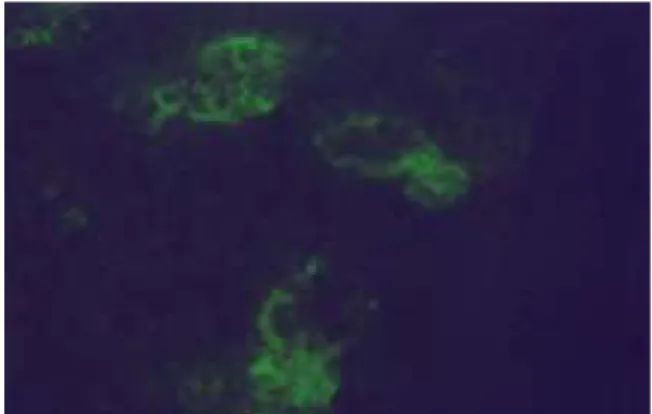
Fig. 11 : Immunofluorescence microscopy of caspase-3 in the submandibular gland of the rat under restraint stress 24 hours (X400) after the application of the stress. Weak immunofluorescent reaction of caspase-3 was observed in gland cells. And destructed gland structures was observed.
Fig. 1. Fig. 2.
Fig. 3. Fig. 4.
Fig. 5. Fig. 6.
Fig. 7. Fig. 8.
Fig. 9. Fig. 10.
Fig. 11.