CONTENTS
Ⅰ. INTRODUCTION
Ⅱ. MATERIALS AND METHODS
Ⅲ. RESULTS
Ⅳ. DISCUSSION
Ⅴ. CONCLUSIONS REFERENCES KOREAN ABSTRACT
Ⅰ. INTRODUCTION
Imaging is the only method of obtaining visual information on the status of the joint tissues short of arthroscopy or open joint surgery. Its primary purpose is to provide information to assist the diagnosis and treatment planning process.
Radiography has long been the primary means for diagnosing organic diseases of the TMJ. However, it has been difficult to determine which radiographic signs are characteristic of individual diseases of the joint. Among the classical radiographic signs of joint disease decreased joint space has been found to be correlated with crepitus, a clinical sign of structural damage to the joint.
4-5)Reduction of the joint space, subcortical sclerosis and flattening of the lateral part of the condyle have
been found to be intercorrelated and frequent among patients with crepitus, pain and joint dysfunction.
6)Despite TMJ imaging has a long history of research and clinical application
58-61), the quality of information gleaned from imaging is often less than desired. The small size of the TMJ, the widely varying fossa and condylar morphology and the surrounding dense osseous structures make clear and undistorted imaging of the joint hard tissue technically difficult. To overcome these obstacles, multiple conventional radiographic technique have been introduced over the years.
7-8)Conventional TMJ radiography has an established role in the detection of structural bone changes and sagittal tomography has been shown to yield the most information.
9)In the study of Tanimoto et al.
11), Autopsy specimens were examined both radiographically and macroscopically to compare direct computed tomography with conventional tomography for their diagnostic yield of the structural bone changes in the temporomandibular joint. They concluded that conventional tomography is superior to computed tomography in the diagnosis of single structural bone changes but comparable for comprehensive diagnosis of TMJ disease.
The Reliability of the Conventional Tomographic Interpretation for the Patients with Temporomandibular
Disorders
Sung-Won Kim, D.D.S., Ki-Suk Kim. D.D.S.,M.S.D.,Ph.D
Department of Oral Medicine, School of Dentistry, and Medical Laser Research Center,
Dankook University
The purpose of this study is to investigate preliminary the reliability of conventional tomographic interpretation for TMJ of patient with temporomandibular disorder in order to perform a further research to depict, by means of conventional tomography, the bone changes that take place in a temporomandibular joint of patient with temporo- mandibular disorder and to correlate these changes to different variables such as condylar angulation, condylar type, condylar position and bone change type.
Ⅱ. MATERIALS AND METHODS 1. Subjects
A series of 256 patients, referred to the Department of Oral Medicine and Orofacial pain and TMJ disorder clinic, Dental Hospital, Dankook University, between July and December 1999 was examined with conventional tomography. From this total, ten subjects were randomly selected for this study.
2. Tomographic equipment
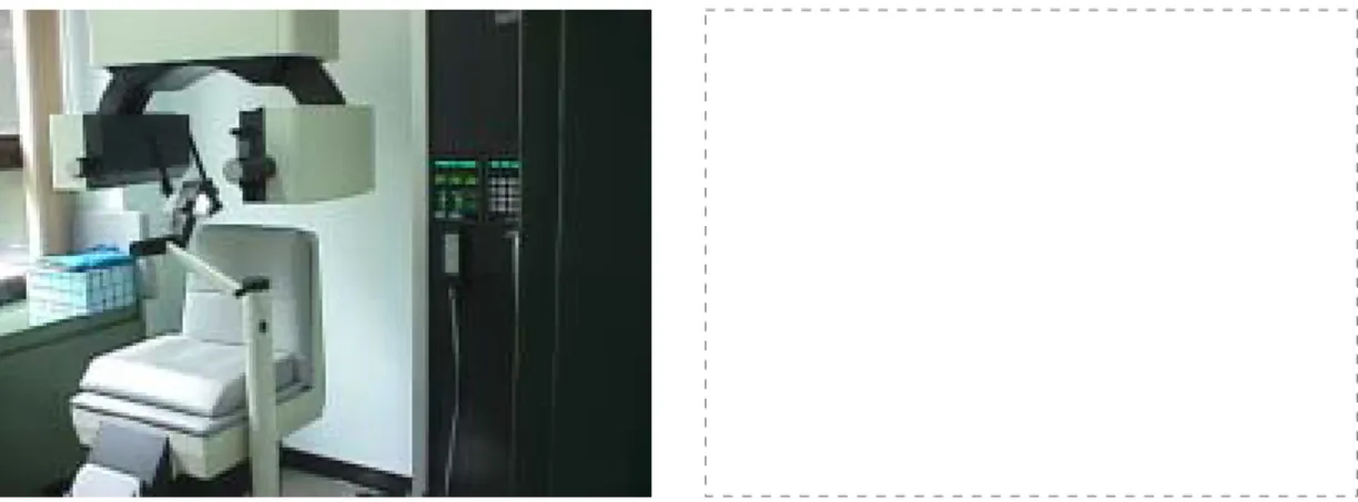
Tomographic imaging was performed using a multidirectional tomograph (SCANORA, Orion Corporation Soredex, Helsinki, Finland). SCANORA is a multifunction x-ray unit designed for
Fig. 1. SCANORA used in tomographic imaging of TMJ
radiographic examination of dento-maxillo-facial regions. The multifunction feature means that the imaging elements include arrangements for examinations utilizing both narrow scanning beam and multidirectional tomography principles.
Although the imaging procedures are computer controlled, and tomographic imaging is included, SCANORA is not a computed tomography device.
Components of SCANORA include an imaging element, patient chair, x-ray generator.
Corrected tomographs were taken of the right and left TMJs in the sagittal plane as part of routine TMJ examination. A submentovertex projection was used to correct for orientation of the condylar heads with respect to the midline and to calculate the depth of cut. All cuts were 4 mm thick and collimated to include only the TMJ area. Four cuts were taken in maximum intercuspation and one cut was taken at maximum opening for each TMJ. The average exposure factors for the SCANORA unit were 72 kV, 3.5 mA, 82 sec (range 57-85 kV).
All radiographs were viewed by two dentists,
who were chosen based on their experience in
evaluating a TMJ x-rays and managing patients
with craniomandibular problems, under standar-
dized conditions and masked to eliminate
extraneous light. From all radiographs, the
examiners investigated bone change severity
(scoring), bone change types, condylar types and
temporomandibular joint spaces such as anterior, superior and posterior spaces. Inter-observer and intra-observer reliability was tested by the use of multiple examinations on the same and on different days. For determination of inter-observer reliability, 10 subjects were examined in one day by two observers, each blind to the other observer's results.
For testing of intra-observer reliability, the subjects were examined twice by each observer respectively blind to his first results. Three days separated the first and second examinations by each observer, in order to minimize both memory of the first results.
3. Scoring of Bone change
Tomograms showing bone change in the frontal and 4 sagittal views of joint were counted and the number was used as a score of bone change for each subject.
4. Bone change type
Radiographic observations of TMJs were recorded according to definitions described in previous report.
11)All tomograms were assessed for the following features:
Concavity: a hollowed-out area on the bony surface of the joint with a well-defined cortical outline.
Cyst: a well-defined, localized area of bone destruction beneath an intact cortical outline of the joint surface.
Round Angled Convex Flat Fig. 2. 4 types of condylar shape
Erosion: a localized area of decreased density of the joint surface and adjacent subcortical bone.
Flattening: a flat bony contour deviating from the convex form.
Osteophyte: a marginal bony outgrowth.
Sclerosis: a localized area of increased density of the cortical bony joint surface extending into the subcortical bone.
5. Condylar shape
Condylar shapes were assessed from frontal view of condyle. Condylar shapes were divided into round, angled, convex and flat type (Fig. 2).
12)6. Joint space
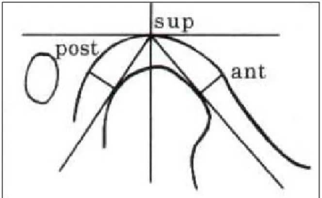
To estimate the joint space, medial 2nd tomographic image among 4 sagittal cuts of joint was used and every image was traced onto acetate overlays with a 0.3 mm diameter lead pencil. The horizontal reference plane defined by the superior glenoid fossa tangent, parallel to the superior border of each tomogram, was assumed parallel to Frankfort Horizontal. The reference planes for joint space were drafted onto the tracing papers. Linear joint spaces were defined as posterior, superior, and anterior in the joint(Fig. 3). Measurements were made manually to the nearest 1/50 mm by vernier caliper.
Fig. 3. The reference plane and three spaces of TMJ
7. Statistic analysis
The relationship between 2 examiners at a simultaneous examination and between 2 examinations of each examiner for bone change, condylar shape and joint space was tested by correlation coefficient. A paired t-test was used to find a difference between two groups.
Ⅲ. RESULTS
Table 1. shows the mean scores and the correlation coefficients for inter- and intra-observer reliability on the condylar type. The correlation of scores were positive for both inter-observer test (r=
0.812, 0.619) and intra-observer test (r=0.955, 0.749).
There were no significant differences between two observers and between two examinations by each
Table 1. The results of correlation coefficient for type of condyle between 2 examiners at 2 different examinations
Examination(n=20)
r(p-value) Paired t-test
Examiner 1st 2nd
A 2.350±1.09 2.350±1.04 0.955(0.000) 1.000 B 2.500±1.10 2.650±1.18 0.747(0.000) 0.419 r(p-value) 0.812(.000) 0.619(0.004)
Paired
t-test 0.330 0.186
Table 2. The results of correlation coefficient for type of bone change between 2 examiners at 2 different examinations
Examination(n=20)
r(p-value) Paired t-test
Examiner 1st 2nd
A 2.45±1.57 2.55±1.64 0.860(0.000) 0.606 B 2.45±1.57 2.55±1.64 0.860(0.000) 0.606 r(p-value) 1.00(0.000) 1.00(0.000)
Paired
t-test 1.00 1.00
observer.
Table 2. shows the mean scores and the correlation coefficients for inter- and intra-observer reliability on the bone change type. The correlation of scores were positive for both inter-observer test (r=1.00) and intra-observer test (r=0.860). There were no significant differences between two observers and between two examinations by each observer.
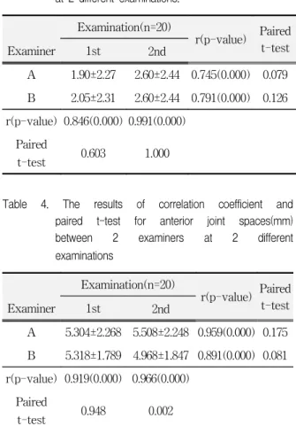
The scores of bone change can be seen in Table 3. There are significant correlations between two observers (r=0.846, 0.991) and two examinations by each dentist (r=0.745, 0.791). At the second examination, mean and standard deviations are identical especially. There aren't significant differences in the joint spaces between two groups respectively.
Table 3. The results of correlation coefficient for score of bone change between 2 examiners at 2 different examinations.
Examination(n=20)
r(p-value) Paired t-test
Examiner 1st 2nd
A 1.90±2.27 2.60±2.44 0.745(0.000) 0.079 B 2.05±2.31 2.60±2.44 0.791(0.000) 0.126 r(p-value) 0.846(0.000) 0.991(0.000)
Paired
t-test 0.603 1.000
Table 4. The results of correlation coefficient and paired t-test for anterior joint spaces(mm) between 2 examiners at 2 different examinations
Examination(n=20)
r(p-value) Paired t-test
Examiner 1st 2nd
A 5.304±2.268 5.508±2.248 0.959(0.000) 0.175 B 5.318±1.789 4.968±1.847 0.891(0.000) 0.081 r(p-value) 0.919(0.000) 0.966(0.000)
Paired
t-test 0.948 0.002
The measurements of joint spaces can be seen in Table 4, 5 and 6. There is significant difference in the anterior joint space only between two observers at 2nd examination as seen in Table 4. There are, however, significant correlation between two observers at two examinations (r=0.919, 0.966) and two examinations by each observer (r=0.959, 0.891).
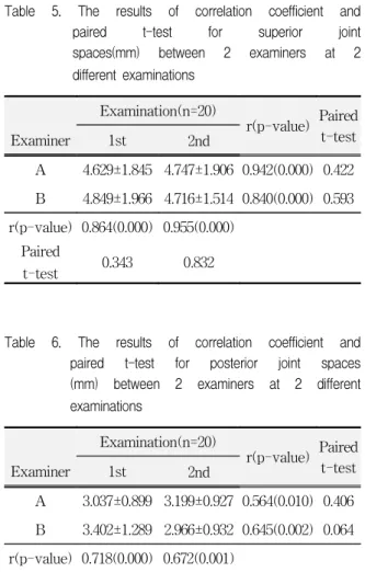
The measurements of superior joint spaces can be seen in Table 5. There are significant correlation in the superior joint spaces between two observers at two examinations (r=0.864, 0.840) and two examinations by each observer(r=0.942, 0.840).
There were no significant differences between two observers and between two examinations by each observer.
Table 6. shows the mean and standard deviations
Table 5. The results of correlation coefficient and paired t-test for superior joint spaces(mm) between 2 examiners at 2 different examinations
Examination(n=20)
r(p-value) Paired t-test
Examiner 1st 2nd
A 4.629±1.845 4.747±1.906 0.942(0.000) 0.422 B 4.849±1.966 4.716±1.514 0.840(0.000) 0.593 r(p-value) 0.864(0.000) 0.955(0.000)
Paired
t-test 0.343 0.832
Table 6. The results of correlation coefficient and paired t-test for posterior joint spaces (mm) between 2 examiners at 2 different examinations
Examination(n=20)
r(p-value) Paired t-test
Examiner 1st 2nd
A 3.037±0.899 3.199±0.927 0.564(0.010) 0.406 B 3.402±1.289 2.966±0.932 0.645(0.002) 0.064 r(p-value) 0.718(0.000) 0.672(0.001)
Paired
t-test 0.085 0.182
of posterior joint spaces. There are, as similar as anterior and superior joint spaces, significant correlation in the posterior joint spaces between two observers at two examinations (r=0.718, 0.672) and two examinations by each observer (r=0.564, 0.645). There were no significant differences between two observers and between two examinations by each observer.
Ⅳ. DISCUSSION
Tomography is body section radiography. Tomo- graphy has become the standard for comprehensive evaluation of the bony components of the TMJ, because it allows visualization of the temporal and condylar component. In addition, it allows the best evaluation of condyle position.
13)Interpretation of a technically correct tomogram is straightforward because its projection can be viewed in standard anatomical planes. Despite its many advantages, full capability tomographic equipment is expensive to use and is not used extensively in dental clinic. Therefore, corrected tomographic equipment (SCANORA multifunction x-ray unit), which is less expensive spiral tomographic system, was used in this study.
Both tomograhic and plain projections have
distortion effects if the angles of the x-ray beam
are not related to the horizontal axis of the condyle
and mandibular fossa. Hence a cephalostat is
required such as head support and chin rest used in
this study. The following radiological principles
should be kept in mind. Projections should be taken
in two or more planes. Axial correction should be
made of the condylar axis by the use of preliminary
submentovertex view followed by orientation with
a cephalostat. Solberg
14)suggested that sagittal
views should be taken in the medial, central and
lateral parts of the joint to represent maxillo-
mandibular positions of clinical relevance. In this
study 4 sagittal views were taken with a 4 mm of
focal thickness from medial pole of the joint to
investigate extensive bone change of the condylar
head. The frontal view is most valuable in
demonstrating condylar remodeling and other changes.
15)Often the changes seen in frontal view are not well identified in sagittal projections. It is reasonable, therefore, to propose that at least the following tomographic views be recommended to examine the TMJ: closed sagittal views (medial, central or 2 central, lateral cuts), maximally open sagittal view (central cut only), frontal plane view with the jaw open, and panoramic survey of the jaw region.
14)This study was preliminary carried out to find a reliability of tomographic interpretation by dentists before a study on a relationship between the bony change of condylar head and one of predisposing factors such as condylar position, condylar type and bony change type is performed in the future.
All radiographic registrations in the past study
43)were made on lateral tomograms, a superior technique for depicting structural TMJ hard tissue changes.
44-46)In that study, frontal tomography was not included because it is suggested that it provides only minor additional information on degenerative TMJ disease when corrected sagittal tomography has been performed.
47-48)This study, however, included frontal tomography to count a score of bone change for condylar head because it was believed four sagittal tomogram did not present all changes of condylar head accurately based on the results of Sato et al.'s report
49)that simultaneous lateral and frontal tomography produces a more accurate radiographic diagnosis of TMJ osteo- arthritis.
Cysts and erosions are considered radiographi- cally significant signs of TMJ pathology, because there is a loss of articular soft tissue in areas corresponding to these signs.
50)The same applies to osteophytes, but only more extensive osteophytes are considered to be indicative of degenerative changes.
51)Cholitgul et al. found sclerosis to be predominantly a false positive findings.
52)In addition, Akerman et al.
50)suggested that sclerosis is not valid for temporal bone. However, sclerosis was previously considered similar in diagnostic value to erosion and osteophyte formation
53)and
there are indications that sclerosis is valid for condyle, sclerosis was included as a component of bone change types in this study.
The significance of condyle-fossa relationship in the temporomandibular joint has not yet been clarified though many efforts have been made by the specialists involved in orthodontics and in the management of TMJ internal derangement and orofacial pain.
54)Although the question of the definition of normal condyle position still needs to be answered, efforts have been made to guide the mandibular condyle into a centric position in the glenoid fossa with the aim of relieving the symptoms in the patients with orofacial pain and TMJ internal derangement.
55-57)Ren et al. concluded that in the joints with normal disk position the condyles were almost randomly distributed in anterior, centric, and posterior positions in glenoid fossa and that posterior condyle position was more prevalent in the joints with anterior disk displacement, approximately half of the joints with anterior disc displacement with reduction and two thirds of the joints with anterior disc displacement without reduction. The present study, therefore, investigated the reliability of joint space measurement in order to determine a condyle position comparing distances of anterior, superior and posterior spaces in a further study.
V. CONCLUSIONS
This study evaluated the inter- and intra- observer reliability on the interpretation and joint space measurement for conventional tomography of TMJ with symptoms of craniomandibular disorders.
Reliability test was performed to determine whether conventional tomography has interpretational precise to allow for consistency in interpretation between different observers and with one observer over time.
Based on the results of this study, it was
concluded that high diagnostic accuracy and
observer agreement can be achieved in conventional
tomography.
REFERENCES
1. Okeson JP (ed): Orofacial Pain : Guidelines for Assessment, Diagnosis, and Management. Chicago, 1996, Quintessence.
2. Chung, S.C., Lee, S.W., Kim, Y.K.: Clinical symptoms and patterns of mandibular movement in the patients with TMJ dysfunction. J Korean Academy of Oral Medicine, 10 : 5-16, 1985.
3. Choi, J.K.: A clinical study on the MPDS patients. J Korean Academy of Oral Medicine, 7 : 47-58, 1982.
4. Kopp S, Rockler G: Relationship between clinical and radiographic findings in patients with mandibular pain or dysfunction. Acta Radiol Diagn, 20 : 465-477, 1979.
5. Park, B.I., Han, K.S.: The relationship between clinical signs and radiolographic findings in temporoman- dibular disorders. J Korean Academy of Oral Medicine, 14 : 57-66, 1989.
6. Kopp S, Rockler B: Relationship between radiographic signs in the temporomandibular joint and hand joints.
Acta Odontol Scand, 37 : 169-175, 1979.
7. Ryu, S.S., Kee, W.C., Choi, J.K.: A relationship between the joint effusion and the presence of pain and disc displacemnet in the temporomandibular joint.
Korean J of Oral Medicine, 25 : 63-71, 2000.
8. Kwon, J.H., Kee, W.C., Choi, J.K.: Configuration of temporomandibular joint articular disc in magnetic resonance images and its relationship to treatment response of anterior disc displacement without reduction. Korean J of Oral Medicine, 25 : 73-85, 2000.
9. Omnell K-A, Petersson A: Radiograhpy of the temporomandibular joint utilizing oblique lateral transcranial projections. Comparison of information obtained with standardized technique and individu- alized technique. Odontol Revy, 27 : 77-92, 1976.
10. Christiansen EL, Thompson JR, Kopp SFO, Hasso AN, Hinshaw DB Jr.: Radiographic signs of temporo- mandibular joint disease: An investigation utilizing x-ray computed tomography. Dentomaxillofac Radiol, 14 : 83-92, 1985.
11. Tanimoto K, Petersson A, Rohlin M, Hansson LG, Johansen CC: Comparison of computed with conventional tomography in the evaluation of temporomandibular joint disease: a study of autopsy specimens. Dentomaxillofac Radiol, 19 : 21-27, 1990.
12. Yale SH, Allison BD, Hauptifuehrer JD: An epidemiologic assessment of mandibular condyle morphology. Oral Surg Oral Med Oral Pathol, 21 :
169-177, 1966.
13. Blaschke DD: Temporomandibular joint. In Goaz P, White S(ed). Oral radiology: principle and interpretation. St. Louis, 1982, CV Mosby, pp508-601.
14. William KS: Temporomandibular disorders: functional and radiological considerations. Br Dent J, 160 : 195-200, 1986.
15. Mahan PA, Gibbs CJ, Mauderli A: Superior and inferior lateral pterygoid activity(abstract). J Dent Res, 61 : 272, 1982.
16. Bell WE: Clinical management of temporomandibular disorders. Chicago, 1982, Year Book Medical Publishers.
17. Atkinson WB, Bates RE: The effects of the angle of the articular eminence on anterior disk displacement.
J Prosthet Dent, 49 : 554-555, 1983.
18. Hall MB, Gibbs CC, Sclar AG: Association between the prominence of the articular eminence and displaced TMJ disks. J Craniomandib Pract, 3 : 237-239, 1985.
19. Sato S, Kawamura H, Motegi K, Takahashi K:
Morphology of the mandibular fossa and the articular eminence in temporomandibular joints with anterior disk displacement. Int J Oral Maxillofac Surg, 25 : 236-238, 1996.
20. Pullinger AG, Bibb CA, Ding X, Baldioceda F:
Contour mapping of the TMJ temporal component and the relationship to articular soft tissue thickness and disk displacement. Oral Surg Oral Med Oral Pathol, 76 : 636-646, 1993.
21. Ren YF, Isberg A, Westesson PL: Steepness of the articular eminence in the temporomandibular joint.
Tomographic comparison between asymptomatic volunteers with normal disk position and patients with disk displacement. Oral Surg Oral Med Oral Pathol Oral Radiol Endod, 80 : 258-266, 1995.
22. Panmekiate S, Petersson A, Akerman S: Angulation and prominence of the posterior slope of the eminence of the temporomandibular joint in relation to disc position. Dentomaxillofac Radiol, 20 : 205-208, 1991.
23. Galante G, Paesani D, Tallents RH, Hatala MA, Katzberg RW, Murphy W: Angle of the articular eminence in patients with temporomandibular joint dysfunction and asymptomatic volunteers. Oral Surg Oral Med Oral Pathol Oral Radiol Endod, 80 : 242-249, 1995.
24. Moffett BC, Johnson LC, McCabe JB, Askew HC:
Articular remodeling in the adult temporomandibular joint. Am J Anat, 115 : 119-142, 1964.
25. Toyama M, Kurita K, Westesson PL, Sakuma S, Ariji E, Rivera R: Decreased disk-eminence ratio is associated with advanced stages of temporomandi- bular joint internal derangement. Dentomaxillofac Radiol, 28 : 301-304, 1999.
26. Kurita H, Ohtsuka A, Kobayashi H, Kurashina K: Is the morphology of the articular eminence of the temporomandibular joint a predisposing factor for disc displacement? Dentomaxillofac Radiol, 29 : 159-162, 2000.
27. Roberts C, Katzberg TW, Tallents RH, et al.: The clinical predictability of internal derangements of the temporomandibular joint. Oral Surg Oral Med Oral Pathol, 71 : 412-414, 1991.
28. Boering G : Temporomandibular Joint Arthrosis. An analysis of 400 Cases. Leiden: Stafleu, 1966.
29. Schiffman E, Anderson G, Fricton J, et al. : Diagnostic criteria for intraarticular TM disorders. Community Dent Oral Epidemiol, 17 : 252-257, 1989.
30. Pullinger A, Seligma D : TMJ osteoarthritis: A differentiation of diagnostic subgroups by symptom history and demographics. J Craniomandib Pract, 4 : 251-256, 1987.
31. Boudewijn S, Lambert GM, Bart van der Kuijl, Geert B : Classification of temporomandibular joint osteoarthrosis and internal derangement. Part I:
Diagnostic significance of clinical and radiographic symptoms and signs. J Craniomandib Pract, 10 : 96-106, 1992.
32. Lieberg J, Rohlin M, Westesson P-L : Observer performance in assessment of condylar position in temporomandibular joint radiograms. Acta Odontol Scand, 43 : 53-58, 1985.
33. Blair GS, Chalmers IM, Leggat TG, Watson Buchanan W : Circular tomography of the temporomandibular joint. Oral Surg, 35 : 416-427, 1973.
34. Kopp S, Rockler B : Variation in interpretation of radiographs of temporomandibular and hand joints.
Dentomaxillofac Radiol, 7 : 95-102, 1978.
35. Zarb GA, Carlsson GE, Sessle BJ, Mohl ND : Temporomandibular joint and masticatory muscle disorders. Copenhagen: Munksgaard, 445-447, 1995.
36. Westesson P-L, Joseph AB, Ross HT, Mark PH : Increased horizontal angle of the mandibular condyle in abnormal temporomandibular joints : A magnetic resonance imaging study. Oral Surg Oral Med Oral Pathol, 72 : 359-363, 1991.
37. Huls A, Schulte W, Voigt K : Neue Aspekte der Myoarhropathien durch die Computertomographie.
Dtsch Zahnarztl Z, 36 : 776-778, 1981.
38. Huls A, Schulte W, Voigt K, Ehrlich-Treuenstatt V : Computed tomography of the temporomandibular joint: new diagnostic possibilities and initial clinical results. Electromedica, 51 : 14-19, 1983.
39. Huls A, Walter E, Schulte W : Konventionelle Rontgendiagnostik und Computertomographie der Kiefergelenke bei Myoarthropathien. Radiologie, 24 : 310-318, 1984.
40. Huls A, Walter E, Schulte W, Freesmayer WB:
Computertomographische Stadieneinteilung des dysfunktionellen Gelenkkopfumbaus. Dtsch Zahnarztl Z, 40 : 37-51, 1985.
41. Huls A, Walter E, Schulte W : Zur Darstellung des Discus articularis im Computertomogramm. Dtsch Zahnartzl Z, 40 : 326-333, 1985.
42. Westesson P-L, Liedberg J : Horizontal condylar angle in relation to internal derangement of the temporomandibular joint. Oral Surg Oral Med Oral Pathol, 64 : 391-394, 1987.
43. Soren Eliasson, Goran Isacsson : Radiographic Signs of Temporomandibular Disorders to Predict Outcome of Treatment. J Craniomandib Disord Facial Oral Pain, 6 : 281-287, 1992.
44. Bean LR, Omnell KA, Oberg T : Comparison between radiologic observations and macroscopic tissue changes in temporomandibular joints. Dentomaxillo- fac Radiol, 6 : 90-106, 1977.
45. Eckerdal O : Tomography of the temporomandibular joint. Acta Radiol, 329, 1973.
46. Omnell KA : Radiology of the TMJ, In Irby WB (ed):
Current Advances in Oral Surgery. vol 3, St. Louis, 1980, C.V. Mosby Co, pp196-226.
47. Rohlin M, Akerman S, Kopp S : Tomography as an aid to detect microscopic changes of the temporo- mandibular joint. An autopsy study of the aged. Acta Odontol Scand, 44 : 131-140, 1986.
48. Tanimoto K, Petersson A, Rohlin M, Hansson LG, Johansson CG : Comparison of computed with conventional tomography in the evaluation of temporomandibular joint disease: A study of autopsy specimens. Dentomaxillofac Radiol, 18 : 151-155, 1989.
49. H. Sato, T. Fujii, N. Yamada and H. Kitamori : The contribution of frontal tomography to the diagnosis of temporomandibular joint osteoarthritis. Dentoma- xillofac Radiol, 21 : 77-80, 1992.
50. Akerman S, Kopp S, Rohlin M : Macroscopic and microscopic appearance of radiologic findings in
temporomandibular joints from elderly individuals.
An autopsy study. Int J Oral Maxillofac Surg, 17 : 58-63, 1988.
51. Sokoloff L : The pathology of osteoarthrosis and the role of aging, in Nuki G (ed): The aetiopathogenesis of Osteoarthrosis. Kent, England, Pitman Medical, 1-15, 1980.
52. Cholitgul W, Petersoon A, Rohlin M, Tanimoto K, Akerman S : Diagnostic outcome and observer performance in sagittal tomography of the temporomandibular joint. Dentomaxillofac Radiol, 19 : 1-6, 1990.
53. Lindvall AM, Helkimo E, Hollender L, Carlsson GE : Radiographic examination of the temporomandibular joint. A comparison between radiographic findings and gross and microscopic morphologic observations.
Dentomaxillofacial Radiol, 5 : 24-32, 1976.
54. Yan-Fang Ren, Annika Isberg, Westesson P-L : Condyle position in the temporomandibular joint:
Comparison between asymptomatic volunteers with normal disk position and patients with disk displacement. Med Oral Pathol Oral Radiol Endod, 80 : 101-107, 1995.
55. Weinberg LA : Definitive prosthodontic therapy for TMJ patients: Part I. Anterior and posterior condyle displacement. J Prosthet Dent, 50 : 544-557, 1983.
56. Weinberg LA : Definitive prosthodontic therapy for TMJ patients: Part II. Posterior and superior condyle displacement. J Prosthet Dent, 50 : 690-699, 1983.
57. Sakuda M, Tanne K, Tanaka E, Takasugi H : An analytic method for evaluating condylar position in the TMJ and its application to orthodontic patients with painful clicking. Am J Orthod Dentofacial Orthop, 101 : 88-96, 1992.
58. Park, G.S., Suh, B.J., Kim, D.Y. : A radiographic study on the horizontal angle of the mandibular condyle in relation to tempooromandibular disorder. J Korean Academy of Oral Medicine, 24 : 25-36, 1999.
59. Seo, M.S., Han, K.S., shin, Min. : Relationship between joint space and craniofacial morphology in patients with craniomandibular disorder. J Korean Academy of Oral Medicine, 17 : 63-73, 1992.
60. Kim, D.Y., Shin, K.B. : A study on the comparison of clinical and radiographic features on patients of craniomandibular disorder. J Korean Academy of Oral Medicine, 16 : 33-47, 1991.
61. Park, B.I., Han, K.S. : The relation between clinical sign and radiographic findings in temporomandibular disorders. J Korean Academy of Oral Medicine, 14 : 57-66, 1989.
Corresponding Author : Ki-Suk Kim, Professor,
Department of Oral Medicine, School of Dentistry, Dankook University, San 7-1, Shinbudong, Cheonan, Choongnam 330-716, Korea