CONTENTS
Ⅰ. INTRODUCTION
Ⅱ. MATHRIALS AND METHODS
Ⅲ. RESULTS
Ⅳ. DISCUSSION
Ⅴ. CONCLUSION REFERENCES KOREAN ABSTRACT
Ⅰ. INTRODUCTION
Dentists often contact patients who have a limitation on mouth opening when they are treated their oral disease. The limitation on mouth opening can be accompany with an infection including temporomandibular disorders, muscle disease, soft tissue scar, tumor, trauma. In those cases, just limited amounts of mouth opening can be measured by the interincisal distance. In the case of normal skeletal structure which has no temporomandibular disorders, movement to open and closing the mouth is composed of translation and rotation of the condyle. The Condyle is placed in a little forward from articular eminence at the maximal mouth opening
1,2)and three fingers could be put between the upper and the lower jaws. Clinicians regard the amount of mouth opening
( 이 논문은 1998년도 전남대학교 학술연구비의 지원에 의하여 연구하였음. This study was financially supported by Chonnam National University in the year of 1998 )
as the very important barometer of tempor- omandibular movement
4). The method to measure the distance between the jaws while the mouth is open is very simple and reliable. Therefore, dentists were used to take this method to evaluate the degree of temporomandibular disorder and the effects of treatment
5,6). Sheppard and Sheppard
4), Nevakari
7)have defined the vertical amount of mouth opening as an interincisal distance and Ingerval
l8)has also defined it as an interincisal distance in addition to overjet. Berrett
9)and Dumas et al.
10)reported that the amount of mouth opening is increasesed as the anterior movement of condyle increases.
The amount of mouth opening had been studied by Sheppard and sheppard
4), Nevakari
7), Ingervall
8)and Agerberg
11,15). They reported that the amount of mouth opening in young children under 13 years is 40mm to 50mm wide and is getting increased as they grow up and there is no difference between that of male and female. The results of Travell's study
16)on men aged 21.7 and women aged 19.6 on the average, Nevakari's study
7)on men and women aged 20 to 25 on the average, and Agerberg's study
12)on men aged 20.5 years old on the average reported that the maximum amount of mouth opening of male is larger than that of female. Agerberg
11,12,15)measured the change of opening amount which is increased by age
The Patterns of Mandibular Movement in Relation to Maxillofacial Skeletal Structure
Byung-Gook Kim, D.D.S., M.S.D., Ph.D., Jae-Hyung Kim, D.D.S.
Department of Oral Medicine, College of Dentistry, Chonnam National University
and reported that the average maximum amount of opening in the average age of 18 months is 37.1mm and in the average age of 6, it is 44.8mm and in the average age of 13, it is 51.2mm and in the average age of 20.5, it is over 56.0mm. But Agerberg's and Oesterber's study
17)suggested that the maximum amount of mouth opening among men in their seventies is decreased to compare with that of men in their twenties.
The study to find a relationship among factors affecting the maximal amount of mouth opening between anthropological measurements and the maximum amount of mouth opening has been performed. Ingervall
14,18), Ageberg
11,12,15), Westling and Helkimo
19)had made an effort to find a relationship of the body index (height or weight) and the maximum amount of opening.
There are many studies that the maximal amount of opening is affected by the flexibility and the border movement of mandible. Westling and Helkimo
19), and Pullinger et al.
20)also reported a way to measure the amount of temporomandibular joint movement with the rotational angle of mandible.
Pullinger et al.
20)reported that the maximum amount of opening of male is larger than that of female in the passive opening but the difference between the rotation angle of mandible and the passive and active mouth opening of female is larger than that of male and the joint movement range of female is wider than that of male and the joint flexibility of female is better than that of male
21). But Westling and Helkimo
19)reported that there is no significant relationship between the maximum amount of opening and the capacity of joint movement in the active or passive mouth opening and the maximal mouth opening rotation angle of mandible has more influence on the facial skeletal structure than the igamental flexibility does. Wright and Hopkins
22), Muto and Kanazawa
23,24)reported that the maximum amount of opening is influenced by mandibular length. Ingervall
14,18)reported that the range of mandible movement has individual differences and in part it is affected by a facial figure and the capacity of mouth opening has a relationship with the
mandible and cranial base length, the ramal length and the length of mandibular inferior border. In order to measure the amount of temporomandibular movement which is not connected with the mandibular length, Dijkstra
25,26)and Gerhardt
27)defined the angular change of mandible to cranial base as the angle of mouth opening and compared the amount of joint movements of other subjects with the use of goniometer.
As mentioned above, many studies on the amount of mouth opening, mandibular movement, physical characteristics and skeletal structure observation had been proceeded. But most of them are under normal conditions of skeletal structure and studies on different skeletal structures in each were rare. The purpose of this study is to figure out a relationship among the amount of mouth opening, the vertical movement of mandible and skeletal structures in each.
Ⅱ. MATHRIALS AND METHODS 1. subjects
172 college students whose age is ranged from 20 to 30 were selected for this study and classified into class I(male:30, female:49), class II(male:18, female:
24) and class III(male:18, female:33), according to Angle's molar relationship. The subjects had not had any experiences of temporomandibular disorder, extraction and orthodontic treatment.
2. method
To measure the maximum interincisal opening (MIO) and the maximum intermolar opening(MMO) and to calculate a ratio between MIO and MMO, a measuring scale was used in the Dept. of Oral Medicine, Chonnam National University Hospital.
The Subjects were taken to be in their upright position and open their mouths as wide as possible.
Impressions of both arches from subjects were
taken with alginate and made into light gypsum
diagnostic models. An arch length was measured as
a distance when a line was drawn vertically on lingual gingival crest between right and left incisors to a line connecting distal surface of the 2nd molar in the right and the left. An arch width was measured as the length of line connecting convex point of lingual surface of the 1st molars in the right and the left side.
Cephaometrics were taken on the maximum opening and centric occlusion by using Verview(J.
morita co., Kyoto, Japan). Conditions were 70-75kvp and 80mmA. Exposing time is 1.2-1.7sec.
TMG/RA-1 film(Kodak, U.S.A.) is used. While the radiograph of the maximum mouth opening was taken, the amount of maximum opening of an interincisal distance was being maintained with a ruler. Distance from a source to film was 150mm and distance from objects to film cassette was 12.5cm.
Cephalometrics were taken and then traced.
Landmarks were identified and analyzed.
Three examiners were taken to a clinical examination, a mandibular movement analysis, the tracing and analyzing of cephalometrics and then finally an average number was considered as the result.
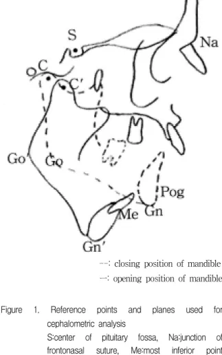
Radiographical landmarks of cephalometrics used in this study were as follows(fig. 1)
1. C : An intersection of perpendicular planes to most anterior, superior and posterior point of condyle
2. C' : A C point of opened mandible
3. GO(Gonion) : An intersection of the posterior border of ramus and the inferior border of mandible
4. Go' : Go': A Go point of opened mandible 5. Gn(Gnathion) : An intersection of Me-Go and
N-Pog
6. Gn' : A Gn point of opened mandible
7. Na(Nasion) : A junction of frontonasal suture 8. Me(Menton) : The most inferior point of
symphysis
9. S(Sella) : The center of pituitary fossa 10. Pog(Pogonion) : The most anterior point of
symphysis
Linear and angular measurements of cephalometrics used in this study were as follows
1. Go-Gn : A mandibular inferior border length 2. C-Go : A ramal length
3. C-Gn : The total mandibular length 4. Na-Me : An anterior facial length 5. C-C' : A distance of condylar movement 6. C-S-C' : An angle of condylar movement 7. Go-Gn to Go'-Gn' : An angle of mouth
opening(AMO)
Figure 1. Reference points and planes used for cephalometric analysis
S:center of pituitary fossa, Na:junction of frontonasal suture, Me:most inferior point of symphysis, Pog:most anterior point of symphysis, Gn:intersection of Me-Go and N-Pog, Go:intersection of posterior border of ramus and inferior border of mandible, C:intersection of perpendicular planes to most anterior, superior and posterior point of condyle, C':C point of opend mandible, Gn':Gn ponit of opened mandible, Go':Go point of opened mandible
--: closing position of mandible ㅡ: opening position of mandible
3. Statistics
In order to confirm differences between male and female on maxillofacial skeletal factors and vertical movement of mandible, the student's-t test or Mann-Whitney rank sum test was taken. To confirm differences among Angle's classifications, a pairwise multiple comparison procedures, Kruskal-Wallis one way analysis of variance on ranks were taken
Ⅲ. RESULTS 1. Facial skeletal structure(Table 1 to 4)
On measuring skeletal structure, Go-GN and C-GN of male were larger than those of female in Angle's Class I, Class II(p<0.001) and Class III(p<0.05). Na-Me of male was larger than that of female in Class II(p<0.01) and Class III(p<0.05).
C-Gn and Na-Me were the largest in Class III among each class and there was no difference between Go-Gn and C-Go. The arch length of mandible and maxilla measured by a diagnosis model was the longest in Class I(p<0.01) and there was no difference between male and female among Class I, II, and III. The arch width of mandible and maxilla was the widest in Class III. The arch with of mandible and maxilla among male was wider than that of female in Class II(p<0.01). The arch with of mandible and maxilla of female was wider than that of male in Class III(p<0.05)
2. Change of the mandible in maximal mouth opening(Table 1 to 4)
The average maximum interincisal opening of male was larger than that of female in Class I(p<0.05), Class II(p<0.01), and Class III(p<0.05).
The average maximum interincisal opening among Class I, Class II and Class III was the largest in Class III(p<0.05) and was 53.58±0.88 mm in Class III. On maximum intermolar opening, male was larger than
female in Class I(p<0.001), Class II(p<0.05), and Class III(p<0.01) and there was no difference among classes. In ratio of the maximum interincisal opening and intermolar opening, there was no difference between male and female and among all classes.
Condyle moves toward anterior and inferior according to articular fossa. The distance of condylar movement was 19.5±0.89 mm in male of Class I and was 17.5±0.43 mm in female of Class and there was no difference. In Class II(p<0.01) the distance of condylar movement of male and female was 23.8±
1.01 mm and 16.5±0.38 mm individually. In Class III(p<0.05) male and female were 21.8±0.17 mm and 14.7 mm ±0.52 individually. The average distance of condylar movement is 18.09±0.41 mm, 18.92±0.92 mm, and 18.33±051 mm each in Class I, Class II and Class III and there was no difference among classes.
There was no difference between the angle of condylar movement of male and female in Class I.
The angle of condylar movement was 37.3±23.9°
and 29.6±1.05°in each male and female of Class II(p<0.01). The angle of condylar movement was 37.7
±21.7°and 28.4±1.89°in each male and female of Class III(p<0.01). In condylar movement, in Male was larger than that of female in Class III. Average angles of condyle movements were 32.46±0.83°, 32.28±1.35°and 31.84±1.72°in each Class I, II and III and there was no difference in classes.
The angle of mouth opening was defined as an intersection angle of mandibular inferior border on the maximal opening and the mandibular inferior border on an centric occlusion. There was no difference between male and female in Class I.
Angles of mouth opening were each 37.75±0.70°and 27.43±2.75°in male and female of Class II(p<0.01).
Angels of mouth opening were 37.42±2.87°and 27.79±0.74°individually in male and female of Class III(p<0.01) and male was larger than that of female.
Average angles of mouth opening were each 31.16±
0.61°, 30.87±2.17°, and 30.27±0.94°in Class I, Class
II and Classs III and there was no difference among
classes
Table 1. Linear(unit:mm) and angular(unit:degree) measurements for facial skeletal factors and mandibular positional changes in Angle's class I
Variables All Male Female P
Maximum Interincisal opening(MIO) 50.37±0.77 54.33±0.73 48.82±0.83 *
Maximum Intermolar opening(MMO) 34.6±0.75 39.1±1.41 32.9±0.56 ***
Ratio between MIO and MMO 1.49±0.03 1.49±0.03 1.49±0.04 NS
Upper arch length 45.05±0.22 44.45±0.37 45.28±0.26 NS
Lower arch length 41.20±0.21 41.33±0.42 41.14±0.25 NS
Upper arch width 37.84±0.29 38.77±0.63 37.47±0.31 NS
Lower arch width 34.20±0.29 35.19±0.67 33.81±0.30 NS
Go-Gn 81.28±0.47 84.5±0.57 80.9±0.54 *
C-Go 65.56±0.63 67.8±0.75 64.5±0.68 NS
C-Gn 119.47±0.833 123.00±0.79 118.39±0.20 **
Na-Me 130.27±0.64 130.42±0.87 130.24±0.74 NS
C-C' 18.09±0.41 19.5±0.89 17.5±0.43 NS
C-S-C' 32.46±0.83 31.2±1.48 33.0±0.99 NS
Go-Gn to Go'-Gn' 31.16±0.61 33.65±1.08 30.19±0.70 NS
C-Go-Gn The values are mean±SEM
NS : not significant, * : p<0.05, ** : p<0.01, *** : p<0.001
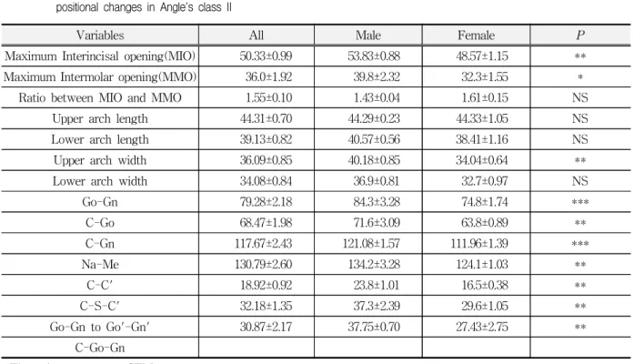
Table 2. Linear(unit:mm) and angular(unit:degree) measurements for facial skeletal factors and mandibular positional changes in Angle's class II
Variables All Male Female P
Maximum Interincisal opening(MIO) 50.33±0.99 53.83±0.88 48.57±1.15 **
Maximum Intermolar opening(MMO) 36.0±1.92 39.8±2.32 32.3±1.55 *
Ratio between MIO and MMO 1.55±0.10 1.43±0.04 1.61±0.15 NS
Upper arch length 44.31±0.70 44.29±0.23 44.33±1.05 NS
Lower arch length 39.13±0.82 40.57±0.56 38.41±1.16 NS
Upper arch width 36.09±0.85 40.18±0.85 34.04±0.64 **
Lower arch width 34.08±0.84 36.9±0.81 32.7±0.97 NS
Go-Gn 79.28±2.18 84.3±3.28 74.8±1.74 ***
C-Go 68.47±1.98 71.6±3.09 63.8±0.89 **
C-Gn 117.67±2.43 121.08±1.57 111.96±1.39 ***
Na-Me 130.79±2.60 134.2±3.28 124.1±1.03 **
C-C' 18.92±0.92 23.8±1.01 16.5±0.38 **
C-S-C' 32.18±1.35 37.3±2.39 29.6±1.05 **
Go-Gn to Go'-Gn' 30.87±2.17 37.75±0.70 27.43±2.75 **
C-Go-Gn The values are mean±SEM
NS : not significant, * : p<0.05, ** : p<0.01, *** : p<0.001
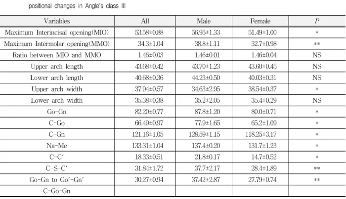
Table 3. Linear(unit:mm) and angular(unit:degree) measurements for facial skeletal factors and mandibular positional changes in Angle's class III
Variables All Male Female P
Maximum Interincisal opening(MIO) 53.58±0.88 56.95±1.33 51.49±1.00 *
Maximum Intermolar opening(MMO) 34.3±1.04 38.8±1.11 32.7±0.98 **
Ratio between MIO and MMO 1.46±0.03 1.46±0.01 1.46±0.04 NS
Upper arch length 43.68±0.42 43.70±1.23 43.60±0.45 NS
Lower arch length 40.68±0.36 44.23±0.50 40.03±0.31 NS
Upper arch width 37.94±0.57 34.63±2.95 38.54±0.37 *
Lower arch width 35.38±0.38 35.2±2.05 35.4±0.29 NS
Go-Gn 82.20±0.77 87.8±1.20 80.0±0.71 *
C-Go 66.49±0.97 77.9±1.65 65.2±1.09 *
C-Gn 121.16±1.05 128.59±1.15 118.25±3.17 *
Na-Me 133.31±1.04 137.4±0.20 131.7±1.23 *
C-C' 18.33±0.51 21.8±0.17 14.7±0.52 *
C-S-C' 31.84±1.72 37.7±2.17 28.4±1.89 **
Go-Gn to Go'-Gn' 30.27±0.94 37.42±2.87 27.79±0.74 **
C-Go-Gn The values are mean±SEM
NS : not significant, * : p<0.05, ** : p<0.01
Table 4. A comparison of linear(unit:mm) and angular(unit:degree) measurements for facial skeletal factors and mandibular positional changes among Angle's class I, II and III and among male and female
Variables Class I Class II Class III P
Maximum Interincisal opening(MIO) 50.37±0.77 50.33±0.99 53.58±0.88*
Maximum Intermolar opening(MMO) 34.6±0.75 36.0±1.92 34.3±1.04 NS
Ratio between MIO and MMO 1.49±0.03 1.55±0.10 1.46±0.03 NS
Upper arch length 45.05±0.22* 44.31±0.70 43.68±0.42
Lower arch length 41.20± 0.21* 39.13±0.82 40.68 ±0.36
Upper arch width 37.84±0.29 36.09 ±0.85 37.94 ±0.57*
Lower arch width 34.20±0.29 34.08±0.84 35.38±0.38*
Go-Gn 81.28±0.47 79.28±2.18 82.20±0.77 NS
C-Go 65.56±0.63 68.47±1.98 66.49±0.97 NS
C-Gn 119.47±0.83 117.67±2.43 121.16±1.05*
Na-Me 130.27±0.64 130.79±2.60 133.31±1.04*
C-C' 18.09±0.41 18.92±0.92 18.33±0.51 NS
C-S-C' 32.46±0.83 32.18±1.35 31.84±1.72 NS
Go-Gn to Go'-Gn' 31.16±0.61 30.87±2.17 30.27±0.94 NS
C-Go-Gn The values are mean±SEM
NS : not significant, * : p<0.05, ** : p<0.01
Ⅳ. DICUSSION
Many studies had been performed to prove relations of mandibular movements, physical characteristics and facial skeletal structures. But most of them were preformed with normal skeletal structures. Therefore cases with different facial skeletal structures in each were rare. For this reason, this study to define a correlation between the amount of mouth opening and the vertical movement of mandible and skeletal structures in groups by the classification of Angle's molar relationship is performed.
Cephalometrics were taken while the maximal mouth opening was being maintained with a ruler.
Interincisal distance of cephalometrics is 1.1 times higher than that of clinical measurements, though this fact was not included in the results.
In the study of position changes in the hyoid bone at the maximal mouth opening, Mutto and Kanazawa
28)reported that the hyoid bone moved to posterior and inferior as the amount of mouth opening increases. They also reported that the inferior drift of hyoid bone on opening and the posterior drift of head were important factors for the maximal mouth opening. So an upright position was being maintained and values were measured in this study.
The amount of maximal mouth opening generally increases until the time of adult and thereafter the amount decreases with age, but reason for that is not clear
4,7,11,12,13,14). The amount of mouth opening in male is larger than that of female. Accordingly the difference by sex is generally derived from different physical characteristics between men and women
11,12,13). Mutto and Kanazawa
23,24)reported that the amount of maximal mouth opening was correlated with the mandible size.
In this study, the maximal interincisal opening in male was larger than that of female. The maximal interincisal opening of male in average was 55 mm and that of female is 49.6 mm. This was not the same as of Agerberg
12)(male:58.6 mm, female:53.3 mm), Travells
16)(male:49 mm, female:53 mm), and
Pullinger's et al.
20)reports(male:57 mm, female:55 mm). This result seemed to be caused by different characteristics of skeletal structures between the orientals and the occidentals. Nevakari
7), Agerberg
11,12), and Landtwing
13)also suggested that the difference of the maximal mouth opening between male and female was in the size of skeleton.
Westling and Helkimo
19)and Muto and Kanazawa
23,24)reported that the amount of mouth opening had not correlation with the body size but with the mandible size. In this study, there was the difference between Go-Gn and C-Gn of male and those of female in Class I and the difference between the maxillary arch width, Go-Gn, C-Go, C-Gn and Na-Me of male and those of female in Class II. In male and female of Class III, the maxillary arch width, Go-Gn, C-Gn, C-Go and Na-Me were different. It was thought that the size of mandible had a correlation with the mouth opening.
The amount of temporomandibular movement influences on the opening capacity. Westling and Helkimo
19)and Pulinger et al.
20)estimated the amount of condylar movement by measuring rotational angle.
Westling and Helkimo
19)reported that the maximal
mouth opening rotation angle of female was higher
than that of male but there was no significant
difference. Westling and Helkimo also reported that
the maximal opening had no correlation with other
articular movements in passive or active opening and
rotation angle of mandible was less dependent on the
ligamentous flexibility but was more influenced by
facial skeletal structures. Pullinger et al.
20)reported
that average range of temporomandibular movement
was 33.3°in men and 35.1°in women. Generally,
epidemiologic studies showed that the range of
temporomandibular movement in female was larger
than that in male. There was no significant
difference between passive and active opening of
male and female. But passive and active opening of
female was larger than that of male. That the
flexibility of ligament of female was larger than that
of male
21)was the reason for this. There was a
difference between measuring method of this study
and that of Westling and Helkimo
19)and Pullinger et
al.
20). In this study, C-S-C' measurement was defined as a value of temporomandibular movement on the maximal opening. There was no difference between C-S-C' of male and female in Class I.
C-S'-C of male was larger than that of female in Class II and Class III. These results were different from those of Pullinger's et al's.
20)The distance and the angle of condylar movement to show the amount of temporomandibular movement had no significant difference among classes and so had no influence on the difference of maximal interincisal opening among classes. But distance of condylar movement was correlated with C-Gn, C-Go, Go-Gn and maxillary arch width in Class I and C-Go, Go-Gn and C-Gn in Class II and the mandibular arch length and the maxillary arch width in Class III.
Dijkstra et al.
25,26)defined the angle of mouth opening(AMO) as an angle displacement of mandible to cranial base to measure the temporomandibular movement which was independent from the mandibular length. Many studies
14,18)on correlations between the amount of maximal mouth opening and the mandibular length had been performed and had meant that the amount of maximal mouth opening was dependent on the mandibular length. Wooten
1)also reported that the distance of condylar movement was not related with the amount of mouth opening.
But Dijkstra et al.
25,26)reported that the amount of maximal mouth opening had strong correlation with the mouth opening angle and the amount of condylar movement had affected on the amount of mouth opening. In this study, the mouth opening angle is correlated with the arch length of mandible and maxilla in Class I and the maxillary arch length and the anterior facial height in Class II and the length of mandibular inferior border, the mandibular arch length and the arching width of mandible and maxilla in Class III.
Westling and Helkimo
19)and Pullinger et al.
20)measured the amount of passive and active opening.
The amount of active opening was measured in this study. The advantage of this method was to measure the amount of TMJ movement, regardless of the
length of mandible. The reason to take this method was that it was difficult for examiners to keep the same force and changes of head position has an effect on the maximal mouth opening.
The maximal interincisal opening showed a significant difference between Class I and III and also a correlation with C-Gn, Na-Me, C-C', C-Go, and Go-Gn in Class I and C-Gn, Na-Me, Go-Gn, and mandibular arch length in Class III. C-Gn and Na-Me showed a significant difference between Class I and III. Therefore, it was thought that the difference of maximal interincisal distance between Class I and III was due to the total mandibular length and the anterior facial height. It was considered that the opening amount of people who has the long mandible and face was larger than that of people with other shapes. This result was identical with the Agerberg's report
11,15). That the difference between maximal mouth opening of Class I and Class II was due to the difference of mandible size was believed to be a reason. This was coincided with the Muto and Kanazawa's report
23,24).
On clinical examining the temporomandibular disorder which had a limitation on the mouth opening, the amount of mouth opening was used for finding degrees of illness and making plans of treatments as an important diagnostic factor. The maximal amount of mouth opening was used for determining the time to finish treatment. Normal recovery of the maximal amount of mouth opening becomes the purpose of treatment and is more important than the improvement of symptom. On treating patients who has a limited mouth opening, especially patients under growing, skeletal factors affecting the amount of mouth opening and the functional orthopedic device are to be confirmed.
V. CONCLUSION
To identify the skeletal factors that had influences
on the vertical movement of mandible and the vertical
displacement on maximal mouth opening according to
Angle's classification, 172(age ranged from 20 to 30)
subjects of college students in Kwangju city without
any experiences of temporomandibular disorder, extraction and orthodontic treatment were selected for this study. The subjects were classified into class I(male:30, female:49), class II(male:18, female:24) and class III(male:18, female:33) according to the Angle's molar relationship. The distance was measured between the incisal edge of maxillary and the mandibular central incisor and between the bottom of central fossa of maxillary and the mandibular 1st molar with a ruler. The arch length and width were measured on diagnostic casts. Cephalometrics were taken and traced. Landmarks were identified and analyzed.
The following results were obtained;
1. The maximum interincisal opening of male is higher than that of female in class I, class II and class III. In each group, the maximum interincisal distance is the largest in class III. The maximum intermolar distance of male is superior to that of female in class I, class II, and class III, but there is no significant difference among them.
2. On the maximum opening movement of Angle's classification, class I and class II, the total mandibular length, the mandibular ramal length, the madibular inferior border length and the upper arch width were important variables and the facial length, showed a reversed correlation with the upper arch length and the lower arch length. On the maximal opening movement of Angle's class III, the upper arch length, the lower arch length and the anterior facial length were important variables, especially when they are compared with class I and II, and showed the negative relationship with the upper arch width.
These results suggest that the maximum opening movement is affected by facial morphology in all of Angle's class I, class II and class III, but each group is affected by different facial skeletal variables.
Accordingly, facioskeletal variables are considered as a diagnosis and a treatment to improve the opening amount of mandible.
REFERENCES
1. Wooten, J.W. : Physiology of temporomandibular joint.
Oral Surg Oral Med Oral Pathol., 21:543-553, 1966.
2. Obwegeser, H.L., Farmand, M., Al-Majali, F., Engelke, W. : Findings of mandibular movement and the position of the mandibular condyles during maximal opening. Oral surg Oral Med Oral Pathol, 63:517-525, 1987.
3. Samat, B.G., Laskin, D.M. : The temporomandibular joint 3rd ed. Springfield, IL, Thomas, pp85, 1979.
4. Sheppard, I.M. and Sheppard, S.M. : Maximal incisal opening - A doagnostic index? J Dent Med, 20:13-15, 1965.
5. Dworkin, S.F., LeResche, L., DeRouen, T., Von Korff, M. : Assessing clinical signs of temporomandibular disorders: Reliability of clinical examiners. J Prothet Dent, 63:574-579, 1990.
6. Carlsson, G.E., Egermark-Eriksson, I., Magnusson, T.
: Intra- and inter-observer variation in functional examination of the masticatory system. Swed Dent J, 4:187-194, 1980.
7. Nevakari, K. : “Elapsio praearticularis" of the temporomandibular joint. A pantomographic study of the so-called physiological subluxation. Acta Odontol Scand 18:123-170, 1960.
8. Ingervall, B. : Range of movement mandible in childern. Scan J Dent Res, 78:311-322, 1970.
9. Berrett, A. : Radiology of the temporomandibular joint. Dent Clin Nor Am, 27:527-540, 1983.
10. Dumas, A., Moaddab, M.B., Willis, H.B., Homayoun, N.M. : A tomographic study of the condyle/fossa relationship in patients with TMJ dysfunction. J Cranio Pract, 2:315-324, 1984.
11. Agerberg, G. : Maximal mandibular movements in children. Acta Odontol Scand., 32:147-159, 1974.
12. Agerberg, G. : Maximal mandibular movements in young men and women. Swed Dent J, 67:81-100, 1974.
13. Landtwing, K. : Evaluation of the normal range of vertical mandibular opening in children and adolescents with special reference of age and stature.
J maxillofac Surg, 6:157-162, 1978.
14. Ingervall, B. : Variation of the range of movement of the mandible in relation to facial mophology in young adults. Scand J Dent Res, 79:133-140, 1971.
15. Agerberg, G. : Maximal mandibular movement in teen-agers. Acta Morphol. Neerl-Scand, 12:79-102, 1974.
16. Travell, J. : Temporomandibular joint pain referred from muscles of the head and neck. J Prosthet Dent, 10:745-763, 1960.
17. Agerberg, G., Osterberg, T. : Maximal mandibular movements and symptoms of mandibular dysfunction in 70-year old men. Swed Dent J, 67;147-63, 1974.
18. Ingervall, B. : Variation of the range of movement of the mandible in relation to facial mophology in children. Scand J Dent Res, 78:535-543, 1970.
19. Westling, L., Helkimo, E. : Maximn jaw opening capacity in adolecents in relation to general joint mobility. J Oral Rehabil, 19:485-494, 1992
20. Pullinger, A.G., Liu, S.P., Low, G., Tay, D. : Differences between sexes in maximum jaw opening when corrected to body size. J Oral Rehab, 14:291-299, 1987.
21. Beighton, P., Solomon, L., Sosklne, C.L. : Articular mobility in an african population. Ann Rheum Diseas, 32:413-418, 1973.
22. Wright, V., Hopkins, R. : The temporomandibular joint. Rheum Diseas, 8:715, 1982.
23. Muto, T., Kanazawa, M. : The relationship between maximal jaw opening and size of skeleton: a cephalometric study. J Oral Rehabil, 23:22-24, 1996.
24. Muto, T., Kanazawa, M. : Linear and angular measurements of the mandible during maximal mouth opening. J Oral Maxillofax Surg, 54:970-974, 1996.
25. Dijkstra, P.U., De Bont, L.G.M., Stegenga, B., Boering, G. : Angle of mouth opening measurement: reliability of a technique for temporomandibular joint mobility assessment. J Oral Rehab, 22:263-268, 1995.
26. Dijkstra, P.U., De Bont, L.G.M., Stegenga, B., Boering, G. : Temporomandibular joint mobility assessment: a comparison between four methods. J Oral Rehab, 22:439-444, 1995
27. Gerhardt, J.J., Rippstein, J.R. : Measuring and Recording of Joint Motion, 2nd ed. Hogrefe Huber Publisher, Toronto, 1990.
28. Muto, T., Kanazawa, M. : Positional change of the hyoid bone at maximal mouth opening. Oral Surg Oral Med Oral Pathol, 77:451-455, 1994.
국문초록
악안면부 골격구조에 따른 하악 개구운동 양상