서 론
우리나라뿐만 아니라 전 세계적으로 노인 인구는 급증하는 추세 이며 연령에 따른 신체적 기능 및 독립성에 대한 요구가 증가하
고 있다. 노인 인구의 약 60%에서 척추 변형을 나타내며 이들에 대한 변형 교정 수술도 점점 증가하고 있다.1-3) 젊은 환자에 비해 노인 환자들을 대상으로 수술을 시행할 경우 다양한 임상적인 요 소들을 고려해야 하는 것은 잘 알려져 있으나 노인 척추 변형 환 자의 변형 교정 수술 시행 시 고려해야 할 사항들에 대해서는 아 직 많은 논의가 되지 않았다. 본 연구에서는 노인 척추 변형 환자 들의 변형 교정 수술 시 합병증을 줄이기 위해 고려해야 할 사항 들과 수술 후 좋은 임상적인 결과를 예측하기 위한 객관적인 인 자들에 대해 고찰하고자 한다.
Copyright © 2019 by The Korean Orthopaedic Association
“This is an Open Access article distributed under the terms of the Creative Commons Attribution Non-Commercial License (http://creativecommons.org/licenses/by-nc/4.0/) which permits unrestricted non-commercial use, distribution, and reproduction in any medium, provided the original work is properly cited.”
The Journal of the Korean Orthopaedic Association Volume 54 Number 1 2019 Received February 1, 2018 Revised May 14, 2018
Accepted September 11, 2018 Correspondence to: Keunho Lee, M.D.
Department of Orthopedic Surgery, Kangdong Sacred Heart Hospital, Hallym University College of Medicine, 150 Seongan-ro, Kangdong-gu, Seoul 05355, Korea TEL: +82-2-2224-2230 FAX: +82-2-489-4391 E-mail: keun118@naver.com ORCID: https://orcid.org/0000-0001-7064-8169
Management of Elderly Patients with Spinal Disease
노인 척추 변형 환자의 수술적 결정
김용찬 • 주형석* • 이근호
†경희대학교 의과대학 정형외과학교실, *서울척병원 정형외과, †한림대학교 의과대학 강동성심병원 정형외과학교실
Surgical Decision for Elderly Spine Deformity Patient
Yong-Chan Kim, M.D., Ph.D., Hyung-Suk Juh, M.D.*, and Keunho Lee, M.D.
†Department of Orthopedic Surgery, Kyung Hee University College of Medicine,
*Department of Orthopedic Surgery, Seoul Chuk Spine Hospital,
†
Department of Orthopedic Surgery, Kangdong Sacred Heart Hospital, Hallym University College of Medicine, Seoul, Korea
Globally, the elderly population is increasing rapidly, which means that the number of deformity correction operations for elderly spine deformity patient has increased. On the other hand, for aged patients with deformity correction operation, preoperative considerations to reduce the complications and predict a good clinical outcome are not completely understood. First, medical comorbidity needs to be evaluated preoperatively with the Cumulative Illness Rating Scale for Geriatrics or the Charlson Comorbidity Index scores. Medical comorbidities are associated with the postoperative complication rate. Managing these comorbidities preoperatively decreases the complications after a spine deformity correction operation. Second, bone densitometry need to be checked for osteoporosis. Many surgical techniques have been introduced to prevent the complications associated with posterior instrumentation for osteoporosis patients. The preoperative use of an osteogenesis inducing agent – teriparatide was also reported to reduce the complication rate. Third, total body sagittal alignment need to be considered. Many elderly spine deformity patients accompanied degenerative changes and deformities at their lower extremities. In addition, a compensation mechanism induces the deformed posture of the lower extremities. Recently, some authors introduced a parameter including total body sagittal alignment, which can predict the clinical outcome better than previous parameters limited to the spine or pelvis. As a result, total body sagittal alignment needs to be considered for elderly spine deformity patients after a deformity correction operation. In conclusion, for elderly spine deformity patients, medical comorbidities and osteoporosis need to be evaluated and managed preoperatively to reduce the complication rate. In addition, total body sagittal alignment needs to be considered, which is associated with better clinical outcomes than the previous parameters limited to the spine or pelvis.
Key words: elderly spine deformity patient, deformity correction operation, osteoporosis, medical comorbidity, total body sagittal alignment
본 론
노인 척추 환자에서 척추 수술의 안정성에 관한 보고들에 의하면 척추 변형 교정 수술과 같이 긴 수술 시간이나 다분절 수술을 시 행한 경우 합병증의 발생을 증가시켰다고 보고하였다. 한 연구에 서는 65세 이상 척추관 협착증 환자 166명을 대상으로 감압술 및 유합술 시행 후 4분절 이상의 다 분절 수술 시 주요 합병증의 위 험성을 증가시킬 수 있다고 하였으며4) Saleh 등5)도 80세 이상 환 자 2,320명을 대상으로 감압술 또는 유합술을 시행하였을 때 긴 수술 시간, 다분절의 유합술 시 합병증이 많이 발생하였다고 하 였다. Wang 등6)은 85세 이상 환자 26명 대상으로 감압술 또는 유 합술을 시행하였을 때 긴 수술 시간이 합병증 증가와 유의한 연 관성을 보였다.
노인 척추 변형 환자에서 변형 교정 수술 시 나이가 증가할수 록 합병증의 발생률이 증가하는 경향을 보였다.7-9) 한 종설(review article)에서 60세 이상의 노인 척추 변형 환자 1,417명 대상으로 평 균 9.5분절의 다분절 유합술을 시행한 결과 약 38%에서 합병증이
발생하였으며 33.3%에서 주요 합병증이 발생하였다고 보고하였 고,7) Acosta 등8)은 노인 척추 변형 환자 21명에 대해 평균 10.5분절 의 유합술을 시행하였을 때 62%의 환자에서 합병증이 발생하였 다고 보고하였다. Daubs 등9)은 또 다른 보고에서 60세 이상 노인 척추 변형 환자 38명을 대상으로 5분절 이상 유합술 시행 시 약 37%에서 합병증이 발생하였고 이 중 69세 이상의 고령 환자에서 합병증이 유의하게 증가하였다고 하였다.
1. 내과적 동반 질환(medical comorbidity)
노인 환자들은 고혈압, 심혈관 질환, 부정맥, 종양, 만성 폐쇄성 폐 질환, 위궤양, 신장 부전증, 당뇨병, 갑상선 질환 등 내과적 동반 질환을 갖고 있는 경우가 흔하다.8) 일부 저자들은 노인 척추 변 형 환자의 동반 질환이 수술 후 사망률 및 합병증과 유의한 상관 관계가 없다고 하였으나9,10) 많은 논문들에서 둘 간에 유의한 상 관관계를 보인다고 보고하였다.11-13) Benz와 Garfin13)과 Smith와 Hanigan14)은 동반 질환의 수가 3개 이상일 경우 그렇지 않은 군에 비하여 유의하게 합병증이 증가하였다고 하였으며 Acosta 등8)은 동반 질환 중 고혈압 동반 시 주요 합병증의 발생률이 10배 증가 하였다고 보고하였다. 이런 동반 질환들을 객관적으로 측정할 수 있는 지표로 Cumulative Illness Rating Scale for Geriatrics (CIRS-G) Table 1. Summary of the Cumulative Illness Rating Scale for
Geriatrics15)
Degree of severity (point) No problem (0)
Mild current problem or past significant problem (1)
Moderate disability or morbidity; requires ‘first line’ therapy (2) Severe or constant significant disability; uncontrollable chronic
problem (3)
Extremely severe (life threatening), end organ failure, severe impairment in function (4)
Body systems evaluated (1) Heart
(2) Vascular (3) Hematopoietic (4) Respiratory
(5) EENT (eyes, ears, nose, throat, larynx) (6) Upper GI tract
(7) Lower GI tract (8) Liver (9) Renal (10) Genito-urinary
(11) Musculoskeletal/integument (12) Neurological
(13) Psychiatric illness (14) Endocrine/metabolic GI, gastrointestinal.
Table 2. Charlson Cormorbidity Index Scores6)
Comorbidity Relative weight
assignment
Metastatic solid tumor 6
Acquired immune deficiency syndrome 6 Moderate-to-severe liver disease 3
Hemiplegia 2
Moderate-to-severe renal failure 2 Diabetes with end organ damage 2
Neoplasia 2
Leukemia/lymphoma 2
Myocardial infarct 1
Congestive heart failure 1 Peripheral vascular disease 1
Cerebrovascular disease 1
Dementia 1
Chronic pulmonary disease 1 Connective tissue disease 1
Ulcer disease 1
Mild liver disease 1
Diabetes 1
The total score was obtained by adding the relative weight of each comorbidity.
(Table 1)15) 및 Charlson Comorbidity Index (CCI) scores (Table 2)6)가 있으며 이들 지표는 노인 척추 변형 환자의 수술 후 합병 증의 발생률을 예측하는 것으로 알려져 있다. Mihailidis 등15)은 CIRS-G의 1 수치가 증가할 경우 총 합병증 발생률은 1.1배, CCI 의 1 수치가 증가할 경우 1.23배 증가한다고 하였다. 특히 CIRS-G 의 경우, 환자가 갖고 있는 내과적 동반 질환이 기능에 심각한 장 애를 가져오는 아주 심하고 생명을 위협하는 상태인지, 조절되 지 않는 심한 상태인지 또는 조절이 잘 되고 있는 경도의 질환인 지에 따라 점수에 차등이 있다. 이에 수술 전 동반 질환을 확인하 여 먼저 조절이 가능할 경우 CIRS-G 수치를 줄여서 합병증의 발 생률을 줄일 수 있다. 이에 노인 척추 변형 환자의 수술적 치료 시 동반 질환들을 고려하여야 하며, 술 후 합병증을 예측하고 합병 증 발생을 예방하여 좋은 임상적인 결과를 얻기 위해 객관적인 지표로서 CIRS-G와 CCI 수치를 평가해야 한다.
2. 골다공증(osteoporosis)
심한 골다공증 환자에서 장분절 유합술을 시행할 경우 기기고정 술 실패, 근위 인접 분절 후만증, 근위 인접 분절 실패, 불유합 등 의 문제를 유발하며 이는 재수술을 초래할 수 있다.16,17) 이는 골 밀도와 비례하는 뽑힘 강도(pull out strength), 돌출 토크(cutout torque), 그리고 삽입 토크(insertional torque) 등이 골다공증 환자 에서 낮기 때문에 생기는 합병증이다.18-22) 성인 척추 변형 환자에 서 변형 교정 수술 시 대부분 기기 고정술을 같이 시행하게 되므 로 노인 척추 변형 환자에서 골다공증이 심한 경우 술 후 합병증
의 발생률을 높일 수 있다.
성인 척추 변형 수술의 고정 실패를 수술 후 6개월 이내인 경우 조기 고정 실패, 그 이후에 생긴 경우 후기 고정 실패로 분류하며 조기 고정 실패는 척추경 또는 척추체의 압박 골절에 의해서 일 어날 수 있는 반면, 후기 고정 실패는 기기 고정술 실패 또는 근위 인접 분절 실패를 동반한 불유합과 밀접한 연관이 있다.16) 이 중 척추 변형 교정 수술 시 주로 사용하는 후방 기기술의 경우 기기 고정술의 실패는 낮은 골밀도 때문에 고정력이 낮아 생기는 경향 이 있으며 이는 나사가 빠지거나 느슨해지는 결과를 일으키게 된 다.23)
골다공증을 동반한 노인 척추 환자에서 기기 고정술 시행할 경 우 더 강한 고정력을 얻고 고정기기의 부하를 줄이기 위한 수술 술기에 대해 많은 연구들이 이뤄졌다.18-22,24,25) 먼저 직경이 큰 척 추경 나사 또는 길이가 긴 나사를 사용할 수 있다. 나사의 직경이 크면 척추경의 더 많은 부분을 채우며 나사선이 척추 피질과 결 합하여 척추경 내에서 나사의 움직임을 최소화할 수 있다. 단, 골 다공증 환자에서 직경이 큰 척추경 나사를 사용할 경우 척추경 골절의 위험성이 증가하는 단점이 있다. 길이가 긴 나사는 뼈와 접촉하는 나사선의 수를 늘려 더 많은 나사선의 표면적을 제공 할 뿐 아니라 척추의 먼 쪽 피질까지 고정할 수 있다. Zindrick 등
18)은 나사를 이용하여 전방 피질을 잡는 것이 나사고정을 향상시 킨다고 보고하였으며 특히 천골에서 길이가 긴 척추경 나사를 이 용하여 먼 쪽 피질까지 고정할 경우 추가 이득을 얻을 수 있다고 하였다. 척추의 피질골이 해면골보다 강하기 때문에 양면 피질골
A
B C
D E
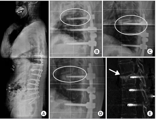
Figure 1. (A) Postoperative simple radio- graphy of a 72-year-old female after 2 weeks of L1–S1 anterior interbody fusion and T10–S1 posterior fixation.
(B) Postoperative simple radiography after 4 weeks with a T10 fracture (Circle indicated recent T10 vertebral fracture with vertebral height loss.). (C) Postoperative simple radiography after 2 months with conservative treatment (teriparatide and brace apply) (Circle indicated no change of healed T10 vertebral fracture.). (D, E) Postoperative simple radiography (Circle indicated no change of healed T10 vertebral fracture.) and computed tomography after 2 months with a healed fracture (Arrow indicated healed T10 vertebral fracture.).
(bicortical) 고정이 단면 피질(unicortical)-해면골 고정보다 더 강 하다.26) Brantley 등27)은 나사의 길이가 나사 경로의 80% 이상으로 긴 경우 고정력이 증가하나 골다공증에서는 고정력 증가가 뚜렷 하지 않다고 보고하였다. 원추 모양의 척추경 나사를 사용할 경 우 더 강한 고정력을 얻을 수 있는 것으로 알려져 있으나 이 경우 나사 삽입 후 뒤로 일부 뺄 경우 고정력이 약화된다는 단점이 있 다.28) 보다 강한 고정력을 얻기 위하여 확장 가능한 나사 및 코팅 된 나사 등 새로운 척추경 나사 디자인이 제시되었다. 확장 가능 한 나사는 척추체 내에 있는 나사 부분만 팽창되어 척추의 해면 골을 압박하게 되며 나사 둘레의 뼈 밀도를 증가시킨다. 코팅된 나사는 현재까지 티타늄 합금 또는 수산화인회석(hydroxyapatite) 으로 얇게 코팅된 스테인레스 스틸(stainless stell) 나사가 있다.29) 또한 수술 고정 분절을 증가시켜 추가적인 척추경 나사 삽입 을 통해 한 분절의 고정물에 걸리는 부하를 줄일 수 있다.30) 65세 이상의 척추 변형 환자에서 변형 교정 수술 시 척추 변형의 첨부 에서 근위부와 원위부 모두 3마디의 고정을 하는 것이 추천되며 또한 추궁판하 강선(sublaminar wire), 후궁 갈고리(laminar hook), 교차 결합(cross link) 등을 사용하여 여러 개의 고정 포인트를 만 드는 것이 척추경 나사의 고정력을 높여준다.31,32) 시멘트를 이용 한 척추경 나사 고정은 하중을 분산시켜 주고, 인접한 골소주와 의 고정력을 높여 나사의 뽑힘 강도를 증가시킨다.33) 시멘트의 최 적량은 유한 요소 분석 결과 2.6 ml,34) 사체를 이용한 연구에서는 1.0-1.5 ml였다.35) 또한, 나사 팁 주위에 시멘트를 집중시키는 것이 효과를 최대화할 수 있다고 알려져 있다.36) 그러나 시멘트 보강은 시멘트 색전의 위험성이 있으며 정맥계나 신경관, 인접한 추간판 으로 유출 등의 합병증을 일으킬 수 있다.37,38)
그 외에 척추경 나사 고정력을 증가시키기 위한 삽입 기술에 대해 많은 연구들이 이루어졌다. 척추경 나사 삽입의 첫 번째 단 계로 파일럿 구멍을 만들게 되는데 이때 특히 골다공증이 심한 환자에서 파일럿 홀의 크기가 과도하게 클 경우 나사 고정력이 불량해질 수 있다. 반대로 파일럿 구멍이 과도하게 작을 경우 삽 입 토크가 증가하고 척추 골절 및 나사 파손의 위험이 높아지게 된다. Battula 등39)은 뽑힘 강도를 극대화하고 의인성 골절을 최소 화하기 위해 파일럿 구멍의 크기는 삽입할 나사 외경의 71.5%보 다 작게 만들 것을 권장하였다. 이러한 이유로 골다공증 환자에 서 고속 버(high speed burr)나 송곳과 같은 보다 정밀한 도구를 사 용하여 파일럿 구멍을 만드는 것이 바람직하다. 최근 연구에서 tapping을 척추경 나사 경로와 같은 크기로 시행하는 것보다는 나 사 경로보다 1 mm 정도 덜 하거나 하지 않는 것이 더 강한 뽑힘 강도를 얻을 수 있다고 보고하였다.20,40)
마지막으로 노인 척추 변형 환자에서 변형 교정 수술하기 전 적극적인 골다공증 치료를 통해 골밀도를 교정하고 수술을 시행 하면 더 좋은 임상결과를 얻을 수 있다.24,41) Yagi 등42)은 척추 변형 교정 수술 후에 예방적으로 골형성 유도제(teriparatide) 사용 시
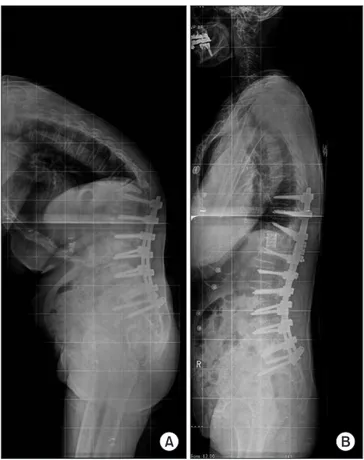
근위 인접 분절 후만증 및 상위 고정 분절의 근위부 분절의 골절 예방 효과를 발표한 바 있다. 또한 척추 변형 교정 수술 이후 생기 는 합병증 중 후만증이 동반되지 않는 근위 분절의 골절에서 골 형성 유도제 사용 및 보조기 착용 등의 보존적 치료로 성공적인 결과를 얻은 바 있다(Fig. 1). 반면 근위 분절의 후만증을 동반한 근위 인접 분절 실패가 발생할 경우 보존적 치료보다 적극적인 수술적 치료를 요한다(Fig. 2).
3. 전 신체 시상학 정렬(total body sagittal alignment) 척추 변형 교정 수술 시 요추 전만의 교정뿐만 아니라 골반 인 자(pelvic parameter)와 C7 시상면 수직축(C7 sagittal vertical axis) 의 중요성이 강조돼 왔다. 특히 척추의 시상면 지표들 중 가장 유의하게 임상결과를 예측하는 지표는 C7 시상면 수직축, 골 반 경사각(pelvic tilt), 골반 입사각-요추 전만 불일치(pelvic inci- dence-lumbar lordosis mismatch)로 보고되고 있다.43-46) 최근에는 몸 전체의 균형을 유지하며 생역학적 기능을 유지하여 편안하게 전방을 주시할 수 있는 전 신체 시상학 정렬이 대두되고 있다.
노인 척추 변형 환자에서 척추의 퇴행성 변화와 변형뿐만 아
A B
Figure 2. (A) Postoperative simple radiography of an 80-year-old female 8 months after L3–S1 anterior interbody fusion and L1–S1 posterior fixation, presents proximal junction failure and kyphosis. (B) The patient underwent a T12 posterior vertebral column resection and T9–L1 posterior rod extension.
니라 고관절, 슬관절 등의 하지 관절의 퇴행성 변화와 변형을 동 반하고 있는 경우가 흔하며 또한 척추의 변형이 보상 작용으로 하지 관절의 변형을 일으키기도 한다. 척추의 퇴행성 변화로 요 추 전만(lumbar lordosis)이 감소되었을 때, 흉추 후만(thoracic kyphosis)의 증가, 골반 후염전(pelvic retroversion), 골반 후방 전 위(posterior pelvic shift), 무릎 굴신(knee flexion), 발목 배굴(ankle dorsiflexion) 등의 보상작용이 나타나게 된다.43,44,47)
따라서 노인 척추 변형 환자에서 수술을 시행할 경우 골반 지 표나 척추에 국한된 과거의 지표로는 전 신체 시상학 정렬을 충 분히 설명할 수 없어 최근에 상부로 두개골, 하부로는 골반 및 하 지의 정렬상태를 포함하여 환자의 수술 후 임상적 결과를 총체적 으로 평가하는 많은 연구가 보고되고 있다.48,49) Kim 등50)은 전인 체 시상면의 선형사슬을 연결하는 5개의 지점(Fig. 3)과 이를 이 용하여 머리의 무게중심에서 내린 시상면 수직선(cranial sagittal vertical axis, CrSVA)의 4개의 거리지표를 제시한 바 있다(Fig. 4).
이들 지표는 임상적 상태를 예측하는 데 기존의 지표보다 상대적 으로 유의하게 강하였으며 특히 이 중 CrSVA-hip이 가장 강한 예 측지표로 보고하였다.
결 론
노인 척추 변형 환자에서 변형 교정 수술을 시행할 경우 내과적 인 동반 질환, 골다공증의 정도를 확인하여 합병증을 줄이기 위 해서 수술 전 미리 교정하는 것이 필요하다. 또한 교정 수술 시 골 반 지표나 척추에 국한된 지표에 비하여 임상적인 상태를 더 잘
예측할 수 있는 전 신체 시상학 정렬을 고려해야 한다.
CONFLICTS OF INTEREST
The authors have nothing to disclose.
REFERENCES
1. Schwab F, Dubey A, Gamez L, et al. Adult scoliosis: preva- lence, SF-36, and nutritional parameters in an elderly volun- teer population. Spine (Phila Pa 1976). 2005;30:1082-5.
2. O’Shaughnessy BA, Bridwell KH, Lenke LG, et al. Does a long-fusion "T3-sacrum" portend a worse outcome than a short-fusion "T10-sacrum" in primary surgery for adult sco- liosis? Spine (Phila Pa 1976). 2012;37:884-90.
3. Diebo B, Liu S, Lafage V, Schwab F. Osteotomies in the treat- ment of spinal deformities: indications, classification, and surgical planning. Eur J Orthop Surg Traumatol. 2014;24 Suppl 1:S11-20.
4. Cassinelli EH, Eubanks J, Vogt M, Furey C, Yoo J, Bohlman HH. Risk factors for the development of perioperative com- plications in elderly patients undergoing lumbar decompres-
1 2
3
4
Figure 4. Schematic diagram showing the four distance parameters for an assessment of total body sagittal alignment. CrSVA-S (1); CrSVA-H (2); CrSVA-K (3); CrSVA-A or ‘global SVA’ (4). CrSVA-S indicates cranial sagittal vertical axis-sacrum; -H, -hip; -K, -knee; -A, -ankle. The CrSVA is a perpendicular line from the cranial center of mass (defined as the midpoint of the nasion-inion line, above and slightly in front of the external auditory meatus). Cited from the article of Kim et al. (Eur Spine J. 2017;26:2167-75).50)
a
b c
d
e
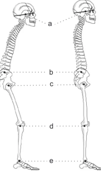
Figure 3. Schematic drawing depicting the 5 landmarks used in the novel parameters for an assessment of the total body sagittal alignment.
Center of gravity of the head (a), sacrum (b), hip (c), knee (d), and ankle (e).
Cited from the article of Kim et al. (Eur Spine J. 2017;26:2167-75).50)
sion and arthrodesis for spinal stenosis: an analysis of 166 patients. Spine (Phila Pa 1976). 2007;32:230-5.
5. Saleh A, Thirukumaran C, Mesfin A, Molinari RW. Compli- cations and readmission after lumbar spine surgery in elderly patients: an analysis of 2,320 patients. Spine J. 2017;17:1106- 12.
6. Wang MY, Widi G, Levi AD. The safety profile of lumbar spi- nal surgery in elderly patients 85 years and older. Neurosurg Focus. 2015;39:E3.
7. Drazin D, Shirzadi A, Rosner J, et al. Complications and out- comes after spinal deformity surgery in the elderly: review of the existing literature and future directions. Neurosurg Focus. 2011;31:E3.
8. Acosta FL Jr, McClendon J Jr, O'Shaughnessy BA, et al. Mor- bidity and mortality after spinal deformity surgery in patients 75 years and older: complications and predictive factors. J Neurosurg Spine. 2011;15:667-74.
9. Daubs MD, Lenke LG, Cheh G, Stobbs G, Bridwell KH.
Adult spinal deformity surgery: complications and outcomes in patients over age 60. Spine (Phila Pa 1976). 2007;32:2238- 44.
10. Ragab AA, Fye MA, Bohlman HH. Surgery of the lumbar spine for spinal stenosis in 118 patients 70 years of age or old- er. Spine (Phila Pa 1976). 2003;28:348-53.
11. Raffo CS, Lauerman WC. Predicting morbidity and mortality of lumbar spine arthrodesis in patients in their ninth decade.
Spine (Phila Pa 1976). 2006;31:99-103.
12. Oldridge NB, Yuan Z, Stoll JE, Rimm AR. Lumbar spine sur- gery and mortality among medicare beneficiaries, 1986. Am J Public Health. 1994;84:1292-8.
13. Benz RJ, Garfin SR. Current techniques of decompression of the lumbar spine. Clin Orthop Relat Res. 2001;384:75-81.
14. Smith EB, Hanigan WC. Surgical results and complications in elderly patients with benign lesions of the spinal canal. J Am Geriatr Soc. 1992;40:867-70.
15. Mihailidis HG, Manners S, Churilov L, Quan GMY. Is spinal surgery safe in octogenarians? ANZ J Surg. 2017;87:605-9.
16. DeWald CJ, Stanley T. Instrumentation-related complica- tions of multilevel fusions for adult spinal deformity patients over age 65: surgical considerations and treatment options in patients with poor bone quality. Spine (Phila Pa 1976).
2006;31:S144-51.
17. Puvanesarajah V, Shen FH, Cancienne JM, et al. Risk factors for revision surgery following primary adult spinal deformity
surgery in patients 65 years and older. J Neurosurg Spine.
2016;25:486-93.
18. Zindrick MR, Wiltse LL, Widell EH, et al. A biomechanical study of intrapeduncular screw fixation in the lumbosacral spine. Clin Orthop Relat Res. 1986;203:99-112.
19. Wittenberg RH, Shea M, Swartz DE, Lee KS, White AA 3rd, Hayes WC. Importance of bone mineral density in instru- mented spine fusions. Spine (Phila Pa 1976). 1991;16:647-52.
20. Halvorson TL, Kelley LA, Thomas KA, Whitecloud TS 3rd, Cook SD. Effects of bone mineral density on pedicle screw fixation. Spine (Phila Pa 1976). 1994;19:2415-20.
21. Okuyama K, Sato K, Abe E, Inaba H, Shimada Y, Murai H.
Stability of transpedicle screwing for the osteoporotic spine.
An in vitro study of the mechanical stability. Spine (Phila Pa 1976). 1993;18:2240-5.
22. Paxinos O, Tsitsopoulos PP, Zindrick MR, et al. Evaluation of pullout strength and failure mechanism of posterior in- strumentation in normal and osteopenic thoracic vertebrae. J Neurosurg Spine. 2010;13:469-76.
23. Gilbert SG, Johns PC, Chow DC, Black RC. Relation of ver- tebral bone screw axial pullout strength to quantitative com- puted tomographic trabecular bone mineral content. J Spinal Disord. 1993;6:513-21.
24. Hart RA, Prendergast MA. Spine surgery for lumbar de- generative disease in elderly and osteoporotic patients. Instr Course Lect. 2007;56:257-72.
25. Hackenberg L, Halm H, Bullmann V, Vieth V, Schneider M, Liljenqvist U. Transforaminal lumbar interbody fusion: a safe technique with satisfactory three to five year results. Eur Spine J. 2005;14:551-8.
26. Lehman RA Jr, Kuklo TR, Belmont PJ Jr, Andersen RC, Polly DW Jr. Advantage of pedicle screw fixation directed into the apex of the sacral promontory over bicortical fixation: a bio- mechanical analysis. Spine (Phila Pa 1976). 2002;27:806-11.
27. Brantley AG, Mayfield JK, Koeneman JB, Clark KR. The effects of pedicle screw fit. An in vitro study. Spine (Phila Pa 1976). 1994;19:1752-8.
28. Bianco RJ, Arnoux PJ, Wagnac E, Mac-Thiong JM, Aubin CÉ.
Minimizing pedicle screw pullout risks: a detailed biome- chanical analysis of screw design and placement. Clin Spine Surg. 2017;30:E226-32.
29. Sandén B, Olerud C, Larsson S. Hydroxyapatite coating enhances fixation of loaded pedicle screws: a mechanical in vivo study in sheep. Eur Spine J. 2001;10:334-9.
30. Hu SS. Internal fixation in the osteoporotic spine. Spine (Phila Pa 1976). 1997;22:43S-8S.
31. Hasegawa K, Takahashi HE, Uchiyama S, et al. An experi- mental study of a combination method using a pedicle screw and laminar hook for the osteoporotic spine. Spine (Phila Pa 1976). 1997;22:958-62.
32. Hasegawa T, Inufusa A, Imai Y, Mikawa Y, Lim TH, An HS.
Hydroxyapatite-coating of pedicle screws improves resistance against pull-out force in the osteoporotic canine lumbar spine model: a pilot study. Spine J. 2005;5:239-43.
33. Choma TJ, Rechtine G, McGuire RA, Brodke DS. Treating the aging spine. Instr Course Lect. 2016;65:269-79.
34. Yan YB, Teo EC, Qiu TX, et al. Finite element study on the amount of injection cement during the pedicle screw aug- mentation. J Spinal Disord Tech. 2013;26:29-36.
35. Paré PE, Chappuis JL, Rampersaud R, et al. Biomechanical evaluation of a novel fenestrated pedicle screw augmented with bone cement in osteoporotic spines. Spine (Phila Pa 1976). 2011;36:E1210-4.
36. Pfeiffer FM, Choma TJ, Kueny R. Finite element analysis of Stryker Xia pedicle screw in artificial bone samples with and without supplemental cement augmentation. Comput Meth- ods Biomech Biomed Engin. 2015;18:1459-67.
37. Kerry G, Ruedinger C, Steiner HH. Cement embolism into the venous system after pedicle screw fixation: case report, literature review, and prevention tips. Orthop Rev (Pavia).
2013;5:e24.
38. Lubansu A, Rynkowski M, Abeloos L, Appelboom G, Dewitte O. Minimally invasive spinal arthrodesis in osteoporotic pop- ulation using a cannulated and fenestrated augmented screw:
technical description and clinical experience. Minim Invasive Surg. 2012;2012:507826.
39. Battula S, Schoenfeld AJ, Sahai V, Vrabec GA, Tank J, Njus GO. The effect of pilot hole size on the insertion torque and pullout strength of self-tapping cortical bone screws in osteo- porotic bone. J Trauma. 2008;64:990-5.
40. Carmouche JJ, Molinari RW, Gerlinger T, Devine J, Patience T.
Effects of pilot hole preparation technique on pedicle screw fixation in different regions of the osteoporotic thoracic and lumbar spine. J Neurosurg Spine. 2005;3:364-70.
41. Kim H, Chang BS, Lee CK. Considerations for surgical treat-
ment of osteoporotic spinal fracture: surgical indication, ap- proach, fixation, and graft material. J Korean Soc Spine Surg.
2016;23:41-53.
42. Yagi M, Ohne H, Konomi T, et al. Teriparatide improves vol- umetric bone mineral density and fine bone structure in the UIV+1 vertebra, and reduces bone failure type PJK after sur- gery for adult spinal deformity. Osteoporos Int. 2016;27:3495- 502.
43. Lafage V, Schwab F, Patel A, Hawkinson N, Farcy JP. Pelvic tilt and truncal inclination: two key radiographic parameters in the setting of adults with spinal deformity. Spine (Phila Pa 1976). 2009;34:E599-606.
44. Schwab FJ, Blondel B, Bess S, et al. Radiographical spinopel- vic parameters and disability in the setting of adult spinal deformity: a prospective multicenter analysis. Spine (Phila Pa 1976). 2013;38:E803-12.
45. Neal CJ, McClendon J, Halpin R, Acosta FL, Koski T, Ondra SL. Predicting ideal spinopelvic balance in adult spinal defor- mity. J Neurosurg Spine. 2011;15:82-91.
46. Smith JS, Shaffrey CI, Lafage V, et al. Comparison of best versus worst clinical outcomes for adult spinal deformity sur- gery: a retrospective review of a prospectively collected, mul- ticenter database with 2-year follow-up. J Neurosurg Spine.
2015;23:349-59.
47. Barrey C, Roussouly P, Perrin G, Le Huec JC. Sagittal bal- ance disorders in severe degenerative spine. Can we identify the compensatory mechanisms? Eur Spine J. 2011;20 Suppl 5:626-33.
48. Lafage R, Schwab F, Challier V, et al. Defining spino-pelvic alignment thresholds: should operative goals in adult spinal deformity surgery account for age? Spine (Phila Pa 1976).
2016;41:62-8.
49. Jalai CM, Cruz DL, Diebo BG, et al. Full-body analysis of age-adjusted alignment in adult spinal deformity patients and lower-Limb compensation. Spine (Phila Pa 1976).
2017;42:653-61.
50. Kim YC, Lenke LG, Lee SJ, Gum JL, Wilartratsami S, Blanke KM. The cranial sagittal vertical axis (CrSVA) is a better ra- diographic measure to predict clinical outcomes in adult spi- nal deformity surgery than the C7 SVA: a monocentric study.
Eur Spine J. 2017;26:2167-75.
노인 척추 변형 환자의 수술적 결정
김용찬 • 주형석* • 이근호
†경희대학교 의과대학 정형외과학교실, *서울척병원 정형외과, †한림대학교 의과대학 강동성심병원 정형외과학교실
전 세계적으로 노인 인구는 급증하는 추세이며 노인 척추 변형 환자에 대한 변형 교정 수술도 점점 증가하고 있다. 그러나 노인 환 자들을 대상으로 척추 변형 교정 수술 시 합병증을 줄이기 위해 고려해야 할 사항들, 그리고 수술 후 좋은 임상적인 결과를 예측하 기 위한 객관적인 인자들에 대해 아직 많은 논의가 되지 않았다. 노인 척추 변형 환자에서 수술 전 고려해야 될 사항으로 Cumulative Illness Rating Scale for Geriatrics 및 Charlson Comorbidity Index scores 등의 지표를 이용하여 내과적 동반 질환을 확인해야 한다.
이들 지표는 술 후 합병증의 발생률과 연관이 있으며 수술 전 내과적인 질환을 조절하여 이 지표 수치를 낮춘 이후 수술을 시행할 경 우 합병증을 줄일 수 있다. 두 번째, 수술 전 골밀도 검사를 시행하여 골다공증의 정도를 확인해야 한다. 심한 골다공증이 동반된 노 인 척추 변형 환자의 경우 기기술과 관련된 합병증을 줄이기 위한 여러 가지 수술적인 기법들이 알려져 있으며 수술 전 골형성 유도 제를 사용하여 골밀도를 교정하는 방법도 시도해볼 수 있다. 세 번째, 전 신체 시상학 정렬을 고려해야 한다. 노인 척추 변형 환자에 서 고관절, 슬관절 등의 하지 관절의 퇴행성 변화와 변형을 동반하고 있는 경우가 흔하며 또한 척추의 변형이 보상작용으로 하지 관 절의 변형을 일으키기도 한다. 최근 전 신체 시상학 정렬이 이전 사용되었던 척추나 골반에 국한된 지표보다 임상적인 상태를 더 잘 예측할 수 있는 것으로 보고되어 변형 교정 수술 시 전 신체 시상학 정렬을 고려하여야 하겠다. 결론으로 노인 척추 변형 환자에서 변 형 교정 수술을 시행할 경우 내과적인 동반 질환, 골다공증의 정도를 확인하여 합병증을 줄이기 위해 수술 전 미리 교정하는 것이 필 요하다. 또한 교정 수술 시 골반이나 척추에 국한된 지표에 비하여 임상적인 상태를 더 잘 예측할 수 있는 전 신체 시상학 정렬을 고 려해야 한다.
색인단어: 노인 척추 변형 환자, 변형 교정 수술, 골다공증, 내과적 동반 질환, 전 신체 시상학 정렬
접수일 2018년 2월 1일 수정일 2018년 5월 14일 게재확정일 2018년 9월 11일 책임저자 이근호
05355, 서울시 강동구 성안로 150, 한림대학교 의과대학 강동성심병원 정형외과학교실
TEL 02-2224-2230, FAX 02-489-4391, E-mail keun118@naver.com, ORCID https://orcid.org/0000-0001-7064-8169
Copyright © 2019 by The Korean Orthopaedic Association
“This is an Open Access article distributed under the terms of the Creative Commons Attribution Non-Commercial License (http://creativecommons.org/licenses/by-nc/4.0/) which permits unrestricted non-commercial use, distribution, and reproduction in any medium, provided the original work is properly cited.”