ⓒ 2020 The Korean Society of Neurogastroenterology and Motility
Introduction
While intestinal microbiomes have been relatively well studied, upper gastrointestinal tract microbiomes have not been thoroughly evaluated. Especially, studies on esophageal microbiomes are rela-tively limited. Traditionally, the esophagus is regarded as devoid of a significant bacterial population.1,2 In addition, microbial flora in a
normal esophagus has been considered transient and translocated from the oropharynx.3 In 1998, Gagliardi et al3 revealed that
Strep-tococcus viridans is the most commonly found microorganism in esophageal cultures, which is also isolated from oropharyngeal cul-tures.
However, next-generation sequencing techniques such as 16S ribosomal RNA (rRNA) gene sequencing have been increasingly
used to open a new horizon for microbial research nowadays.4 The
technique allowed recognition of uncultured bacteria, facilitating easy identification of differences in microbial composition between a normal and diseased esophagus.5 Currently, the esophagus has
been found to contain a diverse microbiome.6,7 Additionally, several
studies evaluated the microbial composition of a normal esophagus as well as various esophageal diseases such as gastroesophageal re-flux disease (GERD), Barrett’s esophagus, esophageal cancer, and eosinophilic esophagitis (EoE).2 Here, we performed a systematic
review on the variation in microbial composition according to the esophageal diseases.
JNM
Journal of Neurogastroenterology and Motility
Received: December 28, 2019 Revised: February 11, 2020 Accepted: February 27, 2020
This is an Open Access article distributed under the terms of the Creative Commons Attribution Non-Commercial License (http://creativecommons. org/licenses/by-nc/4.0) which permits unrestricted non-commercial use, distribution, and reproduction in any medium, provided the original work is properly cited.
*Correspondence: Sang Kil Lee, MD, PhD
Division of Gastroenterology, Department of Internal Medicine, Severance Hospital, Yonsei University College of Medicine, 50-1 Yonsei-ro, Seoul 03722, Korea
Tel: +82-2-2228-1996, Fax: +82-2-393-6884, E-mail: sklee@yuhs.ac
Exploring Esophageal Microbiomes in
Esophageal Diseases: A Systematic Review
Chan Hyuk Park1 and Sang Kil Lee2*1Department of Internal Medicine, Hanyang University Guri Hospital, Hanyang University College of Medicine, Guri, Gyeonggi-do, Korea and 2Division of
Gastroenterology, Department of Internal Medicine, Severance Hospital, Yonsei University College of Medicine, Seoul, Korea
Studies that investigated esophageal microbiomes are limited when compared to those on intestinal microbiomes. Nevertheless, several studies have investigated the relationship between esophageal microbiomes and various esophageal diseases, owing to the advancement of next-generation sequencing techniques. Streptococcus is the most common bacterial taxon in a normal esophagus. Additionally, Haemophilus, Neisseria, Prevotella, and Veillonella are also found. However, gram-negative bacteria, including Prevotella, are more abundant in a diseased esophagus, such as in gastroesophageal reflux disease and Barrett’s esophagus. This systematic review aims to summarize current evidences on esophageal microbiomes in various esophageal diseases.
(J Neurogastroenterol Motil 2020;26:171-179) Key Words
Methods
Search Strategy
We searched for all relevant studies published between January 1980 and February 2020 that examined the human esophageal mi-crobiome using the MEDLINE, EMBASE, and Cochrane Li-brary databases. The following search string was used: ([esophagus] OR [oesophagus] OR [esophageal] OR [oesophageal]) AND ([microbiome] OR [microbiota] OR [microbial] OR [micro-flora] OR [biota] OR [bacterial [micro-flora] OR [bacterial biofilm]). Appendix 1 shows the detailed search strategies in each database.
Inclusion/Exclusion Criteria
The inclusion criteria were as follows: (1) healthy individuals or patients with esophageal diseases including GERD, esophageal cancer, EoE, and achalasia, and (2) composition or any other find-ings about the esophageal microbiome. Non-original studies, non-human studies, abstract-only publications, and studies published in languages other than English were excluded.
Study Selection
First, we reviewed the titles and abstracts of the research papers found during our keyword search. Duplicates from multiple search engines were removed. Next, irrelevant studies were excluded by title and abstract review according to our inclusion and exclusion
criteria. We screened the full text of all remaining studies. Two in-vestigators (C.H.P. and S.K.L.) independently evaluated the studies for eligibility. Any disagreements were resolved through discussion and consensus.
Data Extraction
Data were extracted using a data extraction form that had been developed in advance. Two investigators (C.H.P. and S.K.L.) in-dependently extracted the following information: first author, year of publication, country, study period, population, publication lan-guage, and study outcomes.
Results
Study Selection
Figure 1 shows the study flow diagram for our systematic re-view. Our literature search identified 682 studies. After examining the titles and abstracts, we discarded 200 duplicate articles, which were retrieved through multiple search engines. Another 444 irrel-evant articles were excluded on the basis of their titles and abstracts. After reviewing the full text of the 38 remaining articles, we further excluded 5 articles that did not report the relevant outcomes. Ad-ditionally, 1 non-original article and 2 articles in which full-texts were unavailable were excluded. Finally, 30 studies were included in the systematic review.3,5,6,8-34
The main findings about esophageal microbiome of these studies are summarized in Table.
Included
Eligibility
Screening
Identification
Records after duplicates removed (n = 482)
Records screened (n = 482)
Full-text articles assessed for eligibility
(n = 38)
Studies included in qualitative synthesis
(n = 30) Records identified through
database searching (n = 682)
Additional records identified through other sources
(manual searching) (n = 0)
Irrelevant records excluded by title and abstract review
(n = 444)
Full-text articles excluded: No relevant outcome (n = 5) Non-original article (n = 1) unavailable full-text (n = 2)
Table.
Summar
y of Studies on Esophageal Microbiome
Study Countr y Study period Population Analysis
Main findings about esophageal microbiome
1982, F
inlay et al
8
UK
N/A
12 patients with esophageal cancer
Culture Streptococcus , Coagulase-negative Staphylococcus , and Lactobacillus
were prevalent in patients with esophageal cancer
. 1983, Mannell et al 9 South A frica N/A
50 individuals without esophageal disease, 51 patients with esophageal cancer
Culture Streptococcus viridans , Haemophilus influenzae , and Klebsiella pneumoniae
were abundant in both individuals
with normal esophagus and patients with esophageal cancer
.
1998, Gagliardi et al
3
Brazil
N/A
30 patients with dyspepsia
Culture
Streptococcus viridans
and group D
Streptococcus
were prevalent
in patients with dyspepsia.
2004, P ei et al 6 USA N/A 4 normal individuals 16S rR NA gene sequencing Streptococcus , Prevotella , and Veillonella
were most prevalent
in the esophageal microbiome.
2005, P
ei et al
10
USA
N/A
9 normal individuals, 12 patients with GERD, 3 patients with BE
16S rR
NA
gene sequencing
Bacteroidetes, P
roteobacteria, and F
irmicutes were abundant
in the esophageal microbiome.
2007, Macfarlane et al 11 UK N/A
7 individuals with gastrointestinal symptoms requiring endoscopic examination, 7 patients with BE
Culture, 16S rR
NA
gene sequencing
Campylobacter
was abundant in patients with BE,
while it was not identified in patients with control.
20 07 , Zi lbe rs te in e t al 12 Brazil N/A 10 normal individuals Culture Streptococcus (40.0%), Staphylococcus (20.0%), Cor ynebacterium (10.0%), Lactobacillus (10.0%), and Peptococcus (10.0%)
were identified in the esophagus.
2009, Y
ang et al
13
USA
N/A
12 normal individuals, 12 patients with esophagitis, 10 patients with BE
16S rR
NA
gene sequencing
Type I microbiome, dominated by the
Streptococcus
, cor
related
with normal esophagus, while type II microbiome contained a greater proportion of gram-negative anaerobes/microaerophiles cor
related with esophagitis.
2012, F
illon et al
14
USA
N/A
15 pediatric individuals who scheduled upper endoscopy
16S rR NA gene sequencing Streptococcus , P revotella , and Veillonella
were most prevalent
in the esophageal microbiome.
2013, Blac kett et al 15 Scotland N/A
39 patients with iron deficiency anemia, 37 patients with GERD, 45 patients with BE
Culture
Campylobacter
was prevalent in patients with
GERD or BE compared to the control (patients with iron deficiency anemia).
2013, Liu et al
16
Japan
2008-2009
6 normal individuals, 6 patients with reflux esophagitis, 6 patients with BE
16S rR
NA
gene sequencing
Proteobacteria was most prevalent in normal individuals (49.0%) and patients with reflux esophagitis (43.0%), while F
irmicutes was most prevalent in patients with BE.
20 13 , No rde r Gr us ell et al 5 Sweden 2006-2009
40 individuals without gastrointestinal disease
Culture
Streptococccus V
iridans
was most prevalent in the esophagus.
2014, Amir et al
17
Israel
N/A
15 individuals with normal esophageal mucosa, 13 patients with esophagitis, 6 patients with BE
16S rR
NA
gene sequencing
Enterobacteriaceae was associated with an abnormal esophagus. Proton pump inhibitor treatment changes the composition of esophageal microbiome.
2014, Y
u et al
18
China
2002
192 subjects without esophageal squamous dysplasia, 142 patients with esophageal squamous dysplasia Human Oral Microbe Identification Microar
ray
L
ower microbial richness was associated with the presence of
esophageal squamous dysplasia.
2015, Gall et al 19 USA 1983-2008 12 patients with BE 16S rR NA gene sequencing Streptococcus to Prevotella
species ratio was associated with
progression of Bar
Table. Continued Study Countr y Study period Population Analysis
Main findings about esophageal microbiome
2015, Har
ris et al
20
USA
N/A
25 normal individuals, 8 patients with GERD, 37 patients with EoE
16S rR
NA
gene sequencing
Haemophilus
was significantly increased in untreated EoE
subjects as compared with normal subjects.
Streptococcus
was
decreased in GERD subjects on proton pump inhibitor as compared with normal subjects.
2015, Benitez et al
21
USA
N/A
35 non-EoE pediatric individuals, 33 pediatric patients with EoE
16S rR NA gene sequencing Proteobacteria including Neisseria and Cor ynebacterium was
enriched in patients with EoE, while F
irmicutes was
predominant in non-EoE pediatric individuals.
2016, Y
amamura et al
22
Japan
2005-2013
325 patients with esophageal cancer
Polymerase chain reaction
Fusobacterium nucleatum
in esophageal cancer tissues was
associated with shorter sur
vival.
2017, Elliott et al
23
UK
N/A
20 normal individuals, 24 patients with non-dysplastic BE, 23 patients with dysplastic BE, 19 patients with BA
C
16S rR
NA
gene sequencing
Lactobacillus fermentum
was enriched in esophageal
adenocarcinoma. Microbial diversity in patients with high-grade dysplasia decreased in comparison to control.
2017, P
eters et al
24
USA
N/A
210 normal individuals, 81 patients with EA
C,
25 patients with ESCC
16S rR NA gene sequencing Decreased abundance of Neisseria and Streptococcus pneumoniae
was associated with lower risk of EA
C.
Porphyromonas gingivalis
tended to be associated with higher risk of ESCC.
2018, Deshpande et al
25
A
ustralia
N/A
106 patients with gastrointestinal symptoms
16S rR
NA
gene sequencing
The interaction between
Streptococcus mitis / oralis / pneumoniae and Prevotella spp. was found to be a co -e xclusion interaction. The ratio of Streptococcus to Prevotella is an important defining
characteristic across esophageal community types.
2018, Dong et al 26 China 2015 27 normal individuals 16S rR NA gene sequencing Streptococcus
was more prevalent in the esophagus than in
the oral cavity
. 2018, Nobel et al 27 USA N/A 47 ambulator y patients scheduled to undergo endoscopy 16S rR NA gene sequencing
Increasing fiber intak
e was significantly associated with
increasing relative abundance of F
irmicutes.
2019, Liu et al
28
China
2015-2016
67 patients with ESCC
16S rR
NA
gene sequencing
Mucosal swab specimens and biopsies yielded similar microbial profiles in patients with ESCC.
2019, Ok erek e et al 29 USA N/A 12 patients with BE 16S rR NA gene sequencing Streptococcus
were widespread throughout the esophagus
(from pro
ximal to distal esophagus).
2019, Ok erek e et al 30 USA N/A 17 patients with BE 16S rR NA gene sequencing Streptococcus and Alloiococcus
were more prevalent in
the esophagus compared to the uvular
.
2019, Shao et al
31
China
2015
67 patients with ESCC
16S rR NA gene sequencing The abundance of Fusobacterium was increased, while that of Streptococcus
was decreased in the tumor tissues
compared to non-tumor tissues in patients with ESCC.
2019, Snider et al
32
USA
N/A
16 normal individuals, 14 patients with non-dysplastic BE, 10 patients with dysplastic BE, 4 patients with BA
C
16S rR
NA
gene sequencing
Patients with high-grade dysplasia or adenocarcinoma increased Enterobacteriaceae and
Akk
ermansia muciniphila
and reduced
Veillonella
. P
atients taking proton pump inhibitors increased
Streptococcus
and decreased Gram-negative bacteria.
2019, Y
amamura et al
33
Japan
2001-2016
551 patients with ESCC
Polymerase chain reaction
High burden of F
. nucleatum was associated with
poor recur
rence-free sur
vival in patients with ESCC.
2019, Y
u et al
34
China
2017
17 normal individuals, 32 patients with reflux esophagitis
16S rR
NA
gene sequencing
The richness and diversity of esophageal microbiome tended to be decreased in patients with reflux esophagitis.
N/A, not available; rRNA, ribosomal RNA; GERD, gastroesophageal reflux disease; BE, Bar
rett's esophagus; BA
C, Bar
rett's adenocarcinoma; ESCC, esophageal squamous cell carcinoma; EoE, eosino
Microbiome in a Normal Esophagus
The first study on microbiomes in a normal esophagus, based on bacterial cultures, was conducted by Mannell et al9
in 1983. In their study, S. viridans, Haemophilus influenzae, Neisseria catarrh-alis, Streptococcus group B, Streptococcus faeccatarrh-alis, and Klebsiella pneumonia were commonly isolated in aspirates from the normal esophagus. They also demonstrated that the esophagus is unsterile. The following studies also revealed that various bacteria can be found in a normal esophagus. In 1998, Gagliardi et al3
tried to cul-ture aspirate samples from 30 patients with nonspecific dyspepsia. Among them, S. viridans was most commonly found and isolated from 9 samples (30.0%). Group D Streptococcus, Enterococcus, Staphylococcus aureus, and Klebsiella were also isolated (20.0%, 10.0%, 6.6%, and 6.6%, respectively). In that study, S. viridans as well as Neisseria, non-group D Streptococcus were identi-fied (45.5%, 27.3%, and 18.2%, respectively) in the oropharynx. Although the sample size was limited, the isolated bacteria in the esophagus were similar to those in the oropharynx, but not identi-cal. Recently, Norder Grusell et al5
investigated the bacteria found in both upper and lower esophagus through esophageal biopsy and brush. In their study, the most common cultured bacteria were S. viridans, followed by Fusobacterium, Neisseria, Haemophilus, and Prevotella, regardless of their location in the esophagus.
Since the early 2000s, esophageal microbiomes have been evaluated using culture-independent methods. Pei et al6
examined esophageal biopsy samples obtained from 4 individuals. They per-formed a broad-range 16S rRNA gene polymerase chain reaction (PCR) analysis and obtained 900 PCR cloned products represent-ing 833 unique sequences belongrepresent-ing to 41 genera. A majority of clones belonged to 13 of 41 genera, which were shared by all 4 individuals.6
Specifically, Streptococcus (39.0%), Prevotella (17.0%), and Veillonella (14.0%) were most prevalent.6
In 2012, Fillon et al14
evaluated the esophageal microbiome in 15 individuals to investi-gate the performance of an esophageal string test (Enterotest) as compared to biopsy in the collected esophageal mucosal samples. They investigated the bacterial composition using the 16S rRNA gene sequencing technique. and they showed that the most preva-lent bacterial taxa were Streptococcus, Prevotella, and Veillonella, which were similar with samples obtained through biopsy and those obtained through the esophageal string test.
In summary, the most common bacterial taxa in a normal esophagus include Streptococcus, Haemophilus, Neisseria, Pre-votella, and Veillonella. However, the bacterial composition may dif-fer depending on various factors, even in a normal esophagus. Age
is the best-known factor associated with the esophageal microbi-ome,25 which was positively correlated with Streptococcus, but
neg-atively correlated with Prevotella in the Deshpande et al study25
that investigated the bacterial community in the esophageal microbiome of 106 individuals. It is not yet clear why age affects the composition of esophageal microbiomes. However, the influence of age on the composition of gastric microbiomes has been also known.35
Chronic gastric inflammation and decreased intragastric acidity by aging may change the microbial composition of the stomach. Given that gastric contents can affect the esophageal mucosa, change of gastric microbiome caused by aging may result in change of esophageal microbiomes.
Additionally, proton pump inhibitors (PPIs) may also affect esophageal microbiomes. Amir et al17
showed a significant change of esophageal microbiomes after 8 weeks of PPI treatment (un-weighted UniFrac analysis of similarities R = 0.17, P < 0.05). Decreased acid reflux by PPI administration may affect the esopha-geal microbiomes. Diet can also influence the esophaesopha-geal microbi-omes. In a previous study, dietary fiber intake was associated with increased number of Firmicutes and decreased number of gram-negative bacteria.27
Conversely, low fiber intake was associated with a high number of gram-negative bacteria, including Prevotella, Neisseria, and Eikenella. It has been known that low fiber diet can lead to weight gain,36
while high fiber diet may increase the produc-tion of short-chain fatty acid in the colon and improve systematic insulin sensitivity.37
These changes may be related to the impact of dietary fiber on the esophageal microbiome.
The impact of low fiber intake is similar to that of reflux esophagitis or Barrett’s esophagus on the esophageal microbiome composition, which will be described in the next section.
Reflux Diseases and Esophageal Microbiomes
In addition to demographic factors and medications, various diseases affect the esophageal microbial composition. In a study on gastric microbiomes, bacterial taxa other than Helicobacter pylori were hardly identified in patients infected with H. pylori.38
Highly abundant H. pylori itself may be one of the causes; however, the acidic environment of the stomach is another cause for the decrease in number of other bacteria. In patients with severe atrophy and in-testinal metaplasia, which decreased the intragastric acidity, various bacteria other than H. pylori are found.39
Therefore, the esophageal microbial composition can easily be considered to change in patients with GERD and Barrett’s esophagus.
In 2009, Yang et al13
suggested that the esophageal microbiome could be classified into 2 groups: type I microbiome dominated by
Gram-positive taxa of Firmicutes phylum in normal individuals, and type II microbiome dominated by gram-negative taxa in pa-tients with GERD and Barrett’s esophagus. They concluded that inflammation and intestinal metaplasia are related with esophageal microbiome alteration. The main bacterial taxa in type I microbi-ome was Streptococcus, whereas type II microbimicrobi-omes included Veil-lonella, Prevotella, Haemophilus, Neisseria, Rothia, Granulicatella, Campylobacter, Porphyromonas, Fusobacterium, and Actinomyces. As previously indicated, Haemophilus, Neisseria, Prevotella, and Veillonella are also commonly identified in the normal esophagus. In other words, the type II microbiomes are not exclusively found in a normal esophagus. They have a high probability to be found in an acid-exposed esophagus. Deshpande et al25
classified bacterial taxa into several clusters. Among various bacterial taxa, Streptococ-cus and Prevotella were the representative bacterial taxa of clusters they belonged to.25
Moreover, they revealed that the interaction between Streptococcus and Prevotella was consistently found in a co-exclusion interaction. These findings are consistent with results in the Yang et al study.13 Another study suggested that the
Strepto-coccus-to-Prevotella ratio was also a risk factor for the development of Barrett’s esophagus.19
The difference in esophageal microbiome among the reflux dis-ease status was also shown in the Liu et al study,16
conducted using 16S rRNA gene sequencing. Streptococcus was the most common bacterial taxa in all the following 3 groups: normal esophagus, re-flux esophagitis, and Barrett’s esophagus. However, the proportion of Streptococcus was slightly higher in the normal group than in the reflux esophagitis or Barrett’s esophagus groups. Pasteurella, Haemophilus, Fusobacterium, Prevotella, and Neisseria were more
abundant in the reflux esophagitis group than in the normal group. In another study by Blackett et al15
conducted using a cultural analysis with PCR for specific bacterial taxa, the abundance of Campylobacter was increased in patients with GERD or Barrett’s esophagus. Additionally, a significant increase in IL-18 expres-sion was shown in esophagus colonized by Campylobacter among patients with GERD or Barrett’s esophagus. IL-18 is known as an IFN-γ-inducing factor and plays a primary role in both innate and adaptive immunity.40
Although the causal relationship has not been fully evaluated, an interplay between the esophageal microbiome and inflammatory markers is possible.
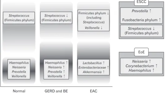
Based on results of these previous studies, the schematic dia-gram on differences in esophageal microbiome composition was observed according to the disease status in Figure 2.
Esophageal Cancer and Esophageal Microbiome
In contrast to changes toward increasing various bacterial taxa in GERD and Barrett’s esophagus, microbial diversity decreased in esophageal adenocarcinoma (EAC) when compared with the control, which enriched acid-tolerant bacteria such as Lactobacillus fermentum.23EAC development may change peritumoral micro-environment including acidity. The production of lactic acid may also further acidify the intraesophageal environment. Additionally, noxious products from these bacteria, including hydrogen peroxide, may directly inhibit the growth of other bacteria and enable Lacto-bacillus to dominate in the lower esophagus.23
A study by Snider et al32
also showed that microbial diversity decreased in patients with EAC. The proportion of Firmicutes phylum (including Strepto-coccus) increased in the low-grade dysplasia, as compared to
high-Streptococcus (Firmicutes phylum) Haemophilus Neisseria Prevotella Veillonella Haemophilus Neisseria Prevotella Veillonella Streptococcus (Firmicutes phylum) Firmicutes phylum (including Streptococcus) Veillonella Lactobacillus Enterobacteriaceae Akkermansia
Normal GERD and BE EAC
ESCC Prevotella Fusobacteria phylum Streptococcus (Firmicutes phylum) EoE Neisseria Corynebacterium Haemophilus
Figure 2. Schematic diagram of
differ-ences in esophageal microbiome compo-sition according to esophageal diseases. GERD, gastroesophageal reflux disease; BE, Barrett’s esophagus; EAC, geal adenocarcinoma; ESCC, esopha-geal squamous cell carcinoma; EoE, eosinophilic esophagitis.
grade dysplasia or adenocarcinoma. In this study, the proportion of Enterobacteriaceae and Akkermansia increased and Veillonella decreased in patients with EAC.
Until recently, characteristics of the esophageal microbiome in patients with esophageal squamous cell carcinoma (ESCC) have not been well known. However, in a recent case-control study including 25 patients with ESCC and 50 matched controls, Prevotella, espe-cially Prevotella nanceiensis, was abundant in patients with ESCC.24
Interestingly, Porphyromonas gingivalis, a periodontal pathogen, tended to increase in patients with ESCC. In a study on the oral mi-crobiome in patients with ESCC, Porphyromonas was abundant in patients with ESCC as compared to those with dysplasia as well as the normal controls.41 An association of Fusobacterium nucleatum,
one of the periodontal bacteria, with the risk of colorectal cancer has been proven.42
Another study by Shao et al31
evaluated the differ-ence in the esophageal microbiome between patients with ESCC and those with gastric cardia adenocarcinoma (GCA). Patients with ESCC showed a high proportion of Fusobacteria phylum (ESCC: 3.9% and GCA: 1.9%). Additionally, the microbiome in esophageal cancer tissue may be used for prediction of patient’s prognosis. In the previous studies, intratumoral F. nucleatum was associated with poor recurrence-free survival as well as cancer-specific survival in patients with esophageal cancer.22,33
Eosinophilic Esophagitis and Esophageal
Microbiome
EoE is a chronic immune/antigen-mediated disorder caused by T helper 2-mediated immune response triggered by food or envi-ronmental allergens.43,44
As an increase in incidence and prevalence of EoE, interest in the esophageal microbiome in patients with EoE has been increasing.43 In patients with EoE, Neisseria and
Coryne-bacterium were enriched as compared to those with non-EoE.21
In another study by Harris et al,20
the bacterial load was increased re-gardless of the treatment status or degree of mucosal eosinophilia in patients with EoE as compared to healthy individuals. Haemophi-lus was significantly abundant in patients with untreated EoE.20
Achalasia and Esophageal Microbiome
Achalasia is a motility disorder presented as dysphagia, re-gurgitation of undigested food, weight loss, and chest pain.45
It is caused by the inability to lower the esophageal sphincter to facilitate relaxation in the setting of absent peristalsis.46
The relationship between achalasia and esophageal microbiome has not been evalu-ated. Although several case reports showed the association between Mycobacterium goodii pulmonary infection and achalasia and
secondary achalasia due to human immunodeficiency viral infec-tion,47,48
evidence that support the association between achalasia and microbial composition in the esophagus of patients with achalasia were limited.
Conclusion
Owing to the advancement of next-generation sequencing tech-niques, associations between the esophageal microbiomes and vari-ous diseases have been widely investigated. Nowadays, the esopha-gus is found to be unsterile, and many bacterial taxa exist depending on the disease status. However, whether the esophageal microbiome induces esophageal diseases remains unknown. Most changes in esophageal microbiome composition may likely be a secondary change due to acid reflux, aggravation of inflammation, and other predisposing factors such as alcohol and smoking. To determine the causal relationship between esophageal microbiome and diseases, well-designed experiments using germ-free animal models are war-ranted. Nevertheless, understanding the esophageal microbiome in various diseases may have a clinical implication because oral micro-biomes are usually correlated with esophageal micromicro-biomes. We will be able to predict various esophageal diseases via oral samples that can be easily obtained compared to esophageal samples. Further re-searches will be conducted on oral and esophageal microbiomes in various esophageal diseases.
Financial support: None.
Conflicts of interest: None.
Author contributions: Chan Hyuk Park performed the litera-ture search and drafted and revised the article; Sang Kil Lee con-ceived and revised the article; and Chan Hyuk Park and Sang Kil Lee approved the final version of the manuscript.
References
1. Yang L, Chaudhary N, Baghdadi J, Pei Z. Microbiome in reflux disor-ders and esophageal adenocarcinoma. Cancer J 2014;20:207-210. 2. Corning B, Copland AP, Frye JW. The esophageal microbiome in health
and disease. Curr Gastroenterol Rep 2018;20:39.
3. Gagliardi D, Makihara S, Corsi PR, et al. Microbial flora of the normal esophagus. Dis Esophagus 1998;11:248-250.
4. Bik EM, Eckburg PB, Gill SR, et al. Molecular analysis of the bac-terial microbiota in the human stomach. Proc Natl Acad Sci USA 2006;103:732-737.
hu-man oral cavity, and the upper and lower esophagus. Dis Esophagus 2013;26:84-90.
6. Pei Z, Bini EJ, Yang L, Zhou M, Francois F, Blaser MJ. Bacterial biota in the human distal esophagus. Proc Natl Acad Sci USA 2004;101:4250-4255.
7. May M, Abrams JA. Emerging insights into the esophageal microbi-ome. Curr Treat Options Gastroenterol 2018;16:72-85.
8. Finlay IG, Wright PA, Menzies T, McArdle CS. Microbial flora in car-cinoma of oesophagus. Thorax 1982;37:181-184.
9. Mannell A, Plant M, Frolich J. The microflora of the oesophagus. Ann R Coll Surg Engl 1983;65:152-154.
10. Pei Z, Yang L, Peek RM, Jr Levine SM, Pride DT, Blaser MJ. Bacterial biota in reflux esophagitis and Barrett’s esophagus. World J Gastroenterol 2005;11:7277-7283.
11. Macfarlane S, Furrie E, Macfarlane GT, Dillon JF. Microbial coloniza-tion of the upper gastrointestinal tract in patients with Barrett’s esophagus. Clin Infect Dis 2007;45:29-38.
12. Zilberstein B, Quintanilha AG, Santos MA, et al. Digestive tract micro-biota in healthy volunteers. Clinics (Sao Paulo) 2007;62:47-54.
13. Yang L, Lu X, Nossa CW, Francois F, Peek RM, Pei Z. Inflammation and intestinal metaplasia of the distal esophagus are associated with altera-tions in the microbiome. Gastroenterology 2009;137:588-597.
14. Fillon SA, Harris JK, Wagner BD, et al. Novel device to sample the esophageal microbiome--the esophageal string test. PLoS One 2012;7:e42938.
15. Blackett KL, Siddhi SS, Cleary S, et al. Oesophageal bacterial biofilm changes in gastro-oesophageal reflux disease, Barrett’s and oesopha-geal carcinoma: association or causality? Aliment Pharmacol Ther 2013;37:1084-1092.
16. Liu N, Ando T, Ishiguro K, et al. Characterization of bacterial biota in the distal esophagus of Japanese patients with reflux esophagitis and Bar-rett’s esophagus. BMC Infect Dis 2013;13:130.
17. Amir I, Konikoff FM, Oppenheim M, Gophna U, Half EE. Gastric microbiota is altered in oesophagitis and Barrett’s oesophagus and further modified by proton pump inhibitors. Environ Microbiol 2014;16:2905-2914.
18. Yu G, Gail MH, Shi J, et al. Association between upper digestive tract microbiota and cancer-predisposing atates in the esophagus and stomach. Cancer Epidemiol Biomarkers Prev 2014;23:735-741.
19. Gall A, Fero J, McCoy C, et al. Bacterial composition of the human upper gastrointestinal tract microbiome is dynamic and associated with genomic instability in a Barrett’s esophagus cohort. PLoS One 2015;10:e0129055.
20. Harris JK, Fang R, Wagner BD, et al. Esophageal microbiome in eo-sinophilic esophagitis. PLoS One 2015;10:e0128346.
21. Benitez AJ, Hoffmann C, Muir AB, et al. Inflammation-associated mi-crobiota in pediatric eosinophilic esophagitis. Microbiome 2015;3:23. 22. Yamamura K, Baba Y, Nakagawa S, et al. Human microbiome
Fusobac-terium nucleatum in esophageal cancer tissue is associated with prognosis. Clin Cancer Res 2016;22:5574-5581.
23. Elliott DRF, Walker AW, O’Donovan M, Parkhill J, Fitzgerald RC. A non-endoscopic device to sample the oesophageal microbiota: a
case-control study. Lancet Gastroenterol Hepatol 2017;2:32-42.
24. Peters BA, Wu J, Pei Z, et al. Oral microbiome composition reflects pro-spective risk for esophageal cancers. Cancer Res 2017;77:6777-6787. 25. Deshpande NP, Riordan SM, Castaño-Rodríguez N, Wilkins MR,
Kaakoush NO. Signatures within the esophageal microbiome are associ-ated with host genetics, age, and disease. Microbiome 2018;6:227. 26. Dong L, Yin J, Zhao J, et al. Microbial similarity and preference for
specific sites in healthy oral cavity and esophagus. Front Microbiol 2018;9:1603.
27. Nobel YR, Snider EJ, Compres G, et al. Increasing dietary fiber intake is associated with a distinct esophageal microbiome. Clin Transl Gastroen-terol 2018;9:199.
28. Liu AQ, Vogtmann E, Shao DT, et al. A comparison of biopsy and mu-cosal swab specimens for examining the microbiota of upper gastrointesti-nal carcinoma. Cancer Epidemiol Biomarkers Prev 2019;28:2030-2037. 29. Okereke I, Hamilton C, Reep G, et al. Microflora composition in the
gastrointestinal tract in patients with Barrett’s esophagus. J Thorac Dis 2019;11(suppl 12):S1581-S1587.
30. Okereke IC, Miller AL, Hamilton CF, et al. Microbiota of the orophar-ynx and endoscope compared to the esophagus. Sci Rep 2019;9:10201. 31. Shao D, Vogtmann E, Liu A, et al. Microbial characterization of
esopha-geal squamous cell carcinoma and gastric cardia adenocarcinoma from a high-risk region of China. Cancer 2019;125:3993-4002.
32. Snider EJ, Compres G, Freedberg DE, et al. Alterations to the esopha-geal microbiome associated with progression from Barrett’s esophagus to esophageal adenocarcinoma. Cancer Epidemiol Biomarkers Prev 2019;28:1687-1693.
33. Yamamura K, Izumi D, Kandimalla R, et al. Intratumoral Fusobac-terium nucleatum levels predict therapeutic response to neoadjuvant chemotherapy in esophageal squamous cell carcinoma. Clin Cancer Res 2019;25:6170-6179.
34. Yu Y, Gao F, Chen X, Zheng S, Zhang J. Changes in the distal esopha-geal microbiota in Chinese patients with reflux esophagitis. J Dig Dis 2019;20:18-24.
35. Park CH, Lee JG, Lee AR, Eun CS, Han DS. Network construction of gastric microbiome and organization of microbial modules associated with gastric carcinogenesis. Sci Rep 2019;9:12444.
36. Ludwig DS, Pereira MA, Kroenke CH, et al. Dietary fiber, weight gain, and cardiovascular disease risk factors in young adults. JAMA 1999;282:1539-1546.
37. Canfora EE, Jocken JW, Blaak EE. Short-chain fatty acids in control of body weight and insulin sensitivity. Nat Rev Endocrinol 2015;11:577-591.
38. Park CH, Lee AR, Lee YR, Eun CS, Lee SK, Han DS. Evalua-tion of gastric microbiome and metagenomic funcEvalua-tion in patients with intestinal metaplasia using 16S rRNA gene sequencing. Helicobacter 2019;24:e12547.
39. Choi S, Lee JG, Lee AR, Eun CS, Han DS, Park CH. Helicobacter pylori antibody and pepsinogen testing for predicting gastric microbiome abundance. PLoS One 2019;14:e0225961.
40. Pages F, Berger A, Lebel-Binay S, et al. Proinflammatory and antitumor properties of interleukin-18 in the gastrointestinal tract. Immunol Lett
2000;75:9-14.
41. Chen X, Winckler B, Lu M, et al. Oral microbiota and risk for esopha-geal squamous cell carcinoma in a high-risk area of china. PLoS One 2015;10:e0143603.
42. Lauritano D, Sbordone L, Nardone M, Iapichino A, Scapoli L, Carinci F. Focus on periodontal disease and colorectal carcinoma. Oral Implantol (Rome) 2017;10:229-233.
43. Dellon ES. The esophageal microbiome in eosinophilic esophagitis. Gas-troenterology 2016;151:364-365.
44. Rothenberg ME. Molecular, genetic, and cellular bases for treating eo-sinophilic esophagitis. Gastroenterology 2015;148:1143-1157.
45. Boeckxstaens GE, Zaninotto G, Richter JE. Achalasia. Lancet 2014;383:83-93.
46. Pandolfino JE, Gawron AJ. Achalasia: a systematic review. JAMA 2015;313:1841-1852.
47. Martínez-González D, Franco J, Navarro-Ortega D, Muñoz C, Martí-Obiol R, Borrás-Salvador R. Achalasia and mycobacterium goodii pul-monary infection. Pediatr Infect Dis J 2011;30:447-448.
48. Wang AJ, Tu LX, Yu C, Zheng XL, Hong JB, Lu NH. Achalasia sec-ondary to cardial tuberculosis caused by AIDS. J Dig Dis 2015;16:752-753.