저작자표시-비영리-변경금지 2.0 대한민국 이용자는 아래의 조건을 따르는 경우에 한하여 자유롭게
l 이 저작물을 복제, 배포, 전송, 전시, 공연 및 방송할 수 있습니다. 다음과 같은 조건을 따라야 합니다:
l 귀하는, 이 저작물의 재이용이나 배포의 경우, 이 저작물에 적용된 이용허락조건 을 명확하게 나타내어야 합니다.
l 저작권자로부터 별도의 허가를 받으면 이러한 조건들은 적용되지 않습니다.
저작권법에 따른 이용자의 권리는 위의 내용에 의하여 영향을 받지 않습니다. 이것은 이용허락규약(Legal Code)을 이해하기 쉽게 요약한 것입니다.
Disclaimer
저작자표시. 귀하는 원저작자를 표시하여야 합니다.
비영리. 귀하는 이 저작물을 영리 목적으로 이용할 수 없습니다.
변경금지. 귀하는 이 저작물을 개작, 변형 또는 가공할 수 없습니다.
약학석사학위논문
Anti-melanogenic Activity of Gagunin D by Modulation of
Tyrosinase Expression and Degradation
티로시나아제의 발현과 분해 조절을 통한 Gagunin D 의 미백 효능 기전 연구
2017 년 2 월
서울대학교 대학원 약학과 천연물과학전공
이 호 연
i
ABSTRACT
Anti-melanogenic Activity of Gagunin D by Modulation of
Tyrosinase Expression and Degradation
Ho Yeon Lee Natural Products Science Major College of Pharmacy The Graduate School Seoul National University
Tyrosinase is the rate-limiting enzyme critical for melanin biosynthesis and controls pigmentation in the skin. Inhibition of tyrosinase has so far been the most common approach in the development of skin-whitening cosmetics. A number of tyrosinase inhibitors are used as cosmetic formulations. However, in this present study, a balance between tyrosinase expression and degradation is considered as a new target for skin whitening agent.
Gagunin D (GD), a highly oxygenated diterpenoid isolated from the marine sponge
ii
Phorbas sp., has exhibited cytotoxicity toward human leukemia cells (K562).
However, the effect of GD on normal cells and the molecular mechanisms of action remain to be elucidated. In this study, The anti-melanogenic activity of GD and its precise underlying mechanisms in mouse originated melan-a cells were identified.
GD significantly inhibited melanin synthesis in the melan-a cells without cytotoxicity and had an effect on a reconstructed human skin model composed of human epidermis and melanocytes.
Further analysis revealed that GD suppressed the expression of tyrosinase and increased the rate of tyrosinase degradation. GD also inhibited tyrosinase enzymatic activity. In addition, GD effectively suppressed the expression of proteins associated with melanosome-transfer.
These findings suggest that GD is a potential candidate for cosmetic formulations due to its multi-functional properties which replaces tyrosinase inhibitor.
Keywords: Gagunin D, Melanogenesis, Tyrosinase, Melan-a Student number: 2015-21893
iii
Table of Contents
Abstract ... i
Table of Contents ... iii
List of Figures ... v
List of Tables ... vii
I. Introduction ... 1
II. Materials and Methods ... 6
A. Materials ... 6
1. Reagents and antibodies ... 6
2. Compounds ... 6
3. Cell culture ... 7
B. Methods ... 9
1. Melanin content assay ... 9
2. MTT cell viability assay ... 9
3. Cell-free tyrosinase activity assay ... 9
4. Cellular tyrosinase activity assay ... 10
5. Western blotting ... 10
6. Real time polymerase chain reaction (PCR) ... 11
7. Immunocytochemistry ... 12
8. Transient transfection and dual luciferase assay ... 12
9. Reconstructed human skin model... 13
10. Statistical analysis ... 13
iv
III. Results ... 15
A. Inhibition of gagunin D (GD) on melanin biosynthesis in melan-a cells . 15 B. Effects of GD on melanogenesis-related proteins ... 18
C. Inhibition of GD on the expression of PAX3 and SOX10 genes ... 21
D. Inhibition of GD on the expression of MITF, tyrosinase, TRP-1 genes .. 23
E. Inhibition of GD on both cell-free and cellular tyrosinase activity ... 28
F. Effects of GD on tyrosinase degradation ... 31
G. Effects of GD on melanosome-transfer-related proteins ... 34
H. Inhibition of GD on melanin biosynthesis in a reconstructed human skin model ... 39
IV. Discussion ... 42
References ... 46
Abstract in Korean ... 51
v
List of Figures
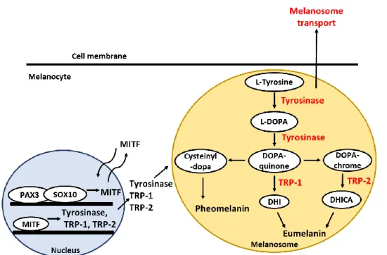
Figure 1. Schematic representation of melanogenesis signaling pathway ... 4
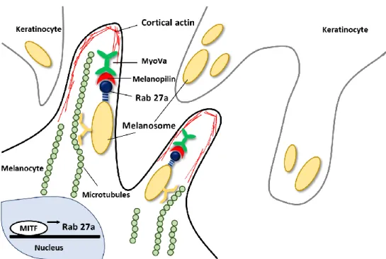
Figure 2. Schematic representation of melanosome-transfer ... 5
Figure 3. Chemical structure of gagunin D (GD) ... 8
Figure 4. Effects of GD on melanin content and macroscopic views ... 16
Figure 5. Effects of GD on cell viability and cellular morphology ... 17
Figure 6. Effects of GD on the expression of melanogenesis-related proteins ... 19
Figure 7. Effects of GD on the time course expression of MITF ... 20
Figure 8. Effects of GD on the expression of PAX3 and SOX10 genes ... 22
Figure 9. Effects of GD on the expression of MITF, tyrosinase and TRP-1 genes ... 24
Figure 10. Effects of GD on transactivation of MITF and tyrosinase in melan-a cells measured by transient transfection and dual luciferase assay ... 26
Figure 11. Effects of GD on cell-free tyrosiase activity ... 29
Figure 12. Effects of GD on cellular tyrosinase activity ... 30
Figure 13. Effects of GD on the time course expression of tyrosinase ... 32
Figure 14. Effects of GD on tyrosinase degradation ... 33
Figure 15. Effects of GD on melanosome-transfer-related proteins ... 35
Figure 16. Expression of rab27a determined by immunocytochemistry ... 36
Figure 17. Expression of melanophilin determined by immunocytochemistry .. 37
Figure 18. Expression of myoVa determined by immunocytothemistry ... 38
vi
Figure 19. Inhibition of GD on melanin biosynthesis in a reconstructed human skin model ... 40
Figure 20. Schematic representation of plausible mechanisms of action of GD in melan-a cells ... 45
vii
List of Tables
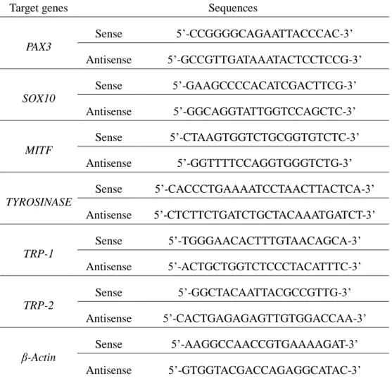
Table 1. Sequences of target gene-specific primers used in real-time PCR ... 14
1
I. Introduction
Melanin plays a protective role against the harmful effect of ultraviolet radiation (UVR) and various environmental oxidative stresses (Michaela Brenner & Vincent J. Hearing, 2008). Melanin is synthesized by melanocytes found in the basal layer of the epidermis (Gertrude-E. Costin & Vincent J. Hearing, 2016). In melanosomes, specialized intracellular organelles of melanocytes, melanin is produced by sequential enzymatic processes. Then matured melanosome moved along the melanocyte dendrites and transferred to neighboring keratinocytes in epidermis for photo-protection (Jennifer Y. Lin & David E. Fisher, 2007; Miri seiberg, 2001). Skin color is determined by the production of melanin and the distribution of melanosomes transferred by each melanocyte to its adjacent keratinocytes (Jennifer Y. Lin & David E. Fisher, 2007).
Tyrosinase, and tyrosinase-related protein 1 (TRP-1) and TRP-2 play key role in melanin synthesis and they are expressed in melanocytes. They are transmembrane glycoprotein produced in the endoplasmic reticulum and moved to a melanosomes (Ghanem Ghanem & Journe Fabrice, 2011). Tyrosinase is the rate-limiting stage in melanin synthesis. It initially hydroxylates tyrosine to 3, 4-dihydroxylphenylalanine (DOPA) and then oxidases DOPA to dopaquinone (Jennife Y. Lin & David E. Fisher, 2007; Shosuke Ito & Kazumasa Wakamatsu, 2008; Hideya Ando et al., 2004). TRP- 1 increases the eumelanin/pheomelanin, ratio facilitating the formation of carboxyl group-containing DHICA oxidase eumelanins (Ines Ferreira dos Santos Videira et al.
2012). TRP-2 known as a dopachrome tautomerase catalyzes a conversion of
2
dopachrome to 5, 6-dihydroxyindol-2-carboxylic acid (DHICA) quickly (Koichiro kameyama et al. 1995). Tyrosinase is endogeneously degraded by proteasomes (Hideya Ando et al., 2009). Ubiquitin-mediated proteasomal degradation plays a important role in the removal of normal protein, as well as in the elimination of misfolded proteins for homeostasis (Michael H. Glickman, 2000). Therefore, a balance between tyrosinase expression and degradation is considered a novel target for skin whitening.
Melanogenesis is regulated by a variety of signal transduction pathway. One of the pathways is related with microphthalmia-associated transcription factor (MITF), which has been demonstrated to upregulate tyrosinase, TRP-1, and TRP-2 through binding to an M box in their promotor sites (Ken-ichi Yasumoto et al., 1997). The expression of MITF is regulated by a lot of upstream transcription regulators, including paired box 3 (PAX3) and SRY-related HMG-box 10 (SOX10) (Nadege Bondurand et al., 2000). Moreover, MITF is also associated with melanosome- transfer by regulating the expression of rab27a, a small GTPase of the melanosome- transfer complex (Christine Chiaverini et al., 2007). The rab27a-melanophilin- myosin Va tripartite complex is essential for the distribution of melanin to basal and suprabasal keratinocytes because the complex aids the association of melanosomes with cortical actin in melanocyte dendrites (Christina Wasmeier et al. 2008;
Masahiro Hara et al. 2000). Myosin Va, which is motor protein, attaches to melanosomes through interaction with melanophilin and rab27a (Masahiro Hara et al. 2000).
In continuous efforts to identify bioactive natural products, Gagunin D (GD) from
3
a Korean sponge was identified as a potential modulator of melanogenesis from a screening of marine natural product-derived compounds. Within naturally derived compounds, marine natural products are chosen due to their promising potential as a future resource.
Marine natural products including sponges have produced various biologically active and structurally unique secondary metabolites (Gerwick, W.H. & Moore, B.S., 2012). Gagunin D (GD), one of 17 gagunins, was isolated from the sponge Phorbas sp. collected from Gagu-Do, Korea, by Shin’s group. GD also shows a unique carbon skeleton with a highly oxygenated functionality on an unusual 10, 13-bis-epi- homoverrucosane (Jung-Rae Rho et al., 2002 & Kyoung Hwa Jang et al., 2008).
Although the unique chemical structure is a quite interesting, biological activities remain to be evaluated.
In this study, Gagunin D (GD), a highly oxygenated diterpenoid, exhibits anti- melanogenic activity in melan-a cells that is associated with the modulation of tyrosinase expression and degradation as well as the regulation of melanosome- transfer.
4
Figure 1. Schematic representation of melanogenesis signaling pathway
5
Figure 2. Schematic representation of melanosome-transfer
6
II. Materials and Methods
A. Materials
1. Reagents and antibodies
Dimethyl sulfoxide (DMSO), thiazolyl blue tetrazolium bromide (MTT), bicinchoninic acid (BCA), trichloroacetic acid (TCA) and copper sulfate were purchased from Sigma-Aldrich (St. Louis, MO, USA). Roswell Park Memorial Institute (RPMI) 1640 medium, fetal bovine serum (FBS), trypsin-EDTA solution (1×), and antibiotic-antimycotic solution (100×) were purchased from Invitrogen (Carlsbad, CA, USA). Gout polyclonal anti-MITF, anti-tyrosinase, anti-TRP-1 and anti-TRP-2 were purchased from Santa Cruz Biotechnology (Santa Cruz, CA, USA).
Rabbit monoclonal anti-myosin Va was from Cell Signaling Biotechnology (Danvers, MA, USA). Goat polyclonal anti-melanophilin and mouse monoclonal anti-Rab27a were from abcam (Cambridge, MA, USA). Complete protease inhibitor cocktail was purchased from Roche Applied Science (Penzberg, Germany). The Fugene® and Dual Luciferase® Reporter Assay System were purchased from Promega (Madison, MA, USA) and New England BioLabs (Lpswich, MA, USA).
2. Compounds
Gagunin D (GD, MW = 606, Figure 3) was isolated from a marine sponge, Phorbas sp. collected from Gagu-Do, Korea by Shin (Jung-Rae Rho et al., 2002 &
Kyoung Hwa Jang et al., 2008). GD was dissolved in 100% DMSO and stored at
7
−20 °C for subsequent experiments.
3. Cell Culture
Melan-a cells (originally established by Dr. Bennett at the University of London) were kindly provided by Skin Research Institute, Amore-Pacific Co., Korea. Melan- a cells were grown in RPMI (Rosewell Park Memorial Institute) 1640 medium supplemented with antibiotic-antimyocotics (penicillin 100 unit/ml, streptomycin 100 unit/ml, amphotericin B 250 ng/ml), 10% fetal bovine serum (FBS), and 20 nM TPA. The cells were incubated at 37 °C in a humidified atmosphere of 10% CO2. Each experiment was carried out at least three times.
8
Figure 3. Chemical structure of gagunin D (GD)
9
B. Methods
1. Melanin content assay
Melan-a cells were plated in 6-well plates at a density of 10 × 10^4 cells/well.
After an additional 72 h incubation, the media were replaced with or without (control) test sample. After an additional 72 h incubation, the adherent cells exposed to the test samples were assayed. The cells were washed with Ca2+ and Mg2+-free phosphate-buffered saline (PBS) and were dissolved in 0.1ml of 1N NaOH containing 10% DMSO for 1 h at 60 °C. The optical density was measured at 475 nm.
2. MTT cell viability assay
MTT solution (final concentration of 500 µg/mL) was added to each well before incubation for 3 h at 37 °C. The medium was removed, dimethyl sulfoxide (DMSO) was added to each well and the absorbance of the dissolved formazan was measured at 570 nm. Percent survival was determined by comparison with a control group.
3. Cell-free tyrosinase activity assay
A crude enzyme source of solubilized tyrosinase was prepared (Victor Chia- Hsiang Lin et al., 2011). Melan-a cells were cultured in 100 mm dishes, and the cells were washed with ice-cold PBS and lysed with 1% (v/v) Triton-X/phosphate- buffered saline (pH 6.8). Cells were disrupted by freezing and thawing and the
10
lysates were clarified by centrifugation at 12,000 rpm for 5 min at 4 °C. The protein concentration was determined by the Bradford method. To determine the inhibitory effect of the in vitro tyrosinase activity, the reaction mixtures contained 0.1 M phosphate buffer (pH 6.8), 1.5 mM of L-DOPA, the tested concentration of GD, and 40 μg of protein in a total volume of 1 mL. After incubation at 37 °C overnight, dopachrome formation was measured at 475 nm.
4. Cellular tyrosinase activity assay
L-DOPA was used as a substrate, and the tyrosinase activity was examined by measuring dopachrome formation at 475 nm. Melan-a cells were cultured in 100 mm dishes and treated with various concentrations of GD for 24 h. The cells were washed with ice-cold PBS and lysed with 1% (v/v) Triton-X/phosphate-buffered saline (pH 6.8). Cells were disrupted by freezing and thawing and the lysates were clarified by centrifugation at 12,000 rpm for 5 min at 4 °C. The protein concentration was determined by the Bradford method. The reaction mixture was composed of 40 μg protein (adjusted to 100 μLwith 0.1 M PBS, pH 6.8) and 100 μL of 5 mM L-DOPA was added to each well of a 96-well plate. After incubation at 37 ◦C for 15 min, the absorbance was measured at 475 nm. The percentage of tyrosinase inhibition was calculated as [1-(sample OD/control OD)]×100.
5. Western blotting
Melan-a cells were treated with different concentrations of GD for the indicated
11
times. The cells were lysed with extraction buffer, and the protein concentrations were determined using the BCA method. The proteins from the cell lysates were loaded onto 10% SDS-polyacrylamide gels and then transferred to PVDF membranes (Millipore, Bedford, MA, USA). Nonspecific binding was blocked with 5% BSA in TBS containing 0.01% Tween-20 for 1 h at room temperature before incubation with diluted primary antibodies (1:1000 or 1:500) overnight at 4 °C. Then, the membranes were incubated with specific secondary antibodies (1:1000) for 1 h and washed three times with TBS-Tween 20. The expression of β-actin was used as an internal standard. Membranes were subjected to WestZol (iNtRON Biotechnology, Gyeonggi-do, Korea) and were visualized using the LAS-4000 imaging system (Fuji film Corp., Tokyo, Japan).
6. Real time polymerase chain reaction (PCR)
Melan-a cells were cultured in 100 mm dishes and treated with GD for 12 h. Total RNA was isolated using TRIzol Reagent (Invitrogen, Carlsbad, CA, USA) and was reverse-transcribed using a Reverse Transcription System (Promega, MI, USA) according to the manufacturer’s instructions. Specific gene primers were designed and synthesized by Bioneer Corporation (Daejeon, Korea). Real-time PCR was conducted using a CFX Connect Real-Time PCR Detection System (Bio-Rad, Hercules, CA, USA). Each PCR amplification included 5 μL of reverse transcription product, iQ SYBR Green Supermix (Bio-Rad, Hercules, CA, USA), and primers in a total volume of 20 μL. The following conditions were used: 95 °C for 20 s prior to
12
the first cycle; 40 cycles of 95 °C for 20 s, 56 °C for 20 s, and 72 °C for 30 s; 95 °C for 1 min; and 55 °C for 1 min. All experiments were performed in quadruplicate, and the analysis was performed using the comparative CT method with β-actin used for normalization. Sequences of primers are listed at table 1.
7. Immunocytochemistry
Melan-a cells were grown on coverglass-bottom dishes coated with 0.2% gelatin.
After treatment with GD, the cells were fixed with 4 % paraformaldehyde in PBS for 15 min. After blocking with 1% BSA in PBS for 30 min at room temperature, the cells were incubated with primary antibody at 4 °C overnight. Following incubation, the cells were incubated with fluorescence-conjugated secondary antibody for 2 h at room temperature. DAPI (0.5 mg/mL) was used to counterstain the nuclei. Images were acquired using a Leica confocal micro system (Wetzlar, Germany).
8. Transient transfection and dual luciferase assay
pMITF-Gluc and pTYR-Gluc reporter systems harboring the promoter region of MITF and TYR were provided by Amore Pacific R&D Center (Seoul, Korea).
pGL3-FL was obtained from S. Oh (Inje University, Busan, Korea). At 40-50%
confluency, the cells were washed with DPBS, and the Gaussia luciferase reporter construct pMITF-Gluc, pTYR-Gluc, and the control firefly luciferase vector (pGL3- FL) were transfected using FuGENE® HD Transfection Reagent (Promega, Madison, USA). After a 24 h incubation with GD, the cell lysates were prepared and the
13
Gaussia and firefly luciferase activity were determined by the Gaussia luciferase assay kit (New England BioLabs, Ipswich, MA) and luciferase reporter assay system (Promega, Madison, WI), respectively, according to the manufacturers’ protocols, using a luminometer (MicroLumat Plus, Berthold Technologies, Dortmund, Germany).
9. Reconstructed human skin model
The reconstructed human skin model, Neoderm®-ME, was purchased from Tego Science (Seoul, Korea) and was analyzed according to the manufacturer’s instructions. The reconstructed human skin was irradiated with 50 mJ/cm2 UVB every three days using BIO-SUN (Vilber Lourmat, Marne, France) for a total of two times to induce melanin production. After treatment of 10 μM GD for 72 h, the Neoderm®-ME was dissolved in 1 N NaOH and sonicated, and the absorbance of the supernatants at 405 nm was measured.
10. Statistical analysis
The data are presented as the mean ± standard deviation (SD) for independently performed experiments. The statistical significance was analyzed using Student’s t- test. One-way analysis of variance (ANOVA) was used to compare the mean responses among treatments.
14
Table 1. Sequences of target gene-specific primers used in real-time PCR
Target genes Sequences
PAX3
Sense 5’-CCGGGGCAGAATTACCCAC-3’
Antisense 5’-GCCGTTGATAAATACTCCTCCG-3’
SOX10
Sense 5’-GAAGCCCCACATCGACTTCG-3’
Antisense 5’-GGCAGGTATTGGTCCAGCTC-3’
MITF
Sense 5’-CTAAGTGGTCTGCGGTGTCTC-3’
Antisense 5’-GGTTTTCCAGGTGGGTCTG-3’
TYROSINASE
Sense 5’-CACCCTGAAAATCCTAACTTACTCA-3’
Antisense 5’-CTCTTCTGATCTGCTACAAATGATCT-3’
TRP-1
Sense 5’-TGGGAACACTTTGTAACAGCA-3’
Antisense 5’-ACTGCTGGTCTCCCTACATTTC-3’
TRP-2
Sense 5’-GGCTACAATTACGCCGTTG-3’
Antisense 5’-CACTGAGAGAGTTGTGGACCAA-3’
β-Actin Sense 5’-AAGGCCAACCGTGAAAAGAT-3’
Antisense 5’-GTGGTACGACCAGAGGCATAC-3’
15
III. Results
A. Inhibition of gagunin D (GD) on melanin biosynthesis in melan-a cells
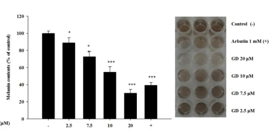
To investigate the effect of GD on melanin biosynthesis, the melanin content was measured. GD significantly decreased the melanin content in a concentration- dependent manner, with an IC50 of 12.7 μM (Figure 4). As shown in Figure 4, the inhibitory activity of 20 μM GD was stronger than that of 1 mM arbutin, a well- known skin-whitening agent. These results showed that GD inhibited melanogenesis in melan-a cells.
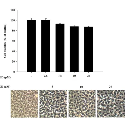
Cell viability was evaluated by MTT assay to determine the cytotoxicity of GD (Figure 5) on murine melanocytes. Melan-a cells were treated with different concentrations of GD for 72 h. GD did not exhibit significant cytotoxicity (cell viability was 89 % at 20 μM GD, Figure 5), and no remarkable morphological changes were observed.
16
Figure 4. Effect of GD on melanin content and macroscopic views
The cells were treated with the indicated concentrations of GD for 72 h. Arbutin (1 mM) was used as a positive control. And macroscopic views of the results. Data are shown as the mean ± standard deviation. *P < 0.05, *** P < 0.001 are considered statistically significant compared to the control group.
17
Figure 5. Effect of GD on cell viability and cellular morphology
Cell viability was determined by MTT assay with the indicated concentrations of GD for 72 h, and the cellular morphology was observed under a phase-contrast microscope (at 100х magnification).
18
B. Effect of GD on melanogenesis-related proteins
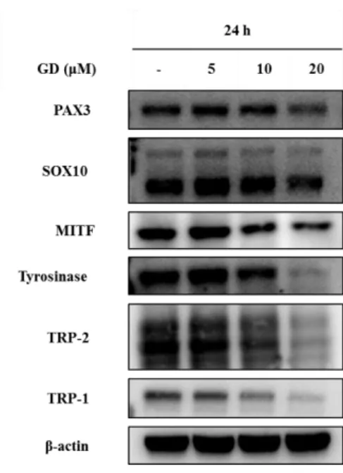
To understand the molecular mechanisms of GD in melan-a cells, expression of melanogenesis-related proteins were examined. Cells were treated with GD for 24 h, and the effects of GD on the expression of melanogenesis-related proteins were analyzed by western blot. GD effectively downregulated the protein levels of PAX3, SOX10, MITF, tyrosinase, TRP-1 and TRP-2 in concentration dependent manner (Figure 6). Expression of MITF was significantly decreased at 20 μM GD.
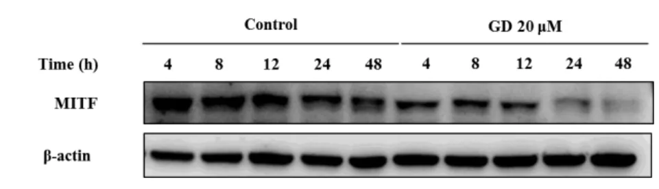
Additional analysis was performed at various time points after treatment with and without 20 μM GD. Starting at 4 h, the expression of MITF was significantly downregulated (Figure 7). Thus GD downregulates the expression of MITF, which is a key factor of melanogenesis signaling pathway.
19
Figure 6. Effects of GD on the expression of melanogenesis-related proteins Melan-a cells were treated with the indicated concentrations of GD for 24 h. The expressions of proteins were examined by western blot analysis. β-actin was used as an internal standard.
20
Figure 7. Effects of GD on the time course expression of MITF
Melan-a cells were treated for the indicated times in the presence or absence of 20 μM GD. β-actin was used as an internal standard.
21
C. Inhibition of GD on the expression of PAX3 and SOX10 genes
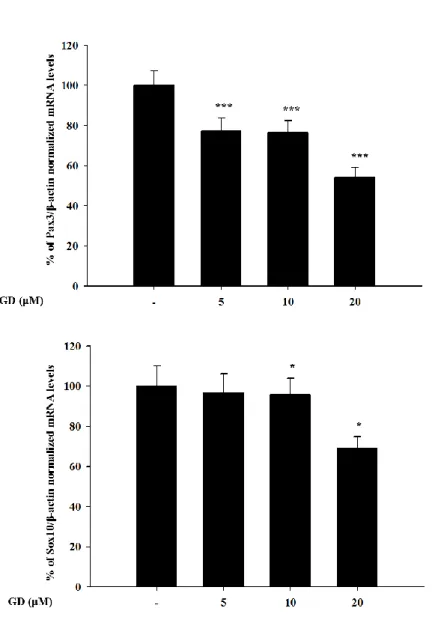
Because GD down-regulated the expressions of melanogenesis-related proteins including MITF, PAX3, SOX10, TRP-1, TRP-2, tyrosinase, it was further hypothesized that GD may affect the gene expression of PAX3 and SOX10, which regulate the expression of MITF. GD effectively downregulated the mRNA expressions of PAX3 and SOX10 at 12 h in a concentration-dependent manner (Figure 8).
This suggests that GD regulated PAX3 and SOX10 at the transcriptional mRNA level as well as the protein level. And the down-regulation of MITF might contribute to the inhibition of both PAX3 and SOX10.
22
Figure 8. Effects of GD on the expression of PAX3 and SOX10 genes
Melan-a cells were treated at various concentrations of GD for 12 h, and the mRNA levels were examined by real-time RT-PCR *P < 0.05, ***P < 0.001 are considered statistically significant compared to the control group.
23
D. Inhibition of GD on the expression of MITF, tyrosinase, TRP-1 genes
To determine whether downstream proteins of PAX3 and SOX10 were also affected by GD at gene level, real time RT-PCR was performed. GD also suppressed the mRNA expression of MITF, tyrosinase and TRP-1 in a concentration-dependent manner (Figure 9). To confirm the effects of GD on the expressions of the target genes, the dual luciferase assay was performed. GD also effectively suppressed the transcriptional activity of MITF and tyrosinase (Figure 10).
24
25
Figure 9. Effects of GD on the expression of MITF, tyrosinase and TRP-1 genes Melan-a cells were treated at various concentrations of GD for 12 h, and the mRNA levels were examined by real-time RT-PCR *P < 0.05, **P < 0.01, ***P < 0.001 are considered statistically significant compared to the control group.
26
Figure 10. Effects of GD on transactivation of MITF and tyrosinase in melan-a cells measured by transient transfection and dual luciferase assay
Melan-a cells were transfected with Gaussia luciferase reporter constructs, pMITF- Gluc and pTyrosinase-Gluc plasmid and treated with the indicated concentrations of GD for 24 h. The cells were co-transfected with the firefly luciferase control vector
27
(pFL) to normalize the transfection rates. *P < 0.05 is considered statistically significant compared to the control group.
28
E. Inhibition of GD on both cell-free and cellular tyrosinase activity
Tyrosinase is the rate-limiting enzyme for melanin synthesis. To investigate whether GD directly effects enzymatic activity, the tyrosinase activity was determined using cell-free lysates from melan-a cells as described in the ‘Materials and Methods’. As shown in Figure 11, GD significantly decreased the tyrosinase activity at concentrations that are higher than 10 µM, which indicated that GD directly inhibited the tyrosinase activity in a cell free system.
In addition, cellular tyrosinase activity was performed and GD inhibited cellular tyrosinase activity in concentration dependent manner (Figure 12).
29
Figure 11. Effects of GD on cell-free tyrosinase activity
To test whether GD had the direct effect on tyrosinase level, tyrosinase activity was measured with melan-a cell extract. Arbutin (1 mM) was used as a positive control.
Tyrosinase activity was measured by dopachrome formation from L-DOPA as a substrate. *P < 0.05, ***P < 0.001 are considered statistically significant compared to the control group.
30
Figure 12. Effects of GD on cellular tyrosinase activity
Effects of GD on cellular tyrosinase activity. Tyrosinase activity was measured by dopachrome formation from L-DOPA as a substrate. **P < 0.01, ***P < 0.001 are considered statistically significant compared to the control group.
31
F.
Effects of GD on tyrosinase degradationWhen the tyrosinase protein levels were monitored for up to 48 h, GD effectively downregulated the expression of tyrosinase protein in a time-dependent manner compared to the control cells (Figure 13). To further analyze whether the effect of GD on the expression of tyrosinase proteins was associated with the proteolytic degradation of the proteins, the levels of tyrosinase expression were monitored for up to 6 h after pretreatment with cycloheximide, a protein synthesis inhibitor (Takeshi Kobayashi & Vincent J. Hearing, 2007). Treatment with GD accelerated the proteolytic degradation of tyrosinase compared to the control groups. Next, to examine the degradation pathway by GD, a 26s proteasome inhibitor MG132 was used for pretreatment, and the tyrosinase level was determined by western blot. As shown in Figure 14, tyrosinase degradation was abrogated by the proteasome inhibitor, a multicatalytic proteinase complex that selectively degrades intracellular ubiquitinated proteins (Hideya Ando et al., 2004; Jayoung Song et al., 2015). Thus, GD accelerated the degradation of tyrosinase.
32
Figure 13. Effects of GD on the time course expression of tyrosinase
Effects of GD on the degradation of tyrosinase using western blot analysis. Melan-a cells were treated for the indicated times in the presence or absence of 20 μM GD.
β-actin was used as an internal standard.
33
Figure 14. Effects of GD on tyrosinase degradation
Effects of GD on the degradation of tyrosinase using western blot analysis. Cells were treated with 10 μg/mL cycloheximide with or without 20 μM GD for the indicated times. Cells were treated with or without 20 μM GD for 18 h after pretreatment with 10 μM MG132 for 1 h. β-actin was used as an internal standard.
34
G. Effects of GD on melanosome-transfer-related proteins
Because GD down-regulated the expression of MITF, it was hypothesized that GD may down-regulate the expression of rab27a and its related proteins. To further examine the effects of GD on melanosome transfer, the expression of melanosome- transfer-associated proteins was determined by western blot. As depicted in Figure 15, GD effectively suppressed the expression of rab27a, melanophilin, and myosin Va, which are the main melanosome-transfer-associated proteins, after treatment of GD for 48 h. The downregulation of rab27a, melanophilin and myosin Va by GD was confirmed by immunocytochemical analysis (Figure 16, Figure 17, Figure 18).
This shows that GD effects the modulation of melanin trafficking as well as synthesis of melanin.
35
Figure 15. Effects of GD on melanosome-transfer-related proteins
Melan-a cells were treated with the indicated concentration of GD for 48 h and melanosome-transfer-related proteins were analyzed by western blot. β-actin was used as an internal standard.
36
Figure 16. Expression of rab27a determined by immunocytochemistry
The cells were treated with 20 µM of GD for 24 h and then stained with rab27a and DAPI. The merged images are shown in the right panels. And effect of GD on the protein levels was determined by immunocytochemistry.
37
Figure 17. Expression of melanophilin determined by immunocytochemistry The cells were treated with 20 µM of GD for 24 h and then stained with melanophilin and DAPI. The merged images are shown in the right panels. And effect of GD on the protein levels was determined by immunocytochemistry.
38
Figure 18. Expression of myoVa determined by immunocytochemistry
The cells were treated with 20 µM of GD for 24 h and then stained with myoVa and DAPI. The merged images are shown in the right panels. And effect of GD on the protein levels was determined by immunocytochemistry.
39
H. Inhibition of GD on melanin biosynthesis in a reconstructed human skin model
To further investigate the effect of GD on melanin biosynthesis in human skin, a reconstructed human skin model, Neoderm®-ME, was employed. Cell viability was evaluated by MTT assay to determine the safety of GD on a reconstructed human skin model. Melan-a cells were treated with GD (10 μM) for 72 h. and cell viability was 90.73% compared with control group. GD did not exhibit a significant cytotoxicity.
The reconstructed human skin was irradiated with 50 mJ/cm2 UVB every three days for a total of two times to induce melanin production (Bum-Ho Bin et al., 2013).
The irradiated human skin was treated with GD for 72 h then the reconstructed human skin was dissolved in 1 N NaOH. The melanin contents were detected by absorbance at 405 nm (0.12, 0.10 respectively, Figure 19). GD significantly inhibited melanin biosynthesis in the UVB-irradiated reconstructed human skin model. Thus, GD could be applied in human skin and be a novel candidate of skin-whitening agent.
40
Figure 19. Inhibition of GD on melanin biosynthesis in a reconstructed human skin model
Cell viability using MTT assay with 10 μM GD for 72 h and the representative
41
images of reconstructed human skin model. The absorbance was measured at 405 nm after dissolving the reconstructed human skins with 1 N NaOH. *P < 0.05 is considered statistically significant compared to the control group.
42
IV. Discussion
Natural product-derived compounds are commonly used in cosmetic formulations (Francisco Solano et al., 2006). In continuous efforts to identify bioactive natural products, this study evaluated the anti-melanogenic activity of natural product and Gagunin D, a marine sponge-derived diterpenoid, was found to be active in the inhibition of melanin synthesis (Jung-Rae Rho et al., 2002; Kyoung Hwa Jang et al., 2008). Melanin biosynthesis is a multistep process, including melanin formation and melanosome transfer (Sharique A Ali et al., 2015). Therefore, there are many targets in the evaluation of skin-whitening activity. Inhibition of tyrosinase, one of the main enzymes in melanin biosynthesis, and modulation of MITF, a transcriptional factor, are common approaches in the search for bioactive sources for anti-melanogenic activity (Francisco Solano et al., 2006). Several natural product-derived compounds, such as arbutin, hydroquinone, niacinamide, have also exhibited anti-melanogenic activity, with targets of tyrosinase and MITF (Nico Smit et al., 2009). In addition, regulation of the expression and degradation of tyrosinase and inhibition of melanosome transfer are plausible targets for skin-whitening agents (Nico Smit et al., 2009; Hyo-Soon Jeong et al., 2013; Miri Seiberg et al., 2000).
Although sponges produce many chemical compounds with widely varying carbon skeletons, the molecular mode of action of most compounds remains unclear (Detmer Sipkema et al., 2004). Gagunin A-G, highly oxygenated diterpenoids isolated from the marine sponge Phorbas sp., were identified by Shin and have
43
exhibited cytotoxicity toward human leukemia cells (Jung-Rae Rho et al., 2002).
Among Gagunin A-G, Gagunin D has the potent anti-melanogenic effect. However, there has been no previous study on its effect on melanogenesis in melanocytes. Thus, the anti-melanogenic effect of GD which does not have the typical structure of conventional hypo-pigmenting agents is characterized.
In the present study, GD effectively inhibited melanin biosynthesis in both cultured melanocytes and in a reconstructed human skin model.
The mechanisms of action in the inhibitory activity of melanin synthesis by GD were also elucidated through analysis of melanogenesis-associated biomarkers. GD significantly suppressed the expression of the transcription factor MITF, which led to the suppression of target proteins tyrosinase, TRP-1 and TRP-2, major enzymes of melanin synthesis in melanocytes. Additionally, the downregulation of MITF was associated with the suppressive expression of transcription factors PAX3 and SOX10, which are well-known transcription factors for MITF. The downregulation of MITF and tyrosinase by GD was confirmed by the inhibitory promoter activities of MITF and tyrosinase through a dual luciferase assay. These findings suggest that GD affects both the expression and activity of MITF in the melanin biosynthetic pathway.
In terms of targeting tyrosinase by GD, GD effectively suppresses the expression of tyrosinase at the mRNA and protein levels and directly inhibits the enzymatic activity both in cell-free and cell-based systems. These dual functions of GD contribute to the anti-melanogenic activity of GD. The mechanisms associated with
44
the regulation of tyrosinase expression by GD in melanocytes were further elucidated.
The downregulation of tyrosinase was correlated with the acceleration of the ubiquitin-dependent proteasomal degradation of tyrosinase. These findings indicate that GD can suppress the level of tyrosinase in cells through the modulation of its expression and enhancement of degradation.
To obtain a successful skin-whitening activity, not only the regulation of de novo melanogenesis but also the modulation of melanin trafficking is an important approach. In general, matured melanin in melanocytes moves into keratinocytes in the skin, in a process called melanosome transfer. To determine whether GD affects melanosome transfer, the biomarkers associated with melanosome transfer were investigated after treatment with GD for 48 h. The protein expression of melanosome-associated biomarkers rab27a, melanophilin and myosin Va was downregulated by GD treatment. These results were confirmed by the immunocytochemical analysis of the corresponding proteins in the cells after treatment with GD. Morphologically, the dendrite-type melanocytes were e altered by GD treatment, indicating an effect on characteristic phenomena of the movement of melanocytes.
Taken together, Gagunin D, a marine natural product, exhibits anti-melanogenic activity in melanocytes through the inhibition of melanogenesis and melanosome transfer activity.
45
Figure 20. Schematic representation of plausible mechanisms of action of GD in melan-a cells
46
References
Bum-Ho Bin, Juyeon Seo, Seung Ha Yang, Eunkyung Lee, Hyunjung Choi, Kyu- Han Kim, Eun-Gyung Cho, & Tae Ryong Lee (2013). Novel inhibitory effect of the antidiabetic drug voglibose on melanogenesis. Experimental dermatology, 22, 541-546.
Christina Wasmeier, Alistair N. Hume, Giulia Bolasco, & Miguel C. Seabra (2008).
Melanosome at a glance. Journal of Cell Science, 121, 3995-3999.
Christine Chiaverini, Laurent Beuret, Enrica Flori, Roser Busca, Patricia Abbe, Karine Bille, Philippe Bahadoran, Jean-Paul Ortonne, Corine Bertolotto, &
Robert Ballotti (2008). Microphthalmia-associated transcription factor regulates rab27a gene expression and controls melanosome transport. The Journal of Biological Chemistry, 283, 12635-12642.
Detmer Sipkema, Maurice C.R. Franssen, Ronald Osinga, Johannes Tramper, &
Rene H. Wiiffels (2004). Marine sponges as pharmacy. Marine biotechnology, 7, 142-162.
Francisco Solano, Stefania Briganti, Mauro Picardo, & Ghanem Ghanem (2006).
Hypopigmenting agents: an updated review on biological, chemical and clinical aspects. Pigment Cell Res, 19, 550-571.
Gerwick, W.H. & Moore, B.S. (2012). Lessons from the past and charting the future of marine natural products drug discovery and chemical biology. Chem. Biol, 19, 85–98.
Gertrude-E. Costin & Vincent J. Hearing (2016). Human skin pigmentatino:
47
melanocytes modulate skin color in response to stress. The FASEB Journal, 21(4), 976-994.
Ghanem Ghanem & Journe Fabrice (2011). Tyrosinase related protein 1 (TYRP1/gp75) in human cutaneous melanoma. Molecular oncology, 5, 150- 155.
Hideya Ando, Hidenori Watabe, Julio C. Valencia, Ken-ichi Yasumoto, Minao Furumura, Yoko Funasaka, Masahiro Oka, Masamitsu Ichihashi, & Vincent J. Hearing (2004). Fatty acids regulate pigmentation via proteasomal degradation of tyrosinase. The Journal of Biological Chemistry, 279(15), 15427-15433.
Hideya Ando, Masamitsu Inchihashi & Vincent J. Hearing (2009). Role of the ubiquitin proteasome system in regulating skin pigmentation. Int.J.Mol.Sci, 10, 4428-4434.
Hyo-Soon Jeong, Hye-Young Yun, Kwang Jin Baek, Nyoun Soo Kwon, Kyoung- Chan Park, & Dong-Seok Kim (2013). Okadaic acid suppresses melanogenesis via proteasomal degradation of tyrosinase. Biol. Pharm. Bull., 36(9), 1503-1508.
Ines Ferreira dos Santos Videira, Daniel Filipe Lima Moura, & Sofia Magina (2013).
Mechanisms regulating melanogenesis. An Bras Dermatol, 88(1), 76-83.
Jayoung Song, Yongseok Kwon, Sanghee Kim, & Sang Kook Lee (2015). Antitumor activity of phenanthroindolizidine alkaloids is associated with negative regulation of met endosomal signaling in renal cancer cells. Chemistry &
Biology, 22, 504-515.
48
Jennifer Y. Lin & David E. Fisher (2007). Melanocyte biology and skin pigmentation.
NATURE, 445, 843-850.
Jung-Rae Rho, Hyi-Seung Lee, Chung J. Sim, & Jongheon Shin (2002). Gagunins, highly oxygenated diterpenoids from the sponge Phorbas sp. Tetrahedron, 58, 9585-9591.
Ken-ichi Yasumoto, Kouji Yokoyama, Kazuhiro Takahashi, Yasushi Tomitas, &
Shiegeki Shibahara (1997). Functional analysis of microphthalmia-associatd transcription factor in pigment cell-specific transcription of the human tyrosinase family genes. The Journal of Biological Chemistry, 272, 503-509.
Koichiro Kameyama, Chie Sakai, Sakae Kuge, Shigeo Nishiyama, Yashusi tomita, Shosuke Ito, Kazumasa Wakamatsu, & Vincent J. hearing (1995). The expression of tyrosinase, tyrosinase-related proteins 1 and 2 (TRP1 and TRP2), the silver protein, and a melanogenic inhibitor in human melanoma cells of differing melanogenic activities. Pigment Cell Res, 8, 97-104.
Kyoung Hwa Jang, Ju-eun Jeon, Sungkwang Ryu, Hyi-Seung Lee, Ki-Bong Oh, &
Jongheon Shin (2008). Polyoxygenataed diterpenes from the sponge Phorbas sp. J.Nat.Prod., 71, 1701-1707.
Masahiro Hara, Mina Yaar, H. Randolph Byers, David Goukassian, Richard E. Fine, Jessica Gonsalves, & Barbara A. Gilchrest (2000). Kinesin participates in melanosomal movement along melanocyte dendrites. J Invest Dermatol, 114, 438-443.
Michael H. Glickman (2000). Getting in and out of the proteasome. Cell &
Developmental Biology, 11, 149-158.
49
Michaela Brenner & Vincent J. Hearing (2008). The protective role of melanin against UV damage in human skin. Photochem Photobiol, 84(3), 539-549.
Miri Seiberg (2001). Keratinocyte-melanocyte interactions during melanosome transfer. Pigment Cell Res, 14, 236-242.
Miri Seiberg, Christine Paine, Elizabeth Sharlow, Patricia Andrade-Gordon, Michael Costanzo, Magdalena Eisinger, & Stanley S. Shapiro (2000). Inhibition of melanosome transfer results in skin lightening. J Invest Dermatol, 115, 162- 167.
Nadege Bondurand, Veronique Pingault, Derk E. Goerich, Nicole Lemort, Elisabeth Sock, Cedric Le Caignec, Michael Wegner, & Michel Goossens (2000).
Interaction among SOX10, PAX3 and MITF, three genes altered in waardenburg syndrome. Human Molecular Genetics, 9(13), 1907-1917.
Nico Smit, Jana Vicanova, & Stan Pavel (2009). The hunt for natural skin whitening agents. Int.J.Mol.Sci., 10, 5326-5349.
Sharique A Ali, Ram K Choudhary, Ishrat Naaz, & Ayesha S Ali (2015).
Understanding the challenges of melanogenesis: key role of bioactive compounds in the treatment of hyperpigmentory disorders. Journal of pigmentary disorders, 2, 11, 1-9.
Shosuke Ito & Kazumasa Wakamatsu (2008). Chemistry of mixed melanogenesis- pivotal roles of dopaquinone. Photochemistry and Photobiology, 84, 582- 592.
Takeshi Kobayashi & Vincent J. Hearing (2007). Direct interaction of tyrosinase with tyrp1 to form heterodimeric complexes in vivo. Journal of Cell Science,
50 120, 4261-4268.
Victor Chia-Hsiang Lin, Hsiou-Yu Ding, Shiou-Yi Kuo, Ling-Wei Chin, Jiumu-Yih Wu, & Te-Sheng Chang (2011). Evaluation of in vitro and in vivo depigmenting activity of raspberry ketone from Rheum officinale. Int. J. Mol.
Sci., 12, 4819-4835.
51
국문초록
티로시나아제의 발현과 분해 조절을 통한 Gagunin D의 미백 효능 기전
연구
서울대학교 대학원 약학과 천연물과학 전공 이호연
피부색을 결정하는 주요 색소인 멜라닌(Melanin)은 표피의 기저층에 존재하 는 멜라닌세포(Melanocyte)의 멜라노솜(Melanosome)에서 단계적 효소 반응 으로 합성된다. 그 후 성숙한 멜라노솜은 주변의 각질세포(Keratinocyte)로 이 동한다. 이동 한 멜라닌은 자외선 등 외부의 환경으로부터 보호하는 역할을 하 며 이러한 일련의 과정을 멜라닌형성(Melanogenesis)이라 한다. 멜라닌형성의 율속 단계인 티로시나아제(Tyrosinase)는 미백 화장품 개발의 타겟으로 연구 되고 있다. 티로시나아제 억제제가 많이 밝혀졌지만 실제로 상용화 되어 있는 물질은 한정적이다. 이는 In vitro 실험 결과와 임상에서의 효과가 일치하지 않
52 고 다양한 타겟을 조절하지 못하기 때문이다.
본 연구에서는 천연물 유래 물질을 스크리닝 하여 티로시나아제 억제와 분해 를 동시에 조절하는 물질을 찾았고 이를 새로운 타겟으로 제시하였다. 대상 물 질인 Gagunin D는 해면 Phorbas 종에서 분리한 Diterpenoid 계열의 화합물로 인간 만성 골수성 백혈병 세포에서 독성을 나타낸 연구가 보고된 바 있다. 그러 나 그 외 새로운 효능은 밝혀지지 않았다. 본 연구에서는 Gagunin D의 미백 효 능을 밝혔고 그 작용기전을 연구하였다.
Gagunin D는 In vitro 상에서 세포 독성 없이 멜라닌의 양을 효과적으로 억 제하였고 표피와 멜라닌 세포로 구성되어 있는 인공 피부 모델(NeodermⓇ)에 서도 멜라닌의 생성을 효과적으로 억제하였다. 티로시나아제와 그 관련 효소인 Tyrosinase-Related Protein-1(TRP-1)과 TRP-2의 발현을 농도 의존적으 로 억제하였고 이들의 전사인자인 Microphthalmia-associated transcription factor(MITF)를 mRNA, 단백질 수준에서 발현을 억제하였고, MITF의 전사인 자 중 Paired box 3와 SRY-related HMG-box 10의 발현도 농도 의존적으로 억제하였다. Gagunin D가 프로테아좀 관련 분해(Proteasomal degradation)를 통해 티로시나아제의 분해를 증가시켰다. 또한 Gagunin D가 티로시나아제의 효 소 활성 또한 억제함으로써 멜라닌 생성을 효과적으로 저해함을 알 수 있었다.
Gagunin D가 멜라노솜 분포의 주요 인자인 Rab27a, Melanophilin, MyoVa의 단백질 발현을 억제하여 이미 존재하는 멜라닌의 분포에 영향을 주는 것을 확 인하였다.
이를 통해 Gagunin D는 티로시나아제의 발현과 분해를 조절하여 멜라닌 형
53
성을 억제하고, 이미 형성 된 멜라닌의 분포를 조절하는 효능으로 다양한 타겟 을 갖는 새로운 미백 후보 물질로서의 가능성을 보였다.
주요어: Gagunin D, Melanogenesis, Tyrosinase, Melan-a 학번: 2015-21893