저작자표시-비영리-변경금지 2.0 대한민국 이용자는 아래의 조건을 따르는 경우에 한하여 자유롭게
l 이 저작물을 복제, 배포, 전송, 전시, 공연 및 방송할 수 있습니다. 다음과 같은 조건을 따라야 합니다:
l 귀하는, 이 저작물의 재이용이나 배포의 경우, 이 저작물에 적용된 이용허락조건 을 명확하게 나타내어야 합니다.
l 저작권자로부터 별도의 허가를 받으면 이러한 조건들은 적용되지 않습니다.
저작권법에 따른 이용자의 권리는 위의 내용에 의하여 영향을 받지 않습니다. 이것은 이용허락규약(Legal Code)을 이해하기 쉽게 요약한 것입니다.
Disclaimer
저작자표시. 귀하는 원저작자를 표시하여야 합니다.
비영리. 귀하는 이 저작물을 영리 목적으로 이용할 수 없습니다.
변경금지. 귀하는 이 저작물을 개작, 변형 또는 가공할 수 없습니다.
이학박사 학위논문
The role of bacteria in the pathogenesis of oral lichen planus and regulatory effect of estrogen on the physical barrier
of oral epithelium
구강편평태선 병인에서 세균의 역할과 에스 트로겐의 구강 상피 장벽 기능 조절 효과
2016년 2월 서울대학교 대학원
치의과학과 면역 및 분자미생물치의학 전공
최윤식
The role of bacteria in the pathogenesis of oral lichen planus and regulatory effect of estrogen on the physical barrier
of oral epithelium
by
Yun Sik Choi
Under the supervision of
Professor Youngnim Choi, D.D.S., Ph. D.
A Thesis Submitted in Partial Fulfillment of the Requirements for the Degree of
Doctor of Philosophy
February 2016
School of Dentistry
The Graduate School
Seoul National University
ABSTRACT
The role of bacteria in the pathogenesis of oral lichen planus and regulatory effect of estrogen on the
physical barrier of oral epithelium
Yun Sik Choi Immunology and Molecular Microbiology Major Department of Dental Science The Graduate School Seoul National University
Background
Oral lichen planus (OLP) is a mucocutaneous disease of unknown etiology that results from T-cell mediated immune responses. Many factors, such as infectious agents, drugs, autoantigens, and dental materials, have been suggested as the specific antigens that are targeted by CD8+ T cells. However, the exact etiopathogenesis of OLP is not known.
The microbial community of the host body is intimately associated with diverse host biological processes. Changes in human microbiota from a healthy state are associated with diverse localized or systemic diseases.
However, the role of bacteria in the etiopathogenesis of OLP is not known.
The role of estrogen in the regulation of epithelial homeostasis, including the regulation of physical barriers and the host immune response, has been suggested by multiple lines of evidence. However, the role of estradiol on the regulation of homeostasis of gingival epithelia has not been studied.
The aim of study was to explore the role of bacteria in OLP and the regulatory effect of 17β-estradiol on gingival homeostasis.
Methods
The sections of oral mucosal biopsies, 36 OLP and 10 controls, were subjected to in situ hybridization, which was performed using a digoxigenin-labeled eubacterial probe targeting bacterial 16S rRNA. The presence of CD4+, CD8+ T cells, and macrophages were determined by the immunohistochemistry, and correlations between the levels of in situ bacteria and the levels of infiltrated cells were determined by Spearman's rho. In addition, dual detection of bacterial signals and CD8 was performed.
Furthermore, the mucosal microbiota was analyzed by pyrosequencing the 16S rRNA and subsequent analysis.
Purified human CD4+, CD8+, and CD14+ cells were infected with 5-(and 6-) carboxy-fluorescein diacetate succinimidyl ester (CFSE)-labeled bacteria, and the presence of bacteria inside those cells was confirmed via confocal microscopy and flow cytometry. In addition, an antibiotics protection
assay was performed. Furthermore, the amounts of secreted chemokines were analyzed by ELISA and multiplex assay.
Confluent monolayers of immortalized human oral keratinocyte (HOK-16B) cells were treated with 17β-estradiol. The transepithelial electrical resistance (TER) was measured at various time points. Furthermore, the levels of TJ proteins were confirmed by real-time RT-PCR or immunofluorescence (IF). In addition, the nuclear factor kappa-light-chain-enhancer of activated B cells (NF-κB) nuclear translocation was confirmed by IF.
Results
In the oral mucosa, positive signals of bacteria were detected within the lamina propria and the epithelia, and the bacterial invasion into lamina propria was significantly increased in OLP patients. The levels of CD4+ and CD8+ T cells but not those of macrophages had a strong positive correlation with the levels of bacteria detected within the lamina propria. In addition, bacterial signals were observed within CD8- and CD8+ T lymphocytes.
Oral bacterial communities in OLP patients were substantially different compared to healthy subjects. At the species levels, the relative abundances of 42 species were significantly different between healthy and OLP patients. C.
gingivalis was significantly associated with increased OLP risk, and induced a significant decrease in TER in a time dependent manner without affecting the viability of HOK-16B cells. All selected bacteria were detected within CD4+,
CD8+, and CD14+ cells after 1 h of infection. However, only C. gingivalis could survive within CD14+ cells for 24h. All selected bacteria-induced chemokines have been implicated in OLP.
Under normal conditions, 17β-estradiol enhanced the epithelial physical barrier and induced increased levels of TJ proteins. Furthermore, pretreatment of 17β-estradiol protected against the disruption of the epithelial physical barrier function through the maintenance of TJ protein expression. In addition, 17β-estradiol inhibited NF-κB nuclear translocation via pro-inflammatory cytokine TNF-α.
Conclusion
In conclusion, increased bacterial invasion into mucosal cells/tissues and altered microbial communities may contribute to the etiopathogenesis of OLP.
Maintaining the epithelial physical barrier could be targeted by 17β-estradiol to prevent bacterial invasion into tissues and reduce the occurrence of OLP.
Keywords: Oral lichen planus, Physical barrier, Inflammation, Bacterial invasion, Estrogen, Anti-inflammatory effect
Student number: 2010-30658
CONTENTS Abstract Contents
Chapter I. Introduction
1.1. Oral lichen planus
- Figure 1. Oral lichen planus involving the buccal mucosa and tongue
1.2. Pilot study: The relationship between bacterial invasion and immune cell infiltration in periodontitis
1.2.1. Periodontitis and dysbiosis of plaque biofilm
- Figure 2. Classification of subgingival microbiota by Socransky 1.2.2. Types of bacteria and virulence factors involved in
periodontitis
1 1 3 4 5 6 1.2.3. Epithelial barrier against invading bacteria
- Figure 3. Epithelial barrier against invading bacteria - Figure 4. Adhering and tight junctions
7 8 10 1.2.4. Invasion of pathogenic bacteria into the oral epithelial cells 1.2.5. Modulation of epithelial TJ-related structures by pathogenic
bacteria
1.2.6. Pathogenesis model of periodontitis
- Figure 5. Decreased levels of TJ proteins ZO-1 by both P.
gingivalis and DSS in the gingival epithelia
- Figure 6. Increased bacterial invasion within the gingival tissues by both P. gingivalis inoculation and DSS treatment
- Figure 7. Decreased levels of TJ proteins and increased bacterial invasion in the periodontal lesions of patients with periodontitis - Figure 8. A strong positive correlation between bacterial invasion
and T cell infiltration
10 11
12 14
17
20
23
- Figure 9. Dysbiosis of oral microbiota by both P. gingivalis inoculation and DSS treatment
- Figure 10. Expression patterns of growth factor receptors in the human gingival epithelia
1.3. Estrogen
1.3.1. Steroid hormone
- Figure 11. Cyclopentanoperhydrophenanthrene 1.3.2. Biosynthesis of steroid hormone
- Figure 12. The pathway for the biosynthesis of steroid hormones 1.3.3. Estrogens
25
27
28 28 28 29 30 30 1.4. Aim of the present study
- Figure 13. Hypothesis for the present study
Chapter II. Materials and Methods
32 32 34 2.1. Study population and sample collection
2.2. In situ hybridization 2.3. Immunohistochemistry
2.4. Dual detection of bacterial signals and CD8 marker 2.5. Image analysis
34 35 36 37 38
2.6. Pyrosequencing 38
2.7. Bacterial culture 39
2.8. Human epithelial cell culture 40
2.9. Measurement of trans-epithelial electronical resistance after bacterial infection
2.10. Purification of primary human CD4+, CD8+, and CD14+
cells
2.11. Bacterial internalization into human cells 2.12. Antibiotics protection assay
2.13. Cytokine ELISA and multiplex assay
2.14. Measurement of TER under the normal condition 2.15. Measurement of TER under the pro-inflammatory cytokine-induced damaged condition
40
42
43 44 44 45 45
2.16. CCK-8 assay
2.17. Immunofluorescence staining
2.18. Real-time Reverse Transcriptional Polymerase Chain Reaction (RT-PCR)
- Table 1. Primer sequences used 2.19. Statistics
Chapter III. Results
46 47 49
50 50 51 3.1. Study population
- Table 2. Clinical information of OLP patients
- Table 3.The detail histopathologic characteristics of total 36 cases - Table 4. List of histological features adapted from Schiødt
3.2. Increased bacterial invasion in OLP lesions
3.2.1. Increased bacterial invasion into the lamina propria in OLP patients
51 51 52 53 56 56 - Figure 14. Increased bacterial invasion into lamina propria in
OLP patients
57
3.2.2. A strong positive correlation between the levels of bacteria within lamina propria and those within the epithelia in the OLP tissues
58
- Figure 15. Different pattern of bacterial detection within epithelia between control subjects and OLP patients
- Figure 16. A strong positive correlation between the levels of bacteria within lamina propria and those within the epithelia in the OLP tissues
59
60
3.3. Bacterial detection within CD4+ and CD8+ T cells 61 - Figure 17. Dual detection of bacterial signals and CD8 marker 62 3.4. A strong positive correlation of the amount of bacteria in the lamina propria with infiltration of CD4+ and CD8+ T cells, but not with macrophages
63
-
Figure 18. A strong positive correlation of the amount of bacteria 64in the lamina propria with infiltration of CD4+ and CD8+ T cells, but not with macrophages
- Figure 19. Presence of bacteria in inflamed mucosal tissues 66 3.5. Dysbiosis of oral mucosal microbiota in the OLP patients 67
- Figure 20. Increased microbial diversity and heterogeneous community in the OLP patients
68 - Figure 21. Comparison of the relative abundance of each taxon
between healthy subjects and OLP patients
70
- Table 5. Species/phylotypes that exhibit the significant changes of relative abundances in OLP compared with healthy subjects
71
3.6. Difference of bacterial invasion, infiltrated immune cells, and microbial community between OLL/OLP and OLP/OLP
72
- Figure 22. Differences of bacterial invasion and infiltrated immune cells between OLL/OLP and OLP/OLP
73
- Figure 23. PCoA plot generated using weighted Unifrac metric 75 3.7. Interaction of selected bacterial species with human oral
epithelial cells and leukocytes
76 - Figure 24. Internalization into HOK-16B cells and modulation of
epithelial physical barrier by C.gingivalis
77
- Figure 25. Bacterial internalization into CD4 +, CD8 +, and CD14+ cells
80
- Figure 26. Bacterial survival within CD4+, CD8+, and CD14 + cells
83
- Figure 27. The levels of chemokines in the medium of infected cells with selected bacteria
85 3.8. Enhanced physical barrier function by 17β-estradiol under
the normal condition
85
- Figure 28. Enhanced physical barrier function by 17β-estradiol under the normal condition
87 - Figure 29. Increased proteins levels of TJ-proteins by
17β-estradiol under the normal condition
90
3.9. Disruption of physical barrier function by TNF-α 91 - Figure 30. Disruption of physical barrier by TNF-α 92 3.10. Protective effect of 17β-estradiol on TNF-α induced-damaged epithelial physical barrier.
93 - Figure 31. Protective effect of 17β-estradiol on TNF-α
induced-damaged epithelial physical barrier
94
-
Figure 32. Protective effect of 17β-estradiol on decreased levels of TJ-proteins by TNF-α96 3.11. Inhibition of NF-κB nuclear translocation by 17β-estradiol in the TNF-α induced-damaged epithelial physical barrier
98 - Figure 33. Inhibition of NF-κB nuclear translocation by
17β-estradiol in the TNF-α induced-damaged epithelial physical barrier
99
Chapter IV. Discussion
100Chapter V. References
109국문초록
Chapter I. Introduction 1.1. Oral lichen planus
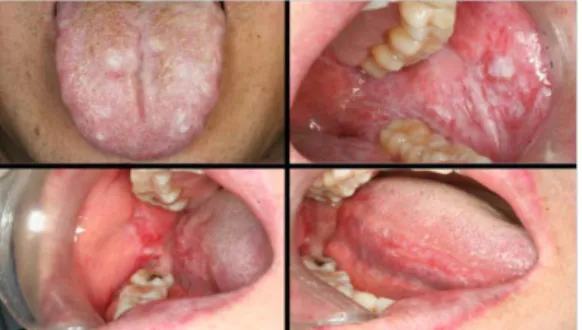
Oral lichen planus (OLP) is a mucocutaneous disease of unknown etiology resulting from T-cell-mediated immune responses. Approximately 2% of the general population are affected by this disease (Thornhill et al., 2006). OLP is observed bilaterally on the buccal mucosa, the gingiva, and the tongue (Fig.1).
OLP is classified into several types, including reticular and erosive OLP. The most common type of OLP is a reticular form that presents white keratotic striations with an erythematous region (Sugerman and Savage, 2002). The second most common type is erosive OLP presenting erythematous region and ulceration surrounded by radiating striae(Sugerman and Savage, 2002). When characteristic skin lesions are present, the diagnosis of OLP can be made with more confidence (Edwards and Kelsch, 2002).
Figure 1. Oral lichen planus involving the buccal mucosa and tongue.
1
The histopathological features of OLP include the liquefaction of the basal layer of the epithelia, band-like lymphocytic infiltration at the interface between the epithelia and submucosa, and degenerating keratinocytes (called as civatte bodies)(Sugerman and Savage, 2002; Thornhill et al., 2006). The infiltrated lymphocytes are mainly CD4+ and CD8+ T cells, and CD8+ T cells are regarded as able to mediate the degeneration/destruction of epithelial cells (Iijima et al., 2003; Sugerman and Savage, 2002). Many factors such as infectious agents, drugs, autoantigens, and dental materials have been suggested as specific antigens that are targeted by the CD8+ T cells (Iijima et al., 2003). Because the etiopathogenesis of OLP remains unclear, corticosteroid is widely used in the treatment of OLP (Edwards and Kelsch, 2002).
Oral lichenoid lesions (OLL) are regarded as variants of OLP and are often indistinguishable in symptoms. OLL is caused by certain medications or dental materials or as a manifestation of graft-vs-host disease (Edwards and Kelsch, 2002; Iijima et al., 2003). OLL lesions disappear after eliminating exposure to etiological factors. The histopathological features of OLL are not specific to one disease (Sugerman and Savage, 2002), and the essential features of OLL are damage to the basal keratinocytes including apoptosis, infiltrated inflammatory cells in the lamina propria, and hyperkeratosis(Ismail et al., 2007; McParland and Warnakulasuriya, 2012). These features are seen
2
in diverse oral diseases such as OLP, OLL, discoid lupus erythematosis (DLE), erythema multiforme, and graft-vs-host disease (Ismail et al., 2007).
The World Health Organization (WHO) presented the criteria of the clinical and histopathological diagnosis of OLP. However, the validity of an OLP diagnosis has not been tested, and there are no agreed upon criteria to distinguish the histopathological features between OLP and OLL (Thornhill et al., 2006). Therefore, making clinical and histological distinctions between OLL and OLP is difficult due to the lack of differential biomarkers.
1.2. Pilot study: Relationship between bacterial invasion and immune cell infiltration in periodontitis
OLP has something in common with periodontitis. First, OLP is a chronic inflammatory disease. Second, OLP involves the degeneration/destruction of epithelial cells, indicating the disruption of the physical barrier. Third, OLP occurs in the oral cavity, which is exposed to more than 1000 bacterial species that form a biofilm. Based on the lessons learned from the study of periodontitis, this study would apply a pathogenesis model that includes the relationship between oral bacteria, the physical barrier, and inflammatory infiltration.
3
1.2.1. Periodontitis and dysbiosis of plaque biofilm
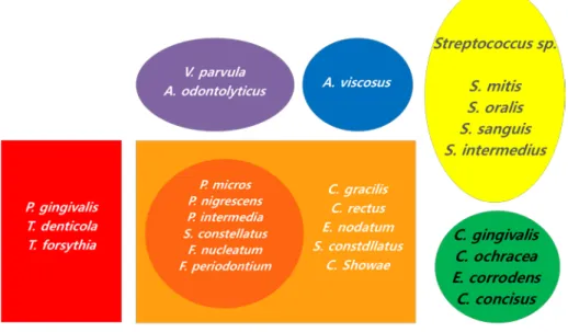
The human oral cavity is colonized by more than 1000 bacterial species (Wade, 2013). The gingival pocket is a unique interface between hard and soft tissue; oral bacteria colonize the hard tissues and form dental plaque in the gingival pocket(Matthews et al., 2007). Although some bacteria can lead to harmony with the host, certain bacteria called periodontal pathogens induce the disruption of homeostasis between bacteria and the host (Feng and Weinberg, 2006; Roberts and Darveau, 2002). Socransky classified these plaque-associated bacteria into six bacterial complexes: purple, yellow, green, orange, red, and Actinomyces(Socransky et al., 1998)(Fig. 1). In addition, Socransky found that the purple, yellow, green, and Actinomyces complexes consist of bacteria that are primary colonizers, the orange complex consists of bacteria that are bridging colonizers, and the red complex consists of bacteria that are late colonizers(Socransky et al., 1998).
4
Figure 2. Classification of subgingival microbiota by Socransky (Socransky et al., 1998).
The microbial community of the human body is intimately associated with diverse host biological process such as tissue development, immune response, and metabolism(Cho and Blaser, 2012; Galimanas et al., 2014;
Pflughoeft and Versalovic, 2012). Changes in human microbiota from a healthy state are associated with diverse localized or systemic diseases(Cho and Blaser, 2012). Periodontitis is a major chronic inflammatory disease caused by the dysbiosis of subgingival microbiota(Galimanas et al., 2014).
The initiation and progression of periodontitis is triggered by two events, and includes an increased number and altered composition of the oral microbiota
5
(Listgarten, 1988; Loe et al., 1978). Increased levels of red complex bacteria including Porphyromonas gingivalis, Tannerella forsythia, and Treponema denticola from dental plaque during the transition from health to periodontitis indicate that they have important roles in the onset and progression of periodontitis (Socransky et al., 1998). In particular, P. gingivalis, the so-called keystone pathogen of periodontitis, has been shown to induce dysbiosis in homeostatic benign microbiota in animal models (Hajishengallis et al., 2011).
1.2.2. Types of bacteria and virulence factors involved in periodontitis
The characteristics of red complex bacteria were widely studied. P. gingivalis produces diverse virulence factors as destructing enzymes, including trypsin like-protease, aminopeptidase, alkaline phospholipase, collagenase, and phospholipase A (Holt et al., 1999; Murakami et al., 2004; Nakayama, 2003).
Lipopolysaccharides (LPS), hemagglutinins, and fimbriae are also virulence factors of P. gingivalis (Holt et al., 1999). P. gingivalis has the ability to evade host immune responses such as the inhibition of phagocytosis, degradation of iron transport proteins, complements, and cytokines(Holt et al., 1999). T. denticola is a well-known periodontal pathogen. Virulence factors of T. denticola are LPS, outer surface proteins, peptidoglycan, and proteolytic
6
enzyme(Ishihara and Okuda, 1999; Masuda and Kawata, 1982; Sela, 2001). T.
denticola can suppress phagocytosis by neutrophils and the proliferation of fibroblasts. In addition, this pathogen can hydrolyze various cytokines and chemokines via dentisilin(Boehringer et al., 1984; Ji et al., 2007). T. forsythia has potent virulence factors, including secreted protein BSA, components of the bacterial surface (S)–layer, hamagglutinin, and trypsin-like proteins (Sharma, 2010). In addition, T. forsythia is resistant to antimicrobial peptide LL-37 and neutrophil phagocytosis(Ji et al., 2007).
1.2.3. Epithelial barrier against invading bacteria
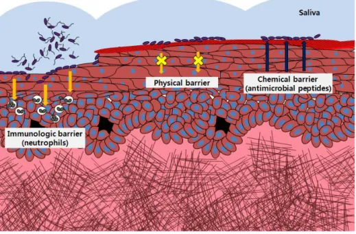
The gingival tissue is in constant close contact with plaque-associated bacteria.
Therefore, the gingival epithelia form a barrier between plaque-associated bacteria and gingival tissue, providing the first line of defense against invading bacteria. The epithelial barrier consists of physical, chemical, and immunological barriers (Fig. 3). First, gingival epithelial cells are adjoined by tight junction (TJ)-related structures, and adhering junctions (AJ) form the unique architectural integrity of the stratified epithelia, which provides a physical barrier(Franke and Pape, 2012; Hatakeyama et al., 2006). Second, a variety of antimicrobial peptides such as human beta defensins and LL-37 that are secreted by epithelial cells form a chemical barrier(Dale and Fredericks,
7
2005). Third, the immunological barrier of gingival epithelia is composed of T cells, dendritic cells, macrophages, mast cells, and neutrophils that are distributed within the epithelia, lamina propria, and/or gingival sulcus(Page, 1986). The importance of chemical barriers is also evident in patients with Kostmann syndrome, who lack LL-37 in their saliva and neutrophils and develop severe periodontitis in young adulthood(Nussbaum and Shapira, 2011). However, the role of physical barriers in the pathogenesis of periodontitis has not been studied.
Figure 3. Epithelial barrier against invading bacteria
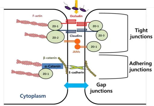
There are two types of cell junctions associated with physical barrier (Giepmans and van Ijzendoorn, 2009)(Fig. 4). AJs initiate cell–cell adhesion,
8
and provide anchoring strength below tight junctions. In addition, they regulate cytoskeleton organization and intracellular signaling. AJs are regulated by the cadherin superfamily, such as E-cadherin and catenin family members including β-catenin, and α-catenin(Hartsock and Nelson, 2008).
Although the assembly of TJs require the formation of AJs, E-cadherin is not required for the organization of TJs (Hartsock and Nelson, 2008; Niessen, 2007). TJs act as a barrier to the paracellular translocation of particles and molecules (Giepmans and van Ijzendoorn, 2009; Niessen, 2007). TJs are regulated by transmembrane proteins such as junctional adhesion molecules (JAMs), occludins, claudins, and associated cytoplasmic proteins zonula occludens (ZOs). ZO-1, ZO-2, and ZO-3 are a membrane-associated guanylate kinase homologs (MAGUK) family that includes binding domains to the proteins of AJs, TJs, and actin cytoskeleton(Hartsock and Nelson, 2008;
Niessen, 2007). ZOs are scaffolding proteins that link other TJ proteins to the actin cytoskeleton (Hartsock and Nelson, 2008; Niessen, 2007). The function of the physical barrier is associated with the expression levels and localization of TJ proteins.
9
Figure 4. Adhering and tight junctions
1.2.4. Invasion of pathogenic bacteria into the oral epithelial cells
Periodontal pathogens such as P. gingivalis, T. forsythia, A.
actinomycetemcomitans, and T. denticola have the ability to invade epithelial cells(Lamont and Yilmaz, 2002; Meyer et al., 1996; Njoroge et al., 1997;
Shin and Choi, 2012). The initial interaction between bacterial ligands and epithelial cells causes a rearrangement of cellular components, which provides pathogen entry into non-phagocytic epithelial cells (Lamont et al., 1995). An invasion of P. gingivalis into host cells is initiated when major fimbriae bind to the integrin β1 integrin receptor, which results in a signaling cascade for the
10
remodeling of the cytoskeleton to enable pathogen entry(Yilmaz et al., 2002).
Another periodontal pathogen T. forsythia invades epithelial cells by using bacterial surface protein (Bsp) A, which requires clathrin-mediated endocytosis and a phosphoinositide 3-kinase (PI3K) signaling cascade (Mishima and Sharma, 2011). In addition to the in vitro intracellular invasion of periodontal pathogens, the presence of bacteria within the gingival tissues of periodontal lesions has been repeatedly reported(Choi et al., 2013; Kim et al., 2010), suggesting an important role of bacterial invasion in the pathogenesis of periodontitis.
1.2.5. Modulation of epithelial TJ-related structures by pathogenic bacteria
Many pathogenic bacteria are known to modulate epithelial physical barriers, particularly TJ proteins such as JAMs, ZOs, occludin, and claudins, to enter host cells and/or tissues (Dickman et al., 2000; Nusrat et al., 2001; Wu et al., 2000). Inflammatory bowel disease, a chronic inflammatory disorder of the gastrointestinal tract caused by a disruptive interaction between the immune system and gut microbes, is associated with barrier dysfunction and increased epithelial permeability (Salim and Soderholm, 2011). Indeed, periodontal pathogens such as P. gingivalis, T. denticola, and A. actinomycetemcomitans
11
induce damage or remodeling of TJs and AJs of gingival epithelial cells in vitro, and the involvement of virulence factors such as gingipain, dentilisin, and cytolethal distending toxin has been shown (Cereijido et al., 2007; Chi et al., 2003; Katz et al., 2002).
1.2.6. Pathogenesis of periodontitis based on the previous study
In the previous studies (Choi et al., 2013; Choi et al., 2014), dextran sulfate sodium (DSS), a TJ-disrupting chemical, was applied onto gingival mucosa of mice in the absence or presence of P. gingivalis to investigate the role of physical barriers in the pathogenesis of periodontitis. The levels of the TJ protein ZO-1, the number of T cells, and the presence of bacteria within the gingival tissue were examined by immunohistochemistry and in situ hybridization, respectively. In addition, oral bacteria communities were analyzed via pyrosequencing.
Previous studies (Choi et al., 2013; Choi et al., 2014) showed that (1) DSS induces periodontitis in mice and that DSS- or P. gingivalis-induced impairment of TJ facilitated the invasion of oral bacteria and T cell infiltration into the gingival tissues. (2) The expression levels of ZO-1 are decreased and, the bacterial invasion of gingival tissue is increased in lesions of periodontitis
12
patients. (3) Both DSS application and P. gingivalis inoculation can induce dysbiosis of oral microbiota in mouse.
Decreased levels of TJ proteins ZO-1 by both P. gingivalis and DSS in the gingival epithelia
The physical barrier function is associated with the expression levels and distribution of TJ proteins. P. gingivalis infection altered the distribution of ZO-1 proteins and induced the rounding of cells, leading to the detachment of cell-to-cell contacts (Fig. 5a-b). DSS treatment also reduced overall levels of ZO-1 expression (Fig. 5a-b). Interestingly, mice from the DSS or P.gingivalis infection showed significantly reduced expression of ZO-1 in the junctional epithelia (Fig. 5c-d).
13
Figure 5. Decreased levels of TJ proteins ZO-1 by both P. gingivalis and DSS in the gingival epithelia. a-b) HOK-16B cells were plated and grown until a confluent monolayer. The cells were treated with vehicle, DSS (dextran sulfate sodium), P. gingivalis (Pg), or DSS+Pg for 4, 6, or 8 h. After fixation, the cells were stained for ZO-1 and examined by confocal microscopy. The fluorescence intensity of ZO-1 was analyzed by Zen 2010 software and normalized to the DAPI intensity. c-d) Six-week-old Balb/c mice received an applications of 5% DSS onto the gingival mucosa (DSS),
14
inoculation with P. gingivalis (Pg), both (DSS+Pg), or vehicles alone.
Gingival tissues from mice were stained for ZO-1 by immunohistochemistry (scale bar = 100 μm at 400x). The mean intensity of ZO-1 per region of interest was analzed by ImageJ software. *, P < 0.05 (Mann-Whitney U test).
15
Increased bacterial invasion within the gingival tissues by both P.
gingivalis inoculation and DSS treatment
Bacterial invasion within gingival tissues is associated with pathogenesis of periodontitis. When the number of invasion was counted, bacterial invasion was increased by P. gingivalis inoculation. Interestingly, DSS treatment alone also induced increased bacterial invasion within gingival tissues (Fig. 6).
16
17
Figure 6. Increased bacterial invasion within the gingival tissues by both P. gingivalis inoculation and DSS treatment. a-b) Gingival tissues from mice were stained with hematoxylin-eosin or in situ hybridized with either P.
gingivalis-specific probe (Pg) or a universial probe (U). Hybridization was also performed with each probe mixed with 10-fold excess unlabeled probe as a negative control (-). Positive signals are marked with arrows. The numbers of positive signals of universal probe were counted. *, P < 0.05 (Mann-Whitney U test).
18
Decreased levels of TJ proteins and increased bacterial invasion in the periodontal lesions of patients with periodontitis
The expression levels of ZO-1 were decreased in the periodontal lesion (Fig.
7a). In addition, the bacterial invasion of gingival tissue was increased in periodontal lesions compared with healthy sites (Fig. 7b). Indeed, bacterial invasion was inversely associated with the levels of ZO-1 in both periodontitis animal model (r = -0.252, P = 0.139) and human periodontitis (r = -0.287, P = 0.07). *, P < 0.05 (Mann-Whitney U test).
19
Figure 7. Decreased levels of TJ proteins and increased bacterial invasion in the periodontal lesions of patients with periodontitis. a) Gingival tissues from healthy subject and patients with chronic periodontitis were stained with ZO-1 by immunohistochemistry (scale bar = 500 μm at 100x, 100 μm at
20
400x). The stained signals per region of interest were analyzed using ImageJ software. b) Gingival tissues from patients with chronic periodontitis were stained with hematoxylin-esoin (H&E) or in situ hybridized with a universal probe (U). The oral epithelium (OE), sulcular/junctional epithelium (SE/JE), and connective tissues (CT) were photographed (scale bar = 50 μm). The stained bacterial signals per region of interest were analyzed using ImageJ software. *, P < 0.05; **, P < 0.01 (Mann-Whitney U test).
21
A strong positive correlation between bacterial invasion and T cell infiltration
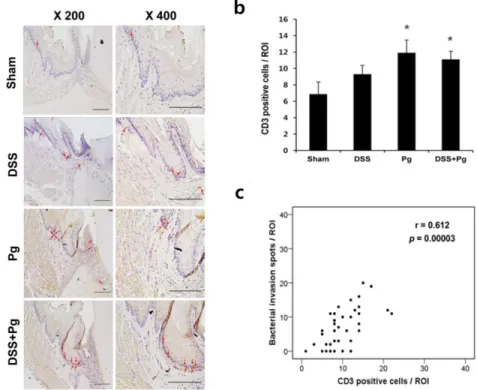
Bacterial invasion within gingival tissues drives inflammatory infiltrate. The numbers of T cells detected in the gingival tissues were increased in all of the experimental groups (Fig. 8). Notably, the number of T cells had a strong positive correlation with the number of bacteria. In addition, bacterial signals from gingival tissues of human periodontitis patients were detected near to the infiltrated immune cells (Fig. 8). Therefore, these data indicate that defect of epithelial physical barrier allows bacterial invasion into the tissues and concurrent chronic inflammation.
22
Figure 8. A strong positive correlation between bacterial invasion and T cell infiltration. a) Gingival tissues from mice were stained for CD3 by immunohistochemistry (scale bar = 200 μm). CD3-positve cells are indicated with arrows. b) The numbers of CD3-positive cells were counted at gingival tissues. *, P < 0.05 (Mann-Whitney U test). c) Two-tailed Spearmas’s rank correlations between the number of CD3-positive cells and bacterial invasion are shown.
23
Dysbiosis of oral microbiota by both P. gingivalis inoculation and DSS treatment
Human periodontitis is associated with a microbial shift in the indigenous oral flora (Galimanas et al., 2014). Although many changes were not observed at the phylum levels, dysbiosis of the bacterial community was evident in all experimental groups (Fig. 9). This data indicates that not only pathogen could trigger dysbiosis of microbiota, but also the altered characteristics of the epithelial environment could induce microbial shift.
24
Figure 9. Dysbiosis of oral microbiota by both P. gingivalis inoculation and DSS treatment. The bacterial DNA obtained from multiple mouse were
25
pooled for each group. The bacterial communities of pooled DNA samples were analyzed by pyrosequencing of amplified 16S rRNA fragments. The composition of oral microbiota at the phylum and species are shown.
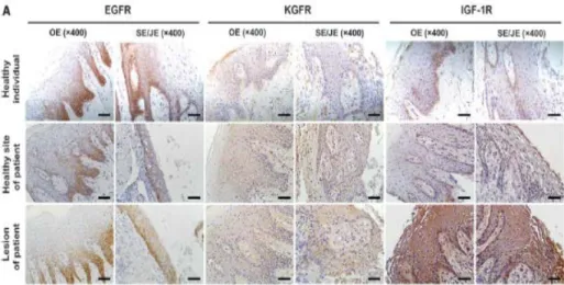
Expression patterns of growth factor receptors in the human gingival epithelia
Because epithelial physical barrier provides first defense line against invading bacteria, the maintenance of epithelial homeostasis is important. Among the growth factors that control epithelium homeostasis, epidermal growth factor (EGF), keratinocyte growth factor (KGF, also known as fibroblast growth factor 7), and insulin-like growth factor-I (IGF-I) have been implicated in the regulation of TJ proteins and paracellular permeability in lung and/or intestinal epithelia. However, the result of pilot study (Choi et al., 2014) show that almost growth factor receptor were expressed in the basal layer of gingival epithelia (Fig. 10).
26
Figure 10. Expression patterns of growth factor receptors in the human gingival epithelia. These gingival tissues were obtained from patients who had undergone periodontal surgery. Gingival tissues from nine patients were acquired during resective periodontal surgeries. When the resective surgery was performed, incision lines had to be extended into the non-inflamed adjacent tissue to gain access into the bottom of deep periodontal pockets. The gingival tissues were separated into healthy sites (n = 10) with no feature of inflammation (pocket depth [PD] ≤3mm and negative bleeding on probing [BOP] and periodontal lesions (n = 10) showing signs of tissue breakdown (mean PD: 5.2 mm) and inflammation, i.e., redness, BOP, and swelling. The expression of EGF receptor (EGFR), KGF receptor (KGFR), and IGF-1 receptor (IGF-1R) were evaluated in gingival tissues from healthy and diseased sites in patients with periodontitis by immunohistochemistry. The
27
oral epithelium (OE) and sulcular/junctional epithelium (SE/JE) were photographed (scale bar = 50 μm).
1.3. Estrogen
1.3.1. Steroid hormoneThere are two types of steroid hormone: adrenocortical and sex hormones (Nussdorfer et al., 1999). Because forms of steroid hormones are lipid, they can penetrate the cell membrane (Yang et al., 2014). Steroid hormones are synthesized in the adrenal cortex and gonads (testes and ovaries) from cholesterol; however, steroid hormones can also be produced by peripheral conversion in local tissues such as the liver and fat (Wierman, 2007). The cyclopentanoperhydrophenanthrene nucleus is the basic structure of these hormones (Fig. 11). Although the overall structure of steroid hormones is similar, each receptor can be highly specific. The basic classification of steroid hormones is classifying them by the number of carbons in their structures (Norman et al., 2004).
Figure 11. cyclopentanoperhydrophenanthrene.
28
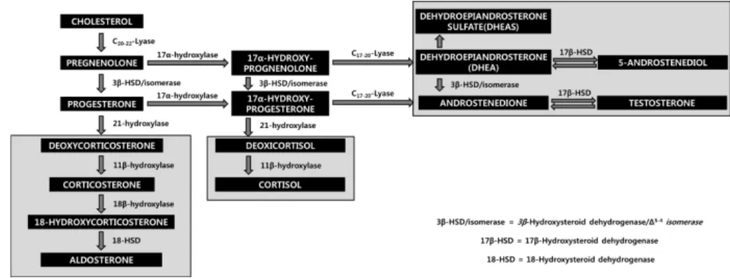
1.3.2. Biosynthesis of steroid hormone
Pregnenolone and progesterone are requisites for the synthesis of all steroid hormones. Cholesterol is converted to pregnenolone via side chain cleavage.
The 3β-hydroxysteroid dehydrogenase and △4,5-isomerase convert pregnenolone to progesterone. The pathway for the biosynthesis of steroid hormones is presented in Figure 12. Progesterone is converted to aldosterone in the adrenal zona glomerulosa cells by the 21β-hydroxylase of the endoplasmic reticulum and 11β- hydroxylase and 18β- hydroxylase of the mitochondria. 17β-hydroxylase and 21β-hydroxylase of endoplasm reticulum with 18β-hydroxylase are required in the synthesis of cortisol.
17-dehydrogenase converts progesterone to testosterone, which is a major product of the Leydig cells of the testis, and is converted to dihydrotestosterone by the 5α-reductase of endoplasmic reticulum and nucleus. Dihydrotestosterone, weak androgen, can be converted to testosterone through androstenedione, and testosterone is converted to estradiol by the aromatase system (Devlin and Wiley-Liss, 2006).
29
Figure 12. The pathway for the biosynthesis of steroid hormones.
1.3.3. Estrogen
Sex steroid hormones including estrogens, androgens, and progestins have a role in normal reproductive function and secondary sex characteristics.
Estrogens are female hormones produced by the ovaries, but they are also produced by local target cells (Wierman, 2007). Estrogens are classified into three common classes: estrone (E1), estradiol (E2), and estriol (E3). Estradiol is the most powerful and common class of estrogen (Devlin and Wiley-Liss, 2006). The functions of estrogen are mediated through two types of nuclear receptors: estrogen receptor-α (ERα) and estrogen receptor-β (ERβ). After binding to estrogens, ER induces gene transcription via the dissociation of receptor-associated proteins (Esposito, 1991). The actions of ERα are involved in classical effects such as the regulation of the menstrual cycle and secondary sex characteristics in females and the maturation of sperm in males,
30
whereas ERβ is involved in a minor role in certain estrogen target tissues (Looijer-van Langen et al., 2011).
Multiple studies have reported that changes of estrogen status in the duration of the menstrual cycle, pregnancy, and puberty are associated with the occurrence of gingivitis (Carrillo-de-Albornoz et al., 2012; CB and NF, 2006; Oh et al., 2002). In addition, estrogen deficiency is considered a risk factor for periodontitis (Haas et al., 2009). Estrogen treatment improves disease status in high-fat diet (HFD)-induced periodontitis animal model (Blasco-Baque et al., 2012). Moreover, the replacement of estrogen leads to a decreased number of periodontal pathogens in post-menopausal women and HFD-induced periodontitis animal model (Blasco-Baque et al., 2012; Tarkkila et al., 2010). Furthermore, estrogen modulates physical barrier functions and inflammatory responses in diverse epithelial cells (Chotirmall et al., 2010; Liu et al., 2005; Looijer-van Langen et al., 2011; Schaefer et al., 2005). However, the role of estrogen on the regulation of gingival epithelial homeostasis has not yet been studied.
Although almost all growth factor receptors were expressed in the basal layer of gingival epithelia (Choi et al., 2014) (Fig. 10), ERβ is expressed throughout all layers of the oral epithelia (Valimaa et al., 2004). Because estradiol is lipid-soluble, its usage would be more effective than growth factors.
31
1.4. Aim of the present study
Based on the lessons learned from the previous study regarding periodontitis, the present study hypothesized that bacterial invasion into the mucosal tissue may be the cause of the immune cell infiltration observed in OLP lesions, and estrogen would regulate gingival epithelial homeostasis (Fig. 13).
Figure 13. Hypothesis for the present study. Bacterial invasion into the mucosal tissue may be the cause of immune cell infiltration in OLP tissues and estrogen would regulate gingival epithelial homeostasis.
32
Under the hypothesis of the present study, six questions were addressed.
First, bacterial invasion within mucosal tissues was determined. Second, the correlation between bacterial invasion and immune cell infiltration was investigated. Third, bacterial composition was characterized in OLP patients and healthy subjects. Fourth, the internalization and survival of oral bacteria within immune cells was studied. Fifth, the effects of 17β-estradiol on the epithelial physical barrier were investigated. Sixth, the anti-inflammatory responses of 17β-estradiol on the gingival epithelial cells were investigated.
33
Chapter II. Materials and Methods 2.1. Study population and sample collection
This study was performed according to the Declaration of Helsinki and conformed to the STROBE guidelines. The protocol was approved by the institutional review board at the Seoul National University Dental Hospital (SNUDH) (CRI 12032). Sixteen patients diagnosed with OLP at the Oral Medicine Clinic, SNUDH in 2013 were enrolled, and an Informed Consent was obtained from all subjects. All enrolled patients had no history of antibiotics or steroid treatment within the last month, and had > 0.1 ml/min unstimulated whole salivary flow rate. Smokers were excluded. REU scoring system was adapted to examine the objective sign. It represented the reticulation/keratosis (R), erythema (E) and ulceration (U) of the lesions and it also correlated well with the pain of patients (Park et al., 2012). The numerical rating scale (NRS) was used for the subjective pain of patients (Park et al., 2012). For the mucosa sampling, a sterilized 20 mm x 20 mm polyvinylidene difluorid membrane was placed on the left or right buccal mucosa of subjects for 30 seconds. A punch biopsy was performed on the lesion with reticulation/keratosis. On histopathologic examination, 3 cases diagnosed with candidiasis or chronic mucositis were excluded, and 13 cases diagnosed with OLP or OLL were included for the study.
34
Additional tissue sections of 23 OLP cases and 10 normal oral mucosa were obtained from the tissue bank at the Department of Oral Pathology, SNUDH, after approval by the IRB at the SNUDH (CRI12023). In total, 36 OLP cases were reviewed again by an oral pathologist and scored for each histopathologic feature of OLP and OLL (Thornhill et al., 2006).
2.2. In situ hybridization
A 70-bp DNA fragment 5'-CAGGTGCTGCATGGCTGTCGTCAGCTCGTGTTGTGAAATGTTGG
GTTAAGTCCCGCAACGAGCGCAACCC-3') chosen for the well conserved area located between V6 and V7 of Escherichia coli 16S rRNA was synthesized as an oligonucleotide and amplified by PCR using the following primers: 5'-CAG GTR CTG CMT GGY-3' and 5'-AGG GTT GCG CTC GTT-3'. Amplifications were performed with following cycling conditions. 40 cycles at 94°C for 30 sec, 60 °C for 30 sec and 72 °C for 1 min 20 s followed by a 5 min extension at 72 °C. After purification, amplified products were labeled with digoxigenin (DIG)-dUTP by random priming using a DIG DNA labeling and detection kit (Roche Applied Science, Penzberg, Germany) to produce DIG-labeled probes.
35
Serial paraffin-embedded sections (4 μm) were subjected to de-paraffinization, re-hydration, and sequential pre-treatment with 0.1N HCl, 1 μg/ml proteinase K, and 0.1 M triethanolamine-HCl. The DIG-labeled probes were diluted in a hybridization buffer (4x SSC, 50% formamide, 1x Denhardt’s solution, 10% dextran sulfate, 0.1% sodium dodecyl sulfate, 0.4 mg/ml salmon sperm DNA), heated at 100 °C, and chilled on ice. After the probe was applied onto tissue sections, the slides were incubated at 90 °C and hybridized in humidified chamber overnight at 45 °C. As a negative control, hybridization was performed with the labeled probe with a 10-fold excess amount of non-labeled probe. After washing with serial SSC, the tissue sections were blocked and hybridized probes were detected with anti-DIG antibody conjugated alkaline phosphatase (Roche Applied Science). The tissues sections were treated with levamisole (Vector Laboratory, Burlingame, CA, USA) to inactivate endogenous alkaline phosphatase and visualized with premixed nitroblue tetrazolium/ 5-bromo-4-chloro-3-indolyl phosphate (NBT-BCIP) solution (Roche Applied Science). The tissue sections were then applied with methyl green as counter stain and mounted.
2.3. Immunohistochemistry
36
Following de-paraffinization, re-hydration, heat-induced antigen retrieval with citrate buffer, quenching of endogenous peroxidase with Dual endogenous peroxide block (DAKO, Santa Barbara, CA, USA), and the tissues sections were blocked with 5% bovine serum albumin (BSA). The tissue sections were then incubated with anti-CD4 antibody clone 4B12 (Monosan, Sanbio, Uden, the Netherlands), anti-CD8 antibody clone 4B11 (Serotec, Blackthorn, Bicester, England), and anti-macrophage-specific antibody clone 3A5 (Serotec). The bounded primary antibodies were detected using DAKO EnvisionTM + Dual Link System-HRP kit (DAKO). The visualized sections were then counterstained with hematoxylin (DAKO), dehydrated, and mounted.
2.4. Dual detection of bacterial signals and CD8 markers
For dual detection of bacterial signals and CD8 markers, the tissue sections were subjected to in situ hybridization of 16S rRNA first. After quenching of endogenous peroxidase with Dual endogenous peroxide block, the tissues sections were blocked with 5% BSA. After incubation of anti-CD8 monoclonal antibody for 1 h, the bounded antibodies were detected using DAKO Envision TM + Dual Link System-HRP kit. The visualized sections were then counterstained with methyl green, dehydrated, and mounted.
37
2.5. Image analyses
The signals of in situ hybridization and immunohistochemistry were quantified using imageJ software (National Institute of Mental Healath, Bethesda, MD, USA). The tissues section were photographed at x200 or x400 magnification. After defining the region of interest (ROI), the positive
signals of 3,3´-diaminobenzidine (DAB) or NBT-BCIP from original images were separated using color deconvolutions (Ruifrok and Johnston, 2001). The mean intensity of signals was then evaluated.
2.6. Pyrosequencing
Genomic DNA of mucosal samples was isolated from the polyvinylidene difluorid membranes using the PowerSoil DNA Isolation Kit (MO BIO Laboratories, Carlsbad, CA, USA). 31 mucosa samples (n=18 and n=13 for healthy subjects and patients with OLP or OLL, respectively) were subjected to pyrosequencing analysis. The amplication and sequencing of 16S rRNA genes were performed at ChunLab Inc. (Seoul, Korea) according to the previously described method (Chun et al., 2010) using a 454 GS FLX Titanum Sequencing System (Roche, Branford, CT, USA).
The basic analysis was performed following the previous description 38
(Chun et al., 2010). After elimination of PCR primer sequences, any reads including two or more ambiguous nucleotides or reads shorter than 300 bp were removed. Chimera sequences were detected by bellerophone method (Huber et al., 2004) and then were also removed. To classify the taxonomy, each read was assigned against EzTaxon database-e (http://eztaxon-e.ezbiocloud.net) (Chun et al., 2007), which includes phylotypes of both cultured and uncultured entries in the GenBank database with complete hierarchical taxonomic classification from the phylum to the species. To calculate the spcies richness and diversity index, Ribosomal RNA database project's pyrosequencing pipeline (http://pyro.cme.msu.edu/) was used. 97% similarity was the cutoff value for for assigning a sequence to the same phylotype. To equalize read size of samples for comparing different read sizes among samples, random subsampling was performed. The overall phylogenetic distance between communities was determined using weighted Fast UniFrac (Hamady et al., 2010) and represented graphically using principal coordinate analysis (PCoA).
2.7. Bacterial culture
Selected bacteria used in this study were from ATCC (American type culture collection) and KCOM (Korean Collection for Oral Microbiology).
39
Streptococcus sanguinis ATCC 804 and S. gordonii ATCC 10558 were cultured in brain heart infusion (BHI) medium at 37 °C under aerobic condition. C. gingivalis KCOM 1581 was cultured in BHI medium supplemented with 5 μg/ml of hemin (Sigma, St Louis, MO, USA) plus 10 μg /ml vitamin K under anaerobic condition (5% H2, 10% CO2, and 85% N2).
Bacteria in the log phase were obtained and washed three times with phosphate buffer saline (PBS). For application of florescence, bacteria were stained with 5-(and 6-) carboxy-fluorescein diacetate succinimidyl ester (CFSE; Molecular probes, Eugene, OR, USA).
2.8. Human epithelial cell culture
The immortalized human oral keratinocytes (HOK-16B) cells originated from retromolar gingival tissues were maintained in keratinocyte growth medium (KGM) supplemented with supplementary growth factor bullet kit (Clonetics, Sandiego, CA, USA) in an atmosphere with 5% CO2 at 37 °C. Cells were subcultured at 70-80% confluence.
2.9. Measurement of trans-epithelial electronical resistance after bacterial infection
40
HOK-16B cells were detached from 100 mm dish by trypsin-EDTA (Gibco, Carlsbad, CA, USA) and washed with pre-warmed KGM medium. The cells (1.0 x 10⁵ cells) were plated onto a 3μm-pore-size polycarbonate filter of a 24-well plate of the transwell two-chamber tissue culture system (SPL life science, Korea). Transepithelial electrical resistance (TER) was assessed using an ERS Volt-Ohm Meter (Millipore Bedford, MA USA). A resistance of epithelial layer > 10 Ω was considered to imply a tight-junctioned epithelial layer (Lux et al., 2001). The cells were cultured for 2 or 3 days with daily medium change until a confluent monolayer reached the peak resistance of about 15 Ω, and then infected with S. sanguini, S. gordonii, and C. gingivalis at the multiplicity of infection (MOI) of 500. TER was measured at 0, 6, 12, and 24 h.
For the assay of cell viability, HOK-16B cells (1.0 x 10⁵ cells) were plated. After formation of confluent monolayer, the cells were infected with S.
sanguinis, S. gordonii, and C. gingivalis at MOI 500 for 24 hours. Next, 20 μl of CCK-8 solution (Dojindo, Kumamoto, Japan) was applied to each well.
The cells were further incubated for 1 h, and then absorbance was measured at 450 nm using a VERSAmax Tunable microplate reader (Molecular devices, Sunnyvale, CA MA, USA). The cell viability was calculated as a relative percentage of vehicle control.
41
2.10. Purification of primary human CD4
+, CD8
+, and CD14
+cells
Peripheral blood purchased from the Red Cross was used and the use of human material was approved by the IRB at Seoul National University, School of Dentistry (IRB No.S-D20150007). Peripheral blood diluted in Dulbecco’s phosphate buffer saline (DPBS) at 1:1 was layered on Ficoll-Hypaque (Amersham biosciences, Uppsala, Sweden) at 720g for 30 min. Buffy coat layer containing lymphocytes and monocytes was separated and washed three times with complete RPMI medium (10% heat inactivated FBS, 2 mM L-glutamine, 25 μM 2-mercaptoethanol, and 100 U/ ml penicillin/streptomycin). Peripheral blood mononuclear cells (PBMCs) were re-suspended in RPMI complete medium. To purify CD14+ cells, isolated human PBMCs were incubated with anti-human CD14 magnetic particles on the BD IMagnetTM (BD) for 30 min. After removing the medium containing negative fraction, CD14 positive fractions are collected and re-suspended in the fresh RPMI medium. To purify CD4 + and CD8+ cells, CD14+ cells-depleted PBMC were further incubated with anti-human CD4 magnetic particles (BD) and anti-human CD8 magnetic particles (BD) for 30 min on the BD IMagnetTM. After removing the medium containing negative fraction, positive fractions for each cell are collected and re-suspended in the fresh
42
RPMI medium. After washing three times with RPMI without antibiotics, the cells were plated.
2.11. Bacterial internalization into human cells
HOK-16B cells plated at a density of 3 x 10⁴ cells cm-2 onto 24-mm diameter glass cover slips (Fisher Scientific, Houston, TX) were infected at 70%
confluence with the CFSE-labeled bacteria at MOI 1000 for 4 h. Purified human CD4+, CD8+, or CD14+ cells (2.5 x 10⁵ cells) in RPMI medium with 10% FBS were infected with the CFSE-labeled bacteria at MOI 1000 without antibiotics for 1 h. After fixation with 4% paraformaldehyde (PFA) for 30 min and permiabilization with 0.3% PBST for 10 min, the infected cells were stained with rhodamine-phalloidin (Molecular probes) and Hochest 33342 (Molecular probes) for 30 min. After washing with distilled water, the leukocytes were attached onto collagen-coated slides. Mounted slides were imaged using a Zeiss LSM700 (Carl Zeiss, Oberkochen, Germany) with serial z-sections.
For flow cytometric analysis, the infected cells were washed with PBS and resuspended in trypan blue (400 mg ml-1 prepared in 0.85% saline solution) to quench the fluorescence of the bacteria bound on the surface. The cells were analyzed using a FACSCalibur (BD biosciences). The cells were
43
gated first on the appropriate population based on the forward vs. side scatters and then on the live cells based on the FL-3 fluorescence of trypan blue. The cells were fixed with 3.7% formaldehyde and infected with the same MOI of CFSE-labeled bacteria served as a negative control.
2.12. Antibiotics protection assay
To examine the persistence of intracellular bacteria, purified human CD4+, CD8+, or CD14+ cells (5.0 x 10⁵ cells) were infected with selected bacterial species at MOI 1000 for 1 and 24h in the presence of gentamycin (50 μg ml-1).
The infected cells were washed with PBS and lysed with sterile distilled water containing 0.5% saponin. After washing with PBS, the lysates were plated onto blood agar plate with hemin and vitamin K under an appropriate atmosphere for 2-3 days.
2.13. Cytokine ELISA and multiplex assay
The levels of chemokines in the culture supernatant of HOK-16B, CD4+, CD8+, and CD14+ cells were measured by ELISA and multiplex assay.
HOK-16B, CD4+, CD8+, and CD14+ cells were infected with the three bacterial species for 1 h and further cultured in the presence of gentamycin
44
(50 μg ml-1) for 23 h. The culture supernatant of infected cells and non-infected control cells was obtained and stored at -80 °C until use. The amounts of CXCL8 (IL-8) secreted into the medium during infection with selected bacteria were measured using ELISA kit (R&D systems, Minneapolis, MN, USA) following manufacture’s introduction. The amounts of CCL3 (MIP-1α), CCL5 (RANTES), and CXCL10 (IP-10) were determined using Multiplex assay kit (R&D systems) according to manufacture’s instruction.
2.14. Measurement of TER under the normal condition
To investigate the effect of 17β-estradiol on the gingival epithelial barrier in the normal condition, HOK-16B cells (4.0 x 10⁴ cells) were plated onto a 3μm-pore-size polycarbonate filter of a 24-well plate of the transwell two-chamber tissue culture system one day before 17β-estradiol treatment.
The cells were treated with 0.2, 2, or 2 nM 17β-estradiol treatment. TER was measured using an ERS Volt-Ohm Meter at 6, 12, 24, 48, 72, and 96 h.
2.15. Measurement of TER under the pro-inflammatory cytokine-induced damaged condition
45
To examine the effect of TNF-α on the gingival epithelial barrier, HOK-16B cells were cultured for 2 or 3 days with daily medium change until a confluent monolayer reached the peak resistance. Tight-junctioned monolayers of HOK-16B were treated with TNF-α (10-250 ng/ml) (R&D systems) for 24 h.
TER was assessed using an ERS Volt-Ohm Meter at 0, 2, 4, 8, and 24 h.
To determine the protective effect of 17β-estradiol on the gingival epithelial barrier under the pro-inflammatory cytokine-induced damaged condition, tight-junctioned monolayers of HOK-16B were pre-treated with either ICI 182,780 (125 μM) (Sigma-Aldrich, St. Louis, MO, USA) for 2 h or 17β-estradiol (2-20 nM) for 4 h. The cells were then treated with TNF-α (100 ng/ml) (R&D systems) for 24 h. TER was measured using an ERS Volt-Ohm Meter at 24 h.
2.16. CCK-8 assay
To investigate the effect of 17β-estradiol on the proliferation of gingival epithelial cells under the normal condition, HOK-16B cells (4 x 10⁴ cells/well) were plated in 96-well plate. The cells were treated with 17β-estradiol for 0, 6, 12, 24, 48, 72, and 96 h.
46
To examine the effect of 17β-estradiol on the viability of gingival epithelial cells under the pro-inflammatory cytokine-induced damaged condition, tight-junctioned monolayers of HOK-16B were treated with TNF-α (10-250 ng ml-1) for 24 h in the absence or presence of 17β-estradiol (2-20 nM) or ICI 182,780 (125 μM).
After treatment for indicated time point, CCK-8 solution was added.
The cells were further incubated for 1 h, and then absorbance was measured at 450 nm using a VERSAmax Tunable microplate reader. The cell viability was calculated as a relative percentage of vehicle control.
2.17. Immunofluorescence staining
To determine the effect of estrogen on the expression levels of ZO-1 and JAM-A under the normal condition, HOK-16B cells were plated at 2 x 10⁵ cells onto 12mm diameter collagen coated cover slips in 24-well plate (Fisher Scientific, Houston, TX, USA).
For the pro-inflammatory cytokine-induced damaged condition, HOK-16B cells (5 x 10⁵ cells) were plated onto 12mm diameter collagen coated cover slips in 24-well plate. Tight-junctioned monolayers of HOK-16B
47
were incubated with TNF-α (20-100 ng ml-1) for 1 or 24 h in the absence or presence of 17β-estradiol (2-20 nM) or ICI 182,780 (125 μM).
After fixation with 4% paraformaldehyde (PFA), the cells were treated with 50 mM ammonium chloride for 10 min to quench autofluorescence.
After permiabilization with 0.3% PBST and blocking with 5% BSA, the cells were incubated with rabbit anti-ZO-1 polyclonal antibody (Invitrogen, Carlsbad, CA, USA), mouse anti-JAM-A antibody clone 2E3-1C8 (Abnova, Taipei, Taiwan), and anti-NF-κB p65 polyclonal antibody (Biolegend, San Diego, CA, USA) overnight at 4°C, and then with Alexa 488 anti-rabbit antibody (Invitrogen) or Alexa 555 anti-mouse antibody (Invitrogen) for 1 h.
After washing with PBS, the cells were counterstained with Hochest 33342 and mounted. For each slides, five areas were photographed at 100x magnification using a Zeiss LSM700 with serial Z-section. Maximum intensity projections were obtained by combining the serial Z-section. The fluorescence intensity of ZO-1 and JAM-A was analyzed using ZEN 2010 (Carl Zeiss) and normalized to the fluorescence intensity of Hochest 33342.
For quantification of nuclear translocation of NF-κB, co-localized signals of nucleus and NF-κB p65 were measured using ImageJ software. Experiments were repeated twice.
48
2.18. Real-time Reverse Transcriptional Polymerase Chain Reaction (RT-PCR)
RNA from HOK-16B cells was obtained using EasyBLUE (iNtRON, Seoul, Korea). Total RNA (2 μg) was subjected to reverse transcription (RT) using oligo dT and the M-MLV Reverese trascrpitae enzyme (Promega, Madison, WI, USA) in a 30 μl reaction mixture at 42°C for 1 h. Real time PCR was performed in a 20 μl reaction including each primer (0.4 μl), SYBR Premix EX Taq (10 μl), ROX II reference dye (0.4 μl), and 2 μl template cDNA.
Sequences of each primer are listed in table 1. Amplification was performed using fluorescence thermocycler (Applied Biosystem 7500 Real-time PCR, Foster City, CA, USA) under the following conditions: initial denature at 95 °C for 4 min, followed by 35 cyles of denatureation at 95 °C for 15 s, annealing at 60 °C for 15 s, and elongation at 72 °C for 33 s. The glyceraldehyde 3-phosphate dehydrogenase (GAPDH) was amplified in parallel with gene of interest. Relative copy numbers of TJ proteins in comparison with GAPDH were calculated using 2-△Ct. Real-time PCR was performed in triplicate for each of cDNA samples.
49
Table 1. Primer sequences used.
Primer Orientation Sequence (5'-3')
GAPDH Forward CAGCCTCAAGATCATCAGCA
Reverse CCATCCACAGTCTTCTGGGT
ZO-1 Forward GGTCAAGGTCAAGCTCAT
Reverse CTGAGTAAGGCAAATGCAG
JAM-A Forward GAACGAGGCATCATCCCTAA
Reverse CCAGCTTCTCGAAGAACCAC
2.19. Statistics
Data were analyzed using Mann-Whitney U test to determine the differences between the healthy subjects and patients with OLP in the clinical study. To the differences between control and experimental groups, two-tailed non-paired Student’s t-test was performed. One-way ANOVA with Tukey’s post hoc was used to assess the difference between groups. Logistic regression analysis was used to examine the possibility of OLP risk by relative abundances of bacterial species in oral mucosa. The associations between two parameters in clinical study were analyzed by Speraman’s rank correlation.
All analyses were performed using SPSS 12.0 (SPSS Inc, Chicago, IL, USA).
50
Chapter III. Results 3.1.
Study populationFor this study, the mucosal bacterial samples and biopsies were obtained from 13 new patients diagnosed as OLP in the Oral Medicine Clinic, SNUDH.
Among the 13 cases, 7 cases were diagnosed as OLL by one or two pathologists based on the histopathologic features of OLL (Table. 3 and 4) (Thornhill et al., 2006). The detail clinical information of the 13 patients is summarized in Table 2. Tissue sections of 23 OLP cases, which present the typical histopathologic features of OLP and no ulceration in the epithelium of biopsied tissues, and 10 normal oral mucosa were additionally obtained from the tissue bank at the Department of Oral Pathology, SNUDH. The detail histopathologic characteristics of total 36 cases are summarized in Table 3.
Table 2. Clinical information of OLP patients
51
Table 3. The detail histopathologic characteristics of total 36 cases
52
Table 4. List of histological features adapted from Schiødt(Thornhill et al., 2006)
Histological features Epithelium – keratinization
1. Hyperorthokeratosis 2. Hyperparakeratosis
Epithelium – thickness and configuration
3. Atrophy; reduction in thickness more than 1/3 of normal area
4. Acanthosis; broadening of rete ridges more than two time normal width for area
5. Simple hyperplasia; thickness more than 1 1/2 time normal thickness for area, excluding stratum corneum
6. Atrophy alternating with hyperplasia 7. Finger-like rete ridges
8. Sawtooth rete ridges Fusobacterium nucleatum Epithelium – other
9. Thick stratum granulosum, more than five cell layers thick
10. Migration by leucocytes; easily visible small groups or heavy infiltration by leucocytes
11. Liquefaction degeneration of basal layer
12. Intraepithelial vesicles; subepithelial vesicles excluded epithelial cellular
53
changes
13. Colloid bodies – civatte bodies
14. Multinucleated epithelial cells; cells containing three or more nuclei 15. Hyperchromatism
16. Epithelial dysplasia; slight, moderate or severe
17. Supra-basilar apoptosisa (connective tissue – superficial inflammatory infiltrate)
18. Band-shaped infiltrate, some areas 19. Band-shaped infiltrate, all areas 20. Not band-shaped infiltrate, some areas 21. Not band-shaped infiltrate, all areas 22. Focal/perivascular infiltrate
23. Intensity of inflammatory infiltrate: moderate or heavy (connective tissue – deep inflammatory infiltrate)
Connective tissue – deep inflammatory infiltrate
24. Deep inflammatory infiltrate; located deep to superficial infiltrate, some or all areas
25. Focal/perivascular infiltrate
26. Intensity of inflammatory infiltrate: none 27. Intensity of inflammatory infiltrate: slight
Connective tissue – cell types of inflammatory infilt