저작자표시-비영리-변경금지 2.0 대한민국 이용자는 아래의 조건을 따르는 경우에 한하여 자유롭게
l 이 저작물을 복제, 배포, 전송, 전시, 공연 및 방송할 수 있습니다. 다음과 같은 조건을 따라야 합니다:
l 귀하는, 이 저작물의 재이용이나 배포의 경우, 이 저작물에 적용된 이용허락조건 을 명확하게 나타내어야 합니다.
l 저작권자로부터 별도의 허가를 받으면 이러한 조건들은 적용되지 않습니다.
저작권법에 따른 이용자의 권리는 위의 내용에 의하여 영향을 받지 않습니다. 이것은 이용허락규약(Legal Code)을 이해하기 쉽게 요약한 것입니다.
Disclaimer
저작자표시. 귀하는 원저작자를 표시하여야 합니다.
비영리. 귀하는 이 저작물을 영리 목적으로 이용할 수 없습니다.
변경금지. 귀하는 이 저작물을 개작, 변형 또는 가공할 수 없습니다.
A Dissertation for the Degree of Doctor of Philosophy
Anti-Cancer Mechanisms of MICA Nanoparticle or PDE4D Inhibitor
in Human Cancer Cells
MICA PDE4D
By
Tae-Wook Kang
August 2016
Program in Zoonotic Animal Disease, College of Veterinary Medicine,
Graduate School of Seoul National University
i
ABSTRACT
Anti-Cancer Mechanisms of MICA Nanoparticle or PDE4D Inhibitor
in Human Cancer Cells
Tae-Wook Kang
Program in Zoonotic Animal Disease, College of Veterinary Medicine,
Graduate School of Seoul National University
Supervisor: Kyung-Sun Kang, D.V.M., Ph.D.
Glioblastoma multiforme (GBM) and breast cancer are the most prevalent malignant tumors in adults, and both exhibit high fatality rates due to their tumorigenic potentials and limitations of cancer therapy. In this study, I investigated on tumor suppressive and immunostimulatory effects using novel small molecule, CG500354, and natural compound, STB-HO.
ii
In the first part of this study, I performed forced differentiation of cancer cells, which is a recently developed approach in developing a cancer therapy. I demonstrated that the novel phosphodiesterase-4 subtype D (PDE4D) inhibitor, CG500354, induces growth arrest and neural differentiation in GBM-derived cells by triggering the activation of cAMP/PKA signaling pathway. Treatment of CG500354 regulated cAMP/PKA signaling pathway by interrupting PDE4 interference and up-regulating phosphorylated protein kinase A (p-PKA) and phosphorylated CREB (p-CREB). Furthermore, PDE4D inhibitor suppressed the expression level of cyclin B1, while up-regulating p21 and p27, which are known to be associated with growth arrest. I next investigated whether PDE4D inhibitor affect neural differentiation of GBM. GFAP, an astrocyte marker, and Tuj-1, a neuronal marker, were significantly increased with p53 expression level in GBM while the expression of nestin, a neural progenitor marker was decreased. These results suggest that CG500354 may play crucial roles in neural differentiation and growth arrest through regulation of cell-cycle-related and neural differentiation markers.
In the second part of this study, I investigated on the regulation of interactions between tumor cells and anti-tumor immune cells by treating with mica nanoparticle, STB- HO. Its efficacy and mechanisms in treating various types of tumor are less verified and the mechanistic link between anti-tumor and immunostimulatory effects has not been elucidated. STB-HO was orally administered into MCF-7 xenograft model. The growth of MCF-7 cell in xenograft model was significantly suppressed, whereas STB-HO did not directly affect the proliferation and apoptosis in vitro. Thus, I observed the interactions of MCF-7 and macrophage, dendritic cells (DCs) and natural killer (NK) cells after STB-HO
iii
treatment to investigate the discrepancy between in vivo and in vitro. The MHC class I, which is known as an inhibitory factor for NK cell-mediated anti-tumor effect, was down- regulated approximately by 10% when treated with STB-HO. Thus, I induced macrophage- like cells from THP-1 and dendritic cells from CD14+ monocytes to analyze the functional alteration of STB-HO. I observed that IL-12, known for its contribution in NK cell cytotoxicity against tumor cell, was consistently increased by STB-HO treatment. I next isolated NK cells from human blood and performed cytotoxicity test by co-culturing NK cells with fluorescence-labeled MCF-7. The dead cell population of MCF-7 rose up to 2%
to 7% and additional STB-HO treatment on co-culture condition rose dead cell proportion up to 16-17% along with the elevation of significant IFN-γ expression. I observed that STB- HO not only increased susceptibility of MCF-7 cells to immune cells, but also stimulated immunocytes in tumor microenviroment to eliminate cancer cells.
Taken together, these findings provide insights that CG500354 activated cAMP/PKA signaling pathway by blocking PDE4D, which triggered neural differentiation and growth arrest of glioblastoma-derived cancer cells. STB-HO regulated the interaction of tumor with its immune microenvironment by enhancing the functions of NK cell, macrophage and DCs to attack tumor cells by secreting IFN-γ and IL-12.
Keywords : Glioblastoma, PDE4D inhibitor, cAMP, Breast Cancer, Mica nanoparticle, Tumor-suppressive effect
Student number : 2012-31103
iv
LIST OF ABBREVIATIONS
BMP4
Bone morphogenetic protein 4cAMP
Cyclic adenosine monophosphateCOX-2
Cyclooxygenase-2CREB
cAMP response element-bindingCSCs
Cancer stem cellsDCs
Dendritic cellsDMSO
Dimethyl sulfoxideEGF
Epidermal growth factorEP
E-series of prostaglandinERα
Estrogen receptorα
FN
FibronectinGBM
Glioblastoma multiformGFAP
Glial fibrillary acidic proteinHIF1α
Hypoxia-inducible factor 1αIL-1
Interleukin-1MACS
Magnetic-activated cell sortingMCF-7
Michigan Cancer Foundation-7v
MHC class I
Major histocompatibility complex class IMNC
Mononuclear cellNF-κB
Nuclear factor-κBNK
Natural killerPDE4
Phosphodiesterase-4PDE4D
Phosphodiesterase-4 subtype DPGE2
Prostaglandin E2PKA
Protein kinase APLO
Poly-ornithineRT-PCR
Reverse transcription-polymerase chain reactionSTAT3
Signal transducer and activator of transcription 3TNF
Tumor necrosis factorTuj-1
Neuron Class III β-tubulinUCB
Umbilical cord bloodvi
TABLE OF CONTENTS
ABSTRACT --- i
LIST OF ABBREVIATIONS --- iv
TABLE OF CONTENTS --- vi
LITERATURE REVIEW --- xii
Chapter I --- Growth arrest and forced differentiation by phosphodiesterase 4 type D inhibitor in human primary glioblastoma multiforme 1 1.1 INTRODUCTION --- 2
1.2 MATERIALS AND METHODS --- 6
1.2.1 Isolation of primary GBM cells --- 6
1.2.2 Chemical --- 6
1.2.3 Clonogenic assays --- 7
1.2.4 Magnetic-activated cell sorting --- 7
1.2.5 Flow cytometry analysis --- 7
1.2.6 cAMP measurement --- 8
vii
1.2.7 siRNA inhibition study --- 8
1.2.8 Quantitative real-time PCR --- 8
1.2.9 Western blot analysis --- 9
1.2.10
Immunocytochemistry --- 9
1.2.11
Animal experiment --- 9
1.2.12
Statistical analysis --- 10
1.3 RESULTS --- 11
1.3.1 CG500354 induces differentiation in human primary GBM-derived neurospheres without cytotoxic effects --- 11
1.3.2 CG500354 induces growth arrest of GBM-derived cell populations by up-regulating p53 --- 15
1.3.3 CG500354 induces the neural differentiation of GBM- derived cells --- 21
1.3.4 CG500354 leads GBM-derived cells to growth arrest by accelerating the cAMP/CREB signaling pathway --- 23
1.3.5 Mimetic substances and si-PDE4D, mimic the effect of CG500354 on the neural differentiation of GBM-derived cells --- 28
1.3.6 In vivo GBM-derived cells from tumor were induced to
neural differentiation by CG500354 injection --- 32
viii
1.4 DISCUSSION --- 35
ix
Chapter II ---
Mica Nanoparticle Eliminates the Human Breast
Carcinoma Cells by Regulating the Interaction of Tumor with its Immune Microenvironment
39
2.1 INTRODUCTION --- 40
2.2 MATERIALS AND METHODS --- 43
2.2.1 Reagents --- 43
2.2.2 MCF-7 xenograft model --- 43
2.2.3 Histological evaluation --- 44
2.2.4 Aluminum staining of dissected tumor sections --- 44
2.2.5 Western blot analysis --- 44
2.2.6 Proliferation assay --- 45
2.2.7 Apoptosis assay --- 45
2.2.8 Flow cytometric assay --- 46
2.2.9 Cytokine production --- 46
2.2.10
Differentiation of THP-1 into macrophage-like cells --- 47
2.2.11
Isolation and culture of human umbilical cord blood (UCB)-derived mononuclear cell (MNC) --- 47
2.2.12
Generation of macrophage from human MNCs --- 48
x
2.2.13
Generation of DCs from human MNCs --- 48 2.2.14
Isolation of NK cells from human MNCs --- 49 2.2.15
NK cell cytotoxicity assay --- 49 2.2.16
Detection of NK cells in mouse spleen and lymph nodes --- 50 2.2.17
Statistical analysis --- 50
2.3 RESULTS --- 51 2.3.1 Tumor growth reduction of MCF-7 xenograft model in the
presence of STB-HO --- 51 2.3.2 STB-HO does not directly regulate the proliferation and
viability of MCF-7 cells --- 56 2.3.3 STB-HO increases the susceptibility of MCF-7 cells to their
microenvironment --- 60 2.3.4 STB-HO skews macrophages and dendritic cells toward
anti-tumor type --- 63 2.3.5 STB-HO up-regulates the NK cell-mediated killing of
MCF-7 cells in vitro and increases the number of NK cells
in vivo --- 66
2.4 DISCUSSION --- 70
xi
GENERAL CONCLUSION --- 74
REFERENCES --- 76
국문초록 --- 102
xii
LITERATURE REVIEW
Benefit and limitation of cancer therapeutics
Cancer is one of the leading causes of death in human. Unlike normal cells, the complexity and diversity of cancer pose many challenges to cancer research and therapy due to its long-term proliferative and dividing capability and resistances. Furthermore, the traditional therapies were designed to target not only rapidly dividing cells but also certain normal cells. To confront these, cancer treatment has evolved from relatively non-specific cytotoxic drugs to selective mechanism-based therapeutics, and many other engineering tools and techniques have been created and studied in vitro and in vivo for new treatment strategies (Gu and Mooney, 2016; Vanneman and Dranoff, 2012). Both traditional- and newly developed- therapeutics are being used in clinical trials worldwide (Fig.1)
Newly developed anticancer therapeutics such as combination therapy, hormone therapy, immunotherapy and targeted therapy (Buckner et al., 2014; Melero et al., 2015;
Sweeney et al., 2014; Vanneman and Dranoff, 2012) not only lead the increase of survival rate in patients, but also improve and redeem the efficacy of traditional cancer treatment such as chemotherapy, radiotherapy and surgical treatment (Nguyen and Tsien, 2013;
Veldeman et al., 2008; Weissleder and Pittet, 2008; Zamboni et al., 2012).
xiii
Targeted therapies such as antibody drugs and small molecule drugs are similar to other standard chemotherapy drugs which often cannot penetrate nor target the core of tumors, and also cannot give selective action only to the cancer cells. But targeted therapies are different in the way to not only affect the outer cell working, but also regulate the inner cell environment by targeting specific genes, proteins or signal transduction. Recently, these therapies are components of treatment for breast cancer, colorectal cancer and melanoma (Baselga, 2001; Gray-Schopfer et al., 2007; Lee-Hoeflich et al., 2008).
Immunotherapy using monoclonal antibodies, interferons, interleukins and checkpoint inhibitors are designed to enhance or train natural immune system function in the tumor microenvironment against kidney cancer, skin cancer and melanomas. Moreover, immunotherapy may offer the possibility for long-term cancer remission and lower the damage to surrounding normal tissue with less side effects such as alopecia, organ dysfunction and mucositis (Gutterman, 1994; Latchman et al., 2001; Pfeffer et al., 1998;
Rosenberg et al., 1988).
The limitations of therapies always exist. To overcome these, novel combination of targeted therapy with immunotherapy is recently being used for several studies (Blank et al., 2011; Hu-Lieskovan et al., 2014). Some targeted therapies manifest immunomodulatory properties and affect diverse aspects of the immune response by improving T cell and NK cell activation. Combining targeted therapies and immunotherapies may offer synergistic responses against cancers and reduce development
xiv
of resistances (Bielekova et al., 2006; Korman et al., 2006; Mellman et al., 2011; Vanneman and Dranoff, 2012).
xv
Figure 1. Current ongoing clinical research trials worldwide
(ClinicalTrials.gov, 2016)xvi
PDE4-cAMP/CREB signaling pathway
Cyclic AMP phosphodiesterase-4 (PDE4) has four PDE4 subfamily genes (A–D), from which are comprised of more than 20 different functional isoforms. And it regulates cAMP through their hydrolytic activity (Houslay and Adams, 2003; Li et al., 2011). PDE4 has been suggested as an excellent target because of its cAMP regulating role (Fig.2). PDE4 is selectively eliminated from cancers as a targeted gene in cancer therapy and it indicates that this PDE4 targeted therapy might be a promising approach to tumor therapy (Drees et al., 1993; Merz et al., 1998; Persani et al., 2000; Sengupta et al., 2011; Sette et al., 1994).
The cell growth and proliferative role of cyclic adenosine monophosphate (cAMP) and cAMP-dependent protein kinase (PKA) have been reported (Gottesman and Fleischmann, 1986). It also has been reported that cAMP/CREB signaling pathway related genes act as oncogenes or tumor suppressor (Kirschner et al., 2000; Landis et al., 1989).
Moreover, the connection between cAMP level and tumor, and the critical way to regulate cAMP expression towards anti-cancer effect was investigated (Furman and Shulman, 1977;
Savai et al., 2010).
The cyclic AMP response element binding (CREB) protein in cAMP pathway and its expression is involved in the regulation of cell replication and differentiation in many tumor types like glioblastoma, breast cancer, melanoma (Chhabra et al., 2007; Dumont et al., 1989; Goldhoff et al., 2008; Melnikova et al., 2010; Zhao et al., 2004). Several papers
xvii
investigated a new approach of cancer treatment which suggested that inducing phenotypical alteration of cancers from oncogenic state into a less oncogenic and more differentiated state might be a breakthrough to overcome cancer (Chang and Szabo, 2000;
Filleur et al., 2005; Kitamura et al., 1999).
xviii
Figure 2. PDE inhibition lead to the increase of cAMP
(Medina, 2011)xix
Immune microenvironment in tumor
The relationship between the tumor and immune system has been a confounding issue. It has been proposed only for a decade that the inflammation plays a critical role in tumorigenesis, and molecular mechanisms based on several evidence have been elucidated (Coussens and Werb, 2002; Grivennikov et al., 2010; Karin, 2006). A role for inflammation in tumorigenesis is partly unraveled, and it has become evident that an inflammatory microenvironment is an essential component of all tumors and could be a key to target cancers, because of its intrinsic target specificity, adaptability and the immune response (Fisher et al., 2014; Mantovani et al., 2008).
The tumor microenvironment contains innate immune cells such as mast cells, neutrophils, basophils, eosinophils, cytotoxic cytokines related natural killer cells and IL- 12 cytokine related macrophage and dendritic cells (Fig.3) (de Visser et al., 2006;
Grivennikov et al., 2010). Macrophages have been identified as the key factors of neoplastic progression through the secretion of cytokines including tumor necrosis factor (TNF) and interleukin-1 (IL-1), and appropriately activated macrophages can derive cancer-destructive inflammatory responses towards an anti-tumor function (Kulbe et al., 2007; Szlosarek and Balkwill, 2003; Voronov et al., 2003; Yu et al., 2007; Zhu et al., 2006).
Indeed, several papers have reported that innate immune cells, especially natural killer cells, play a crucial role targeting tumor by their cytotoxic activity against cancer
xx
(Caligiuri, 2008; Trinchieri, 1989; Vivier et al., 2008). Moreover, the crosstalk of natural killer cells with other immune cells such as dendritic cells is critical for anti-tumor response.
And the anti-tumor response of natural killer cells by macrophages regulation via bidirectional interactions has been demonstrated (Cooper et al., 2004; Klezovich-Benard et al., 2012; Moretta, 2002; Nedvetzki et al., 2007; Tjwa et al., 2012; Walzer et al., 2005).
xxi
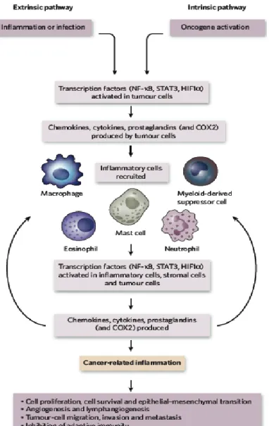
Figure 3. Pathways that connect inflammation and cancer
xxii
Figure 3. Pathways that connect inflammation and cancer
Cancer and inflammation are connected by two pathways: the intrinsic pathway and the extrinsic pathway. The intrinsic pathway is activated by genetic events that cause neoplasia.
These events include the activation of various types of oncogene by mutation, chromosomal rearrangement and the inactivation of tumor-suppressor genes. Cells that are transformed in this manner produce inflammatory mediators, thereby generating an inflammatory microenvironment in tumors for which there is no underlying inflammatory condition. By contrast, in the extrinsic pathway, inflammatory or infectious conditions augment the risk of developing cancer at certain anatomical sites. The two pathways converge, and result in the activation of transcription factors, mainly nuclear factor-κB (NF-κB), signal transducer and activator of transcription 3 (STAT3) and hypoxia-inducible factor 1α (HIF1α), in tumor cells. These transcription factors coordinate the production of inflammatory mediators, including cytokines and chemokines, as well as the production of cyclooxygenase 2 (COX2). The cytokines activate the same key transcription factors in inflammatory cells, stromal cells and tumor cells, resulting in more inflammatory mediators being produced and a cancer-related inflammatory microenvironment being generated (Mantovani et al., 2008).
1
Chapter I
Growth arrest and forced differentiation by
phosphodiesterase 4 type D inhibitor in human primary
glioblastoma multiforme
2
1.1 INTRODUCTION
Glioblastoma is the most prevalent and aggressive primary brain tumor in patients, with a median survival of less than a year after diagnosis due to its chemoresistance and radioresistance potential (Bao et al., 2006; Liu et al., 2006; Stupp et al., 2009). It has recently been accepted that undifferentiated cancer cells, called cancer stem cells (CSCs), in various tissues comprise only a small subpopulation of the tumor, but each single cell play a crucial role in the initiation and the progression of cancers which cause relapse and metastasis by giving rise to a new tumor (Beck and Blanpain, 2013). Regarding the biological characteristics of CSCs, recent studies have reported evidence that CSCs are similar to normal stem cells in self-renewal and multi-lineage differentiation potential, but they differ in their long-term proliferative capability.
Similar to tissue-specific stem cells (somatic or adult stem cells), there are no universal biological markers for CSCs. Nonetheless, the cell surface marker CD133 has been frequently used for characterization of tissue-specific stem cells. Over the last several years, CD133 expression has been detected extensively in various stem/progenitor cells, especially in the human neural systems, including the fetal brain, the post-mortem retina and embryonic stem cell-derived neural progenitors (Carter et al., 2009; Peh et al., 2009;
Uchida et al., 2000). Moreover, CD133 has been most frequently applied in brain tumors as a putative CSCs biomarker (Singh et al., 2004). Recent studies have suggested that CD133 expressing cell subpopulation in GBM is thought to be a CSCs population (Bao et
3
al., 2006; Collins et al., 2005; Eramo et al., 2008; Singh and Dirks, 2007). This CD133 expressing subpopulation reveals its increased tumorigenic potential higher than CD133 negative subpopulation (Beier et al., 2007; Galli et al., 2004; Reya et al., 2001; Yuan et al., 2004). Furthermore, a reliable study demonstrated that targeting the CSCs subpopulation could be the key in GBM therapy (Lathia et al., 2010). There have been many attempts to develop therapeutic strategies targeting tumorigenic cell populations, but significant results have not yet been achieved.
Other than extermination of CSCs subpopulation, forced differentiation of tumor cells to limit their long-term proliferative potential is a new approach in the search for alternative cancer therapies. Piccirillo has demonstrated that bone morphogenetic protein 4 (BMP4) induces the neural differentiation in human primary GBM cells. He showed that BMP4 plays a role as a growth inhibitor on CD133-expressing GBM-derived cells in vitro and that BMP4 treatment interrupts tumorigenic activity in vivo (Piccirillo et al., 2006).
Other studies have reported induced terminal differentiation using a chemical combination of retinoic acid and arsenic trioxide in leukemic cells and demonstrated that the depletion of the leukemic CSCs by using a chemical combination is involved with cAMP signaling pathway (de The and Chen, 2010; Guillemin et al., 2002; Nasr et al., 2008). Although multiple drugs were found to affect the phenotype of the tumor mass, the efficacy of these drugs was limited and did not affect the tumor-initiating cells.
A few studies have investigated whether phosphodiesterase 4 (PDE4) plays a pivotal role in tumor inhibition (Goldhoff et al., 2008; Moreno et al., 2006; Yang et al., 2007). PDE4 inhibitors have been frequently used to enhance cAMP/CREB signaling
4
pathway because PDE4 is well known for its role to reduce cAMP level in cells. The cAMP/CREB signaling pathway is participates in many biological functions and seems to play an important role in differentiation (Mayr and Montminy, 2001; Mayr et al., 2001).
Moreover, the cAMP/CREB signaling pathway can induce the differentiation of human cancer cells and mesenchymal stem cells (Sato et al., 2006; Siddappa et al., 2008; Zhao et al., 2004). In cancer biology, it has been demonstrated that cAMP/CREB signaling pathway related genes play as oncogenes or oncogene inhibitors (Kirschner et al., 2000; Landis et al., 1989; Lania et al., 2001; Lucchi et al., 2011; Persani et al., 2000). Rolipram was recently used in a GBM research, due to its promoting cAMP signal transduction (Li et al., 2011).
Rolipram is a specific PDE4 inhibitor which commonly known for memory enhancer (Otmakhov et al., 2004; Romano et al., 1996) owing to its role in up-regulating the level of cAMP and the phosphorylation of CREB (Park et al., 2012; Schneider, 1984). The Forskolin induces up-regulation of cAMP level and leads gap junctional intercellular communication enhancement, which is mostly inhibited in cancer cells, and to neuronal differentiation of SVG cells (Dowling-Warriner and Trosko, 2000). Although recent reports have suggested the pivotal role of cAMP/CREB signaling pathway in the differentiation of cancer cells, the cellular mechanism of forced differentiation process by cAMP and CREB is remains to be unraveled.
Several studies reported about the relationship between tumor suppression and neural differentiation in GBM. Nevertheless, the tumor-suppressive potential of PDE4D inhibitors have barely been studied. There are four PDE4 subfamilies (A to D), which are comprised of more than 20 different isoforms (Li et al., 2011) that regulate the cellular
5
cAMP level through their hydrolytic activity (Houslay and Adams, 2003; Marko et al., 2000). All the variants of PDE4 and their anti-cancer effects are not clearly known yet.
However, the specific variant PDE4D inhibitor, CG500354, may provide a closer step forward to unveiling the therapeutic issues to target aggressive glioblastoma.
In this study, I demonstrate that the novel small molecule, CG500354, acts as a tumor-suppressor in human primary GBM by increasing p53, expression which subsequently compel growth arrest of GBM. I determined that CG500354 forces induction of the neural differentiation in GBM-derived cells through cAMP/CREB signaling pathway.
In particular, I suggested that the well-known tumor suppressors, Rolipram and Forskolin, are also involved in cAMP/CREB signaling pathway as predicted and can induce the neural differentiation of GBM-derived cells. According to my data, CG500354, the novel small molecule, reveals its tumor suppressive potential and its specific mechanism which accompanied with p53 up-regulation and increased cAMP/CREB activity.
6
1.2 MATERIALS AND METHODS
1.2.1 Isolation of primary GBM cells
Following informed consent and in accordance with the appropriate institutional review boards, GBM was obtained from a patient undergoing surgery at the Samsung Medical Center (Seoul, Republic of Korea).Tumors were classified as GBM based on WHO criteria by examination of pathologists51. Within one hour of surgical resection, the tumor mass was mechanically and enzymatically dissociated into single cells. GBM was briefly maintained in NBE neurosphere culture medium: Neurobasal-A medium (Invitrogen, Carlsbad, CA, USA) supplemented with recombinant human basic fibroblast and epidermal growth factors (50 ng/ml each; Invitrogen), N2 and B27 supplements (Invitrogen), 2mM glutamine (Invitrogen), 100 units/ml penicillin and 100 mg/ml streptomycin (Invitrogen). I used human primary cells from three different GBM origins:
GBM559, GBM592 and GBM626.
1.2.2 Chemical
CG500354 (Patent No. 61/470884) was provided by Crystal Genomics, Inc.
(Seongnam, Republic of Korea). Rolipram (C16H21NO3; 4-[3-(cyclopentyloxy)-4- methoxyphenyl]-2-pyrroli-dinone) and Forskolin (C22H34O7; 7b-acetoxy-8,13-epoxy- 1a,6b,9a-trihydroxylabd-14-en-11-one) were purchased from Sigma Aldrich (Saint Louis, MO, USA). For the cell treatments, Rolipram and Forskolin were used at a concentration
7 of 10
μ
M.1.2.3 Clonogenic assays
Dissociated sphere cells (5 × 10⁵) were cultured in ultra-low attachment dishes.
The cells were treated with DMSO or CG500354 for 72 hours. The number of spheres with a diameter of > 100 mm were counted randomly in triplicate cultures.
1.2.4 Magnetic-activated cell sorting
To isolate the CD133-positive GBM-derived cells from the DMSO- or CG500354-treated cells, I resuspended the treated cells with 300 ml/108 buffer (PBS with 0.5% FBS) and added 50 ml/108 of FcR Blocking Reagent and 50 ml/108 CD133 MicroBeads (#130-050-801, Miltenyi Biotec) for 30 minutes at 6 °C to 12 °C. Then, I isolated CD133-positive cells using through Column and stained with anti-CD133/2-PE antibody (Miltenyi Biotec) to further analyze.
1.2.5 Flow cytometry analysis
GBM-derived cells were treated with DMSO or CG500354 for 72 hours and stained with an anti-CD133/2-PE antibody (#130-090-853, Miltenyi Biotec, Bergisch Gladbach, Germany) for 30 minutes. The CD133-stained GBM derived cells were analyzed with a FACS Calibur flow cytometer (Becton Dickinson).
8
1.2.6 cAMP measurement
GBM cells were treated with DMSO or CG500354 (3 μM/ml) for 72 hours. The cAMP level was measured from the collected supernatants using a cyclic AMP EIA Kit (#581001, Cayman Chemical Company, Ann Arbor, MI, USA) according to the manufacturer’s instructions. The cAMP values were calculated using a multifunction microplate reader (Tecan, San Jose, CA, USA).
1.2.7 siRNA inhibition study
To specifically inhibit PDE4D expression, siRNA-mediated gene knockdown studies were performed using commercial siRNAs targeting the PDE4D transcript (ON Target plus SMART pool, Dharmacon, Lafayette, CO, USA) along with a non-targeting siRNA (#D-001810-01, ON Target plus SMART pool, Dharmacon). The siRNA transfections were performed according to the manufacturer’s instructions. Briefly, the cells were seeded at a concentration of 5 × 10⁵ per well in 6-well plates. When the cells reached 60–70% confluence, a mixture of 50 nM siRNA and transfection reagent was added to the cell culture medium without antibiotics. The cells were incubated for 24 hours before evaluating mRNA expression by RT-PCR analysis or for 48 hours before evaluating protein expression by western blot analysis.
1.2.8 Quantitative real-time PCR
The relative gene expression levels were calculated using the 2-∆∆Ct method after normalization to the endogenous expression level of GAPDH.
9
1.2.9 Western blot analysis
For western blot analysis, the following primary antibodies were used: mouse monoclonal anti-GFAP (ab4648; Abcam, Cambridge, UK), mouse monoclonal anti-Tuj1 (MMS435; Covance, Princeton, NJ, USA), mouse monoclonal anti-P53 (2524; Cell Signaling Technology Inc., Boston, MA, USA), rabbit monoclonal anti-p-CREB (9198;
Cell Signaling Technology Inc.), rabbit monoclonal anti-p-PKA (5661; Cell Signaling Technology Inc.) and mouse monoclonal anti-GAPDH (MAB374; Millipore, Billerica, MA, USA).
1.2.10 Immunocytochemistry
The primary antibodies used for immunocytochemistry were: mouse monoclonal anti-nestin (MAB5326; Millipore), mouse monoclonal anti-GFAP (ab4648; Abcam), mouse monoclonal anti-Tuj1 (MAB1195; R&D Systems, Minneapolis, MN, USA) and mouse monoclonal anti-P53 (2524; Cell Signaling Technology Inc.). Both fluorescent secondary antibody conjugates, goat anti-mouse Alexa Fluor 488 (Invitrogen) and goat anti-rabbit Alexa Fluor 488 (Invitrogen), were used at a 151000 dilution. DAPI was used for counterstaining. The coverslips were mounted with fluorescent mounting solution (DAKO, Carpinteria, CA, USA) and observed by confocal microscopy (Eclipse TE200, Nikon, Japan).
1.2.11 Animal experiment
NOD-SCID mice were purchased from Jackson Laboratories (Bar Harbor, ME,
10
USA). All experiments were performed in accordance with the guidelines and regulation, which were approved by the Institute of Laboratory Animals Resources (SNU-101013-5, Seoul National University, Republic of Korea). Four-week-old female mice were subcutaneously injected with 1 × 10⁷ GBM-derived cells, which were suspended in 0.1 ml PBS. When tumors appeared, the mice were randomly divided into two groups and treated daily with intraperitoneal injections of 3 mg/kg CG500354 (n = 4) or DMSO as the vehicle control (n = 3) for 10 days. The mice were sacrificed, and the tumors were separated from the host for histological evaluation.
1.2.12 Statistical analysis
All experiments were conducted at least in triplicate, and the results are expressed as the mean ±SD. The statistical analyses were conducted using analysis of variance (ANOVA) followed by a Duncan’s multiple range test or Student’s t-test. The following P values were considered significant: *, P < 0.05;
**, P < 0.01; ***, P < 0.001.
11
1.3 RESULTS
1.3.1 CG500354 induces differentiation in human primary GBM-derived neurospheres without cytotoxic effects
I first determined the effects of a novel small molecule, CG500354, on the oncogenic phenotype of human primary GBM-derived cells. CG500354 has been designed, synthesized and evaluated by Crystal Genomics, Inc. using its structure-based drug discovery platform which employs the integrated technologies of the SPSTM (Soluble Protein Solution), technology for obtaining soluble forms of disease related proteins that are usually insoluble when overexpressed in heterologous systems, SCPTM (Structural Chemo Proteomics), technology for rapidly generating novel leads using the 3-D structure information of the target proteins, and SDFTM (Structural-based Drug Factory), technology to optimize novel leads to drug candidate compounds using the 3-D structure information of the complexes of target proteins and inhibitors. The synthetic compound, CG500354, is an inhibitor of cAMP-specific 3′.5′-cyclic phosphodiesterase 4D (PDE4D), which plays a role in cAMP homeostasis by hydrolyzing the second messenger cAMP (Fig.
1A). Human primary cells derived from three different GBM origins, GBM559, GBM592 and GBM626, were grown as neurospheres under floating cell culture conditions (Fig. 1B).
A very small portion of human GBM cells expresses CD133, which is a putative biomarker of neural stem cells and CSCs. It is known that this CD133 expressing subpopulation plays a crucial role in the proliferation and survival of GBM. To classify human primary GBM
12
samples, I isolated the CD133 subpopulation from the GBM-derived cells using magnetic- activated cell sorting (MACS). Then, I performed flow cytometry to detect the CD133 expressing GBM-derived cells and verified the present of CD133-expressing subpopulation (Fig. 1C). In a xenograft mouse model the GBM-derived cells were able to form a glioblastoma. This formed tumor showed two typical histopathological features of GBM:
pseudopalisading necrosis and endothelial proliferation (Fig. 1D).
To identify the effects of CG500354 in vitro, I performed a clonogenic assay with treatment of CG500354 or dimethyl sulfoxide (DMSO) as a vehicle control for 72 hours on GBM-derived neurospheres cultured in non-adherent culture conditions. In the result of clonogenic assay, the CG500354-treated neurospheres revealed the smaller neurosphere numbers compare to the DMSO-treated neurospheres (Fig. 1E). While I seeded neurospheres on the PLO/FN-coated dishes for immunocytochemistry, I could observed that most CG500354-treated neurospheres turn into adherent and differentiated state, but many DMSO-treated neurospheres still remain as a sphere (Fig. 1F). However, in viability assay, number of GBM cells was not prominently changed in response to 72hours of CG500354 treatment (Fig. 1G). These results suggest that the small molecule, CG500354, induces a dramatic change in the morphology of human GBM-derived neurospheres without cellular cytotoxicity.
13
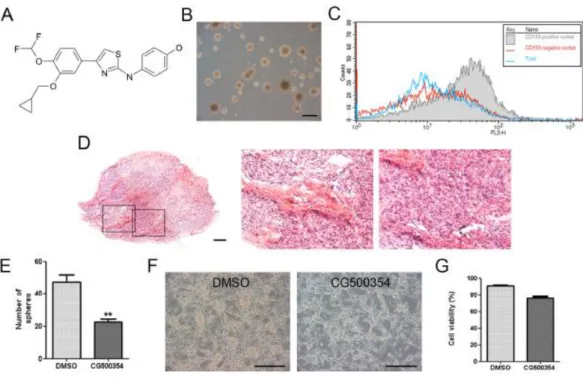
Figure 1. Identification of human primary GBM-derived cells.
(A) Chemical structure of the novel small molecule CG500354. (B) Phasecontrast morphological image of GBM-derived spheres in non-adherent culture conditions. Scale bar = 1 mm. (C) Characterization of CD133 expressing GBM-derived cells after magnetic- activated cell sorting by using flow cytometry analysis. (D) Characterization of pseudopalisading necrosis (middle) and endothelial proliferation (right) after hematoxylin and eosin staining in glioblastoma tumor which isolated from in a xenograft mouse model.
Scale bar = 400 μm. (E) The number of neurospheres significantly decreased by 72hours of CG500354 treatment in a clonogenic assay. (F) Representative phase-contrast images of GBM-derived cells after DMSO or CG500354 treatment. Scale bar = 1 mm. (G) A trypan
14
blue exclusion test was performed to calculate the number of viable cells compared to the total cell number in the CG500354 (3 μM) or DMSO-treated GBM-derived cell population.
DMSO was used as a vehicle control.
15
1.3.2 CG500354 induces growth arrest of GBM-derived cell populations by up- regulating p53
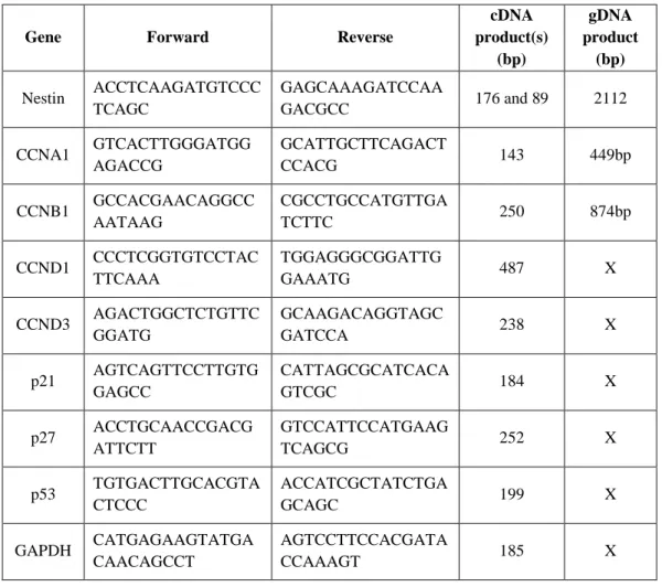
I performed a cell cycle analysis to identify the growth-suppressive potential of CG500345 in human primary GBM-derived neurospheres. In the presence of CG500354, the G1 fraction of the total neurosphere cells was increased approximately 10% while the G2 fraction was not detected (Fig. 2A). Moreover, I performed a quantitative RT-PCR analysis for additional evidence of the cell cycle arrest (Table. 1). Cell cycle regulators compose a complex regulatory system and include cyclins and cyclin-dependent kinases (Cdks). Cdks are inactivated unless bound to phase-specific cyclins. Here, I assessed the mRNA expression of cyclin A1 for S phase, cyclin B1 for M phase and cyclin D1 and D3 for G1 phase were significantly decreased in the presence of CG500354 (Fig. 2B). I further analyzed the gene expression level of two Cdk inhibitor proteins, p21 and p27, and found that both were significantly up-regulated (1.95-fold and 1.35-fold, respectively) in CG500354-treated GBM-derived neurospheres (Fig. 2C). These results indicate that CG500354 induces growth arrest in GBM derived neurospheres by inhibiting cell cycle regulators.
The tumor suppressor protein p53 is a transcription factor that has crucial effects on cell cycle arrest, DNA repair, apoptosis, senescence and angiogenesis (Brown et al., 2009; Riley et al., 2008). Thus, I analyzed p53 and its pro-apoptotic targets following CG500354 treatment. In our results, CG500354 treatment with 1 μM and 3 μM increased the protein expression level of p53 in the GBM-derived neurospheres (Fig. 2D). In immunocytochemistry, CG500354 treatment increased the number of p53-expressing cells
16
from 55.2% to 86.3% by 1 μM CG500354 and to 93.1% by 3 μM CG500354 (Fig. 2E–F).
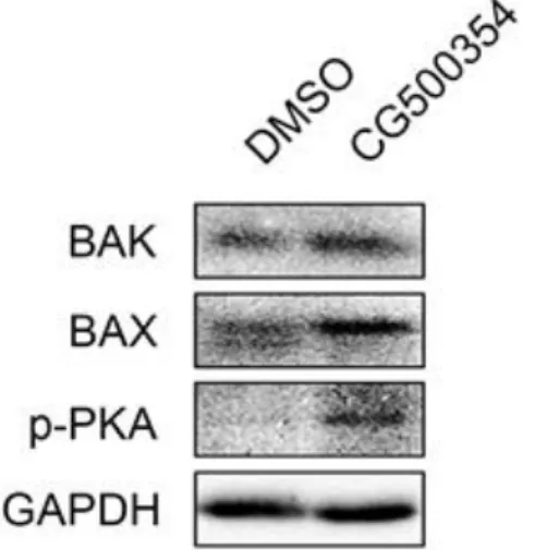
Western blot analysis shows increased expression level of pro-apoptotic targets, BAK and BAX (Fig. 3). These data suggested that the growth arrest was triggered by p53 and its downstream targets, such as p21, p27 and pro-apoptotic targets, whereas the forced neural differentiation was induced by cAMP/CREB signaling pathway.
17
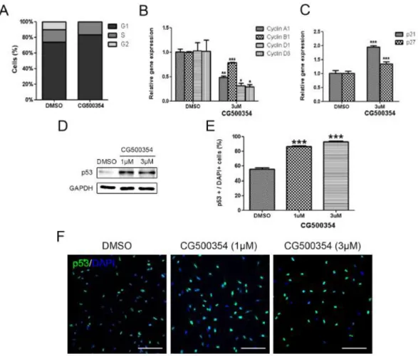
Figure 2. Growth arrest of human primary GBM-derived cells through p53 overexpression.
(A) Increased percent of the G1 phase was detected after CG500354 treatment in cell cycle analysis. (B–C) In the presence of CG500354, Cdk inhibitor proteins, p21 and p27 mRNA levels were significantly increased while the cell cycle regulators cyclin A1, B1, D1 and D3 were significantly down-regulated in quantitative RT-PCR analysis. (D) Significantly increased protein expression level of p53 was detected by western blot analysis in both two
18
different concentration (1 μM or 3 μM) of CG500354. (E–F) GBM-derived cells treated with both concentrations (1 μM and 3 μM) of CG500354 were stained with a p53 antibody.
The number of p53-expressing cells (green) were significantly increased after in both concentrations (1 μM and 3 μM). Nuclei (blue) were counterstained with DAPI. Scale bar
= 0.5 mm.
19
Table 1. Primer list for
quantitative RT-PCR analysisGene Forward Reverse
cDNA product(s)
(bp)
gDNA product
(bp) Nestin ACCTCAAGATGTCCC
TCAGC
GAGCAAAGATCCAA
GACGCC 176 and 89 2112 CCNA1 GTCACTTGGGATGG
AGACCG
GCATTGCTTCAGACT
CCACG 143 449bp
CCNB1 GCCACGAACAGGCC AATAAG
CGCCTGCCATGTTGA
TCTTC 250 874bp
CCND1 CCCTCGGTGTCCTAC TTCAAA
TGGAGGGCGGATTG
GAAATG 487 X
CCND3 AGACTGGCTCTGTTC GGATG
GCAAGACAGGTAGC
GATCCA 238 X
p21 AGTCAGTTCCTTGTG GAGCC
CATTAGCGCATCACA
GTCGC 184 X
p27 ACCTGCAACCGACG ATTCTT
GTCCATTCCATGAAG
TCAGCG 252 X
p53 TGTGACTTGCACGTA CTCCC
ACCATCGCTATCTGA
GCAGC 199 X
GAPDH CATGAGAAGTATGA CAACAGCCT
AGTCCTTCCACGATA
CCAAAGT 185 X
20
Figure 3. Expression level of pro-apoptotic targets.
Protein expression level of two apoptotic markers and phosphorylated PKA in GBM- derived cells were increased after a CG500354 treatment.
21
1.3.3 CG500354 induces the neural differentiation of GBM-derived cells
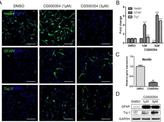
To verify the neural differentiation potential of CG500354 in human primary GBM-derived cells, 2 × 10⁴ cells were seeded on PLO/FN-coated coverslips and treated with two different concentration (1 μM or 3 μM) of CG500354. After 72 hours of CG500354 treatment, the cells exhibited the neuronal morphology thus I investigated the expression of the neural progenitor cell marker nestin, the neuronal marker Tuj1 and the astrocyte marker GFAP by using immunocytochemistry (Fig. 4A). In the vehicle control, 92.4% of the cells expressed nestin, and this percentage was significantly reduced to 20.3%after 3 μM CG500354 treatment, whereas 1 μM CG500354 did not affect nestin expression (Fig. 4B). Analysis of nestin expression at the mRNA level showed identical results as predicted (Fig. 4C). In opposition to nestin, the number of GFAP- and Tuj1-expressing cells was significantly increased upon CG500354 treatment. In comparison to the vehicle control, the cell numbers rose up to 6-fold in GFAP and 5-fold in Tuj1 with 3 μM CG500354 (21.4% and 79.6%, respectively). In addition, western blot analysis showed similar expressing pattern of GFAP and Tuj1 with immunocytochemistry (Fig. 4D). I observed that the neural progenitor/precursor marker expression was changed in a dose dependent manner. From the above, the concentration of 3 μM CG500354 was most efficient in the neural differentiation of human GBM-derived cells. Altogether, these results indicate that CG500354 induces the neural differentiation with GFAP and Tuj1 up- regulations.
22
Figure 4. Induction of neural differentiation by CG500354 treatment.
(A–B) Representative immunochemical images of primary GBM-derived cells show that CG500354 treatment decreased the number of nestin-positive neural progenitor cells and increased the number of GFAP- and Tuj1-positive neural subtypes. The effect of CG500354 on the neural differentiation of GBM was more effective in concentration of 3 μM than 1 μM CG500354. Nuclei (blue) were counterstained with DAPI. Scale bar = 0.5 mm. (C) Quantitative RT-PCR analysis shows that the relative expression level of nestin was significantly reduced by 3 μM CG500354. (D) Western blot analysis shows that the expression of the GFAP and Tuj1 proteins was up-regulated by CG500354 treatment.
23
1.3.4 CG500354 leads GBM-derived cells to growth arrest by accelerating the cAMP/CREB signaling pathway
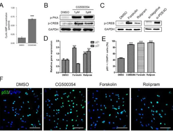
As previously described, CG500354 is an inhibitor of PDE4D, which regulates the cAMP signaling. In the cAMP/CREB signaling pathway, the accumulation of cAMP induces the diffusion of a subunit of cAMP dependent phosphorylated protein kinase A (PKA) into the nucleus and activates cAMP response element-binding protein (CREB). In several studies, CREB is described to stimulate the differentiation of human cancer cells and mesenchymal stem cells (Sato et al., 2006; Siddappa et al., 2008). Here, I examined the CG500354-mediated activation of the cAMP/CREB signaling pathway. After CG500354 treatment, the supernatants from the cell culture of GBM cells were harvested and measured the secreted cAMP concentration. These measurements revealed an approximately 4.5-fold increase in the level of cAMP secretion in the CG500354-treated GBM-derived cells compared to the control cells (Fig. 5A). I then performed western blot analysis of the major downstream targets PKA and CREB to verify the activation of the cAMP signaling pathway. CG500354 treatment effectively increased the expression of both phosphorylated PKA and CREB, and this up-regulation was detected from both CG500354 doses (Fig. 5B).
To compare the potential of CG500354 in GBM with two known mimetic substances, I treated GBM-derived cells with the cAMP regulator Forskolin and the PDE4 inhibitor Rolipram. Then, I confirmed both mimetic substances increased the level of phosphorylated CREB as CG500354 (Fig. 5C). Quantification of the western blot analysis showed that the levels of phosphorylated CREB in the Forskolin-treated cells were
24
approximately 11-fold higher than that in the control cells, but not as much as CG500354.
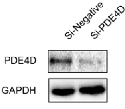
In addition, I performed the direct suppression of PDE4D expression using a siRNA, targeting the PDE4D gene (si-PDE4D). This si-PDE4D reduced 73.6% of the PDE4D expression and increased the level of phosphorylated CREB in the human primary GBM- derived cells as expected (Fig. 5C and Fig. 6).
To explore whether these two mimetic substances can also induce growth arrest like CG500354, I treated GBM-derived cells with CG500354, Forskolin or Rolipram for 72 hours, and the treated GBM-derived cells were analyzed using immunocytochemistry (Fig. 5E–F). Similar to the CG500354-treated cells, the Forskolin- and Rolipram-treated cells showed significant increases in p53 protein expression. The percentages of p53- expressing cells in both of the groups treated with mimetic substances were as high as that observed in the group treated with CG500354. Moreover, treatment with Forskolin and Rolipram induced the gene expression of the p53 downstream targets p21 and p27 (Fig.
5D). The Rolipram significantly increased the expression of both p21 and p27, which inhibit cell cycle progression. Therefore, CG500354 and its mimetic substances Forskolin and Rolipram are able to induce growth arrest in GBM-derived cells. From the analyses of cAMP signaling and cell cycle regulators, I concluded that CG500354 appeared more effective than both mimetic substances and it takes a novel therapeutic potential to handle the GBM growth arrest.
25
Figure 5. Activation of cAMP/CREB signaling pathway by CG500354 and mimetic substances.
(A) Measurement of the extracellular cAMP level was performed with a Cyclic AMP EIA Kit. The supernatant from the CG500354-treated GBM cells showed a significantly higher level of cAMP than that from the DMSO-treated GBM cells. (B) Western blot analysis shows that the expression of phosphorylated PKA and CREB was strongly induced by CG500354 treatment. (C) Both mimetic substances (Forskolin and Rolipram) and si- PDE4D also increased the expression of phosphorylated CREB. (D) Quantitative RT-PCR analysis reveals that both mimetic substances induced p21 expression and only Rolipram also increased p27 expression. (E–F) GBM-derived cells treated with CG500354,
26
Forskolin or Rolipram were stained with a p53 antibody. The numbers of p53-expressing cells (green) in the three groups were all equivalently high. Nuclei (blue) were counterstained with DAPI. Scale bar = 0.5 mm.
27
Figure 6. Knockdown of PDE4D expression using a PDE4D-siRNA.
Direct suppression of PDE4D protein expression in GBM-derived cells was examined after PDE4D-siRNA treatment by western blot analysis.
28
1.3.5 Mimetic substances and si-PDE4D, mimic the effect of CG500354 on the neural differentiation of GBM-derived cells
I next investigated the effect of these mimetic substances on the neural differentiation in human primary GBM-derived cells. After Forskolin and Rolipram treatment, the neural differentiation markers GFAP and Tuj1 showed the up-regulation in the western blot analysis (Fig. 7A). I further performed the immunocytochemistry to analyze the nestin, GFAP and Tuj1 expressions (Fig. 7B–C). Similar to CG500354, Forskolin- and Rolipram-treatment cells were detected 8 to 10-fold more GFAP-expressing cells and 5-fold more Tuj1-expressing cells in comparison to the vehicle control. Unlike CG500354, the number of nestin-expressing cells did not change after treatments of two mimetic substances. These results suggest that Forskolin and Rolipram induce the differentiation of GBM derived cells but do not deplete the cell population remaining in the neural progenitor state.
To further investigate the neural differentiation phenomena induced by CG500354, I performed gene silencing of PED4D using siRNA and it induced the overexpression of GFAP and Tuj1 in the GBM-derived cells as expected. The protein expression levels of GFAP and Tuj1 were significantly up-regulated in the si-PDE4D transfected GBM-derived cells (Fig. 7D). Moreover, the immunocytochemical analysis showed that the decease of nestin-expression and increase of GFAP- and Tuj1-expression (Fig. 7E–F). Both our immunocytochemistry and western blot analyses revealed that si- PDE4D affects the expression of neural differentiation markers similar to CG500354. From the above, I could indicate that novel CG500354 have shown higher potential in cell cycle
29
regulation than Rolipram while activating cAMP signaling pathway as Forskolin.
Therefore, our data on the mimetic substances and si-PDE4D provide direct evidence that CG500354 works as a multi-controller that induces neural differentiation and growth arrest in human primary GBM-derived cells.
30
Figure 7. Induction of neural differentiation of human GBM by mimetic substances and si-PDE4D.
(A), (D) Western blot analysis shows that the expression of the neural subtype markers GFAP and Tuj1 was up-regulated upon Forskolin, Rolipram and si-PDE4D treatment. (B–
C), (E–F) Representative immunochemical images of primary GBM-derived cells reveal that Forskolin, Rolipram and si-PDE4D induced an increase in the number of the GFAP-
31
and Tuj1-positive cells. Upon Forskolin or Rolipram treatment, the nestin-expressing cell population did not decrease (B), whereas this population was reduced by si-PDE4D as expected (E). Nuclei (blue) were counterstained with DAPI. Scale bar = 0.5 mm.
32
1.3.6 In vivo GBM-derived cells from tumor were induced to neural differentiation by CG500354 injection
To validate the in vivo effects of CG500354, I performed subcutaneous xenotransplantation of GBM-derived cells into NOD/SCID mice. After GBM tumor formation, I treated mice with CG500354 or DMSO via intraperitoneal injection for 10 days. Then, I sacrificed the mice and isolated GBM tumors from the host for hematoxylin and eosin staining (Fig. 8A). These GBM tumors were characterized with pseudopalisading necrosis, endothelial proliferation and irregular nuclear contours. Most part of the tumor showed a small nuclear size and 29.1% of this part appeared to be Tuj1-positive (Fig. 8B).
But, the other part of the tumor showed a large nuclear size and 11.4% of this part appeared to be GFAP-positive by immunohistochemistry (Fig. 8C). These results indicated that approximately 40% of the GBM tumor was induced to differentiate into neural subtypes by CG500354 intraperitoneal injection.
33
Figure 8. In vivo neural differentiation of human primary GBM-derived cells after xenotransplantation.
(A) After GBM tumor formation, tumors were isolated and stained with hematoxylin and eosin. A composited image (second from left) reveals an entire section of a tumor and two high-resolution images indicates pseudopalisading necrosis (second from right) and endothelial proliferation (right). (B–C) Representative immunochemical images of sections
34
from GBM-derived tumors show that the cells inside of the tumors were forced to differentiate into Tuj1- and GFAP-expressing neural subtypes. (D) Schematic diagram of the mechanism of CG500354-triggered cAMP/CREB signaling pathway. Likewise, both of the mimetic substances Forskolin and Rolipram are involved in this signal transduction pathway.
35
1.4 DISCUSSION
In this study, I investigated the dual effects of CG500354 as a PDE4D inhibitor on human primary GBM. First, I showed that CG500354 induces growth arrest and attenuates stemness in GBM derived cells. In human GBM cells, this small molecule regulates the expression of the tumor suppressor p53 and its downstream target p21 as well as p27. The increased expression of both Cdk inhibitors led to the decreased expression of a number of phase-specific cyclins and a reduction in the clonogenic potential of the GBM- derived cells. Second, CG500354 accelerated the neural differentiation, which were expressed Tuj-1 and GFAP in GBM-derived cells. Our findings suggest that the cAMP/CREB signaling pathway is involved in this neural differentiation via the phosphorylation of PKA and CREB following the CG500354-mediated up-regulation of cAMP.
In the preliminary study, I have done all experiments with three different GBM origins, including GBM559, GBM592 and GBM626, to analyze CG500354 effects. Those three GBM origin-derived cell types have shown similar results. Coincidently, a quantitative RT-PCR analysis showed reduced gene expressions of SOX2 and nestin, and an increased gene expression of p53 in GBM559-, GBM592- and GBM626-derived cells.
I have representatively shown the results of GBM559 in this study.
Recent studies have demonstrated that the regulation of GBM CSCs is approached from inhibition of the DNA-damage checkpoint kinases CHK1 and CHK2 using small molecules (Bao et al., 2006). Using this strategy indicated an improved radiotherapy for GBM CSCs. Another alternative strategy of GBM treatment is the induction of a
36
differentiated state of CSCs. A reliable report has demonstrated that GBM CSCs could be differentiated into endothelial cells, which could suppress the vasculature development (Ricci-Vitiani et al., 2010). However, the tumor-derived endothelial cells exist at a small portion in GBM tumor mass and its clinical relevance is questionable (Rodriguez et al., 2012). Zheng and colleagues demonstrated that the p53 together with PTEN could regulate the Myc expression in human primary GBM (Zheng et al., 2008). These GBM-derived cells after Myc suppression induced the down-regulation of nestin expression while they differentiated into Tuj1- and GFAP-expressing neural cell types. In this study, I assessed the GBM cell population that remained in the neural progenitor state, in which the nestin- expressing GBM CSCs reside. Interestingly, this cell population was diminished by CG500354 treatment but was not altered by treatment with mimetic substances (Fig. 4B and Fig. 7B). These results show that CG500354 plays a better role in human primary GBM and functions even better than Forskolin and Rolipram with respect to its potential anti- cancer effects.
From our findings on the mechanism of action of CG500354, I argue that p53 is implicated in the phenotype changes of the GBM derived cells. Several studies have commonly identified mutations of tumor suppressor genes, such as p53, PTEN and RB, in brain tumor (Chow et al., 2011; Verhaak et al., 2010). As shown in Figures 2F and 3B, I determined that the level of p53 expression was significantly increased after CG500354 treatment and was followed by differentiation into neural subtypes. I used QIAGEN’s Ingenuity Pathway Analysis (IPA, QIAGEN Redwood City, www.qiagen.com/ingenuity) software to analyze molecular interactions between PDE4D and p53. Based on IPA,
37
representative regulatory networks were constructed that PDE4D directly activates PKA and cAMP, and indirectly activates p21 and p27 as well as p53. Another reason for the dual effects of CG500354 is that the PDE4D inhibitor CG500354 could affect the cAMP/CREB signaling pathway, which is widely known as a signal dependent gene regulatory pathway that is involved in many biological functions, including the cell cycle, cell survival and cell differentiation (Mayr and Montminy, 2001; Mayr et al., 2001). In the cAMP-dependent signal transduction pathway, PDE4D plays a suppressive role, as it causes a decrease in the level of cAMP. In a recent report on the role of cAMP, PDE4 was indicated as the most important target for the induction of apoptosis and differentiation in leukemia cells (Copsel et al., 2011; Karin, 1994). In another study, PDE4D was demonstrated to be abundantly expressed in glioblastoma cells and its inhibitor Rolipram was found to induce tumor regression (Goldhoff et al., 2008). Likewise, our data in Figure 4 reveals that CG500354, Forskolin and Rolipram could regulate the level of cAMP and its downstream target CREB.
Together with data described above, our results indicate that CG500354 up-regulates the cAMP/CREB signaling pathway activity and induces the differentiation of human primary GBM.
In conclusion, I have shown that the protein level of p53 and the extracellular level of cAMP are significantly increased upon CG500354 treatment. I further indicated that CG500354 can alter the phenotypes of human primary GBM cells from an oncogenic state into a less oncogenic and more differentiated state. During these tumor-suppressive events, p21- and p27-mediated growth arrest and CREB-mediated neural differentiation also occurred. Moreover, the effects of CG500354 on the neural differentiation of the cells were
38
more significant with respect to its reduction of the nestin-expressing population than the mimetic substances Forskolin and Rolipram. Therefore, the novel small molecule CG500354, which targets PDE4D, might be important in the development of new drugs for human GBM.
39
Chapter II
Mica Nanoparticle Eliminates the Human Breast Carcinoma Cells by
Regulating the Interaction of Tumor with its Immune
Microenvironment
40
2.1 INTRODUCTION
Breast cancer is one of the most common cancers developed in women, with high occurrence and fatality rates. Annually, approximately 1.38 million women worldwide are diagnosed with this disease, which is the second leading cause of cancer deaths among women in the US. (Jemal et al., 2011). The surgery, chemotherapy, radiation therapy and immunotherapy are the most prevalent types of cancer therapy (Alama et al., 2012;
Blagosklonny, 2003; Dey et al., 2008). However, the primary treatment is based on surgery or chemotherapy, which still have the issues of enhancing metastasis, systemic toxicity and drug resistance. These therapeutic limitations led researchers to develop targeted cancer therapies. Drugs or other natural compounds have been developed as targeted therapy for cancer to suppress the proliferation and metastasis. Therefore, several natural compounds such as plant extracts, minerals, vitamins or the combination of these compounds, have been suggested as alternative anti-tumor medicines (Feldman et al., 2014; Liu et al., 2013;
Vekariya et al., 2012).
Mica has been demonstrated to have the anti-tumor and the immunostimulatory potential. Cho and colleagues reported that mica is revealed as a chemopreventive agent against colorectal cancers (Cho et al., 2013). Moreover, mica nanoparticles have been used as feed supplements in domestic animals to enhance immune activity owing to its immune stimulation responses against virus infection (Jung et al., 2013; Jung et al., 2010; Jung et al., 2015a). Recently, Jung et al. reported macrophage activation through up-regulation of lysosome and phagosome pathway which are caused by mica nanoparticles. (Jung et al.,
41
2015b). Although the immunostimulatory ability can be efficiently used to suppress tumor, most studies using mica have investigated only the anti-tumor or the immunostimulatory effects separately. None have tried to incorporate these two effects by elucidating mechanistic action of mica on immune cells constituting tumor microenvironment. In addition, till now, a study regarding the anti-tumor effect of mica has been performed against only one type of cancer, even though there are more than 100 types of cancers have been isolated and identified including breast cancer cells (Brody, 2011).
Natural Killer (NK) cells are innate immune cells that play a pivotal role in anti- tumor response by activating strong cytotoxicity against cancer cells (Trinchieri, 1989).
Upon activation, NK cells eliminate target cells by secreting cytolytic enzymes or cytokines including interferon (IFN)-γ (Caligiuri, 2008; Vivier et al., 2008). Moreover, the crosstalk of NK cells with other immune cells is important for anti-tumor responses. For example, DCs can activate resting NK cells, which in turn, induce DC maturation (Cooper et al., 2004; Moretta, 2002; Walzer et al., 2005). Recently, it has been demonstrated that macrophages also regulate the anti-tumor responses of NK cells via bidirectional interactions (Klezovich-Benard et al., 2012; Nedvetzki et al., 2007; Tjwa et al., 2012).
Bellora et al. reported that immunostimulatory type of macrophages (M1) induces strong activation of resting NK cells and the polarization of macrophages toward M1 phenotype can restore immunomodulatory type of macrophages (M2), the general phenotype in cancer tissues (Bellora et al., 2010). These findings indicate that the crosstalk between NK cells and other immune cells might be an important target of mica to promote anti-tumor immunity. Therefore, the present study was designed to investigate the anti-tumor effect of
42
STB-HO (mica nanoparticles) against MCF-7, the human breast carcinoma cell line.
Furthermore, in an attempt to verify the underlying mechanisms, I explored the direct effects of STB-HO on MCF-7 as well as the indirect, immunity-mediated effects.
43
2.2 MATERIALS AND METHODS
2.2.1 Reagents
LPS was purchased from Invivogen (San Diego, CA). IFN-γ was purchased from PeproTech (Rocky Hill, NJ) and IL-12 from R&D Systems (Minneapolis, MN).
2.2.2 MCF-7 xenograft model
Athymic nude mice (female, 5wk old) were obtained from SLC (Hamamatsu, Japan) and mice were group-housed under specific pathogen-free conditions in the animal facility of Seoul National University. All experiments were performed in accordance with the guidelines and regulation, which were approved by the Institute of Laboratory Animals Resources (SNU-140103-5, Seoul National University, South Korea). 2 × 10⁶ MCF-7 cells mixed with 0.1 mL matrigel (Corning, Corning, NY) were subcutaneously injected into the right flank of athymic nude mice. After one week of stabilization period, the mice were divided into three groups and orally administrated with 35 mg/kg STB-HO (n = 8) or 70 mg/kg STB-HO (n = 7) or normal saline as the positive control (n = 7) daily for 12 weeks.
Tumor size was measured twice a week using a caliper. Tumor volume (V) was calculated as the formula: V (mm3) = length*width2/2. After sacrifice, tumors were isolated for size measurement, histological evaluation and signaling analysis on protein level.
44
2.2.3 Histological evaluation
Tumor samples were collected, fixed in 10% formalin, subjected to consecutive steps of alcohol-xylene changes, and embedded in paraffin. Sections of 3 μm thickness were prepared and stained with H&E or aluminum staining.
2.2.4 Aluminum staining of dissected tumor sections
Paraffin sections were deparaffinized and stained with Einarson’s reagent overnight. Stained sections were placed in 0.5% phloxine B for 3min and the excessive dye was removed by washing. Then, the sections were immersed in 5% phosphotungstic acid for 1min and subsequent 1% glacial acetic acid for 2min, followed by washing with 80%
ethanol till the sections appear translucent. Sections were further replaced in 1% glacial acetic acid for 1min and counterstained with 0