저작자표시-비영리-변경금지 2.0 대한민국 이용자는 아래의 조건을 따르는 경우에 한하여 자유롭게
l 이 저작물을 복제, 배포, 전송, 전시, 공연 및 방송할 수 있습니다. 다음과 같은 조건을 따라야 합니다:
l 귀하는, 이 저작물의 재이용이나 배포의 경우, 이 저작물에 적용된 이용허락조건 을 명확하게 나타내어야 합니다.
l 저작권자로부터 별도의 허가를 받으면 이러한 조건들은 적용되지 않습니다.
저작권법에 따른 이용자의 권리는 위의 내용에 의하여 영향을 받지 않습니다. 이것은 이용허락규약(Legal Code)을 이해하기 쉽게 요약한 것입니다.
Disclaimer
저작자표시. 귀하는 원저작자를 표시하여야 합니다.
비영리. 귀하는 이 저작물을 영리 목적으로 이용할 수 없습니다.
변경금지. 귀하는 이 저작물을 개작, 변형 또는 가공할 수 없습니다.
1
변제익 석사 학위논문
복부 수술을 받은 초극소 저체중 출생아에서 진단에 따른 예후 비교 연구
Comparison of outcomes of extremely-low-birth-weight neonates with abdominal surgery according to differential diagnosis
2018 년 12 월
서울대학교 대학원 의학과, 외과
변 제 익
2
복부 수술을 받은 초극소 저체중 출생아에서 진단에 따른 예후 비교 연구
지도교수 정 성 은
이 논문을 의학석사 학위논문으로 제출함 2018년 12월
서울대학교 대학원 의학과 외과전공
변 제 익
변제익의 석사 학위논문을 인준함 2018 년 12 월
위 원 장 (인)
부위원장 (인)
위 원 (인)
3
1. 국문요약(국문초록)
요약
(국문초록
)신생아 집충치료의 발달로 인해 초극소 저체중 출생아(<1000g) 의 생존율이
상승하면서 복부 합병증의 빈도가 높아지고 있다. 복부 합병증 중 수술적 치료를 요하는 경우는 다음 4가지 진단이 있을 수 있다; 신생아괴사성장염 (NEC), 자발적 장천공 (SIP), 태변관련장마비 (MRI), 그리고 태변과 무관한 장마비 (MNRI). 이 논문은 본 병원에서 진료받은 초극소 저체중 출생아 중 복부 수술을 받은 환아들에 있어 진단에 따른 특성과 임상경과를 비교 분석하고자 한다. 2003년부터 2015년까지 805명의 초극소 저체중 출생아 중 복부수술을 받은 65명의 환아를 대상으로 하였고 그 중 NEC는 29명, SIP는 18명, MRI 13명, 그리고 MNRI 5명 이었다. 장루 형성술은 61명 (93.8%) 에서 시행되었고, 4명 (6.2%) 에 있어서 일차문합술이 시행 되었으나 수술 후 문합부 천공으로 4명 모두 2차 수술이 필요하였다. 퇴원 전 Clavien‑Dindo classification III 이상의 합병증은 14례가 있었으며 30일 사망은 7례, 그 중 6례는 NEC 로 진단받은 경우였다. 수술 받은 환아의 5년 생존율은 83%로 수술받지 않은 환아의 5년 생존율인 84%와 유의한 차이를 보이지 않았다. 생존율에 영향을 미치는 인자는 NEC 로
진단받은 경우였다. (p=0.033) 결론적으로 초극소 저체중 출생아에서 급성 복증이 있는 경우 수술적 치료는 좋은 결과를 보였고 수술 방법으로는 장루형성술이 추천된다. NEC 는 다른 진단과 비교하여 부정적인 임상경과를 보였다.
………
주요어 : 초극소 저체중 출생아, 자발적 장천공, 태변관련장마비, 태변과 무관한 장마비, 복부수술
학 번 : 2013-23486
4
2. 외국어초록(Abstract)
Abstract
Comparison of outcomes of extremely-low-birth-weight neonates with abdominal surgery according to differential diagnosis and surgical procedure
Jeik Byun College of Medicine The Graduate School Seoul National University
Background: Improvements in peri-natal intensive care made extremely low birth weight (ELBW, <1000g) neonates to survive and the chance of acute abdominal problem has increased. Differential diagnosis resulting in abdominal surgery can be categorized into following four entities; necrotizing enterocolitis (NEC), spontaneous intestinal perforation (SIP), meconium related ileus (MRI), and meconium non-related ileus (MNRI).
Purpose: To review our experience in abdominal surgery for ELBW neonates and evaluate characteristics and prognosis according to differential diagnosis.
Methods: Medical records of ELBW neonates treated in Seoul National University Children’s Hospital between 2003 and 2015 were retrospectively reviewed.
Results: Of 805 ELBW neonates, 65 (8.1%) received abdominal surgery. The number of cases within four disease categories was 29 NEC, 18 SIP, 13 MRI, and 5 MNRI. Ostoma formation of any fashion was performed in 61 (93.8%), and primary anastomosis without ostoma was performed in 4 (6.2%). All patients without ostoma formation encountered re-perforation of the anastomosis site and underwent additional surgery for diversions. There were 14 post- operative complications of Clavien‑Dindo classification over grade III before hospital discharge. 30 days post-operative mortalities were in 7 cases, and 6 were NEC diagnosed.
5
Long-term survival outcome of surgical and non-surgical group among ELBW neonates were not statistically different. Diagnosis of NEC was a poor prognostic factor for survival outcome (p=0.033).
Conclusion: Abdominal surgery for ELBW neonates is a feasible procedure, and ostoma formation can lead to reduced complication compared to primary anastomosis. High morbidity and mortality was noticed in NEC diagnosed patients.
………
Keywords : ELBW; Extremely low birth weight, NEC; Necrotizing enterocolitis, SIP; Spontaneous intestinal perforation, MRI; Meconium related ileus, MNRI; Meconium non-related ileus, Abdominal surgery
Student Number : 2013-23486
6
Introduction
Over the past 20 years, improvements in neonatal intensive care units have resulted in better survival outcomes for ELBW neonates.(1-4) As a result, the incidence of acute abdominal problems has increased, leading the gastrointestinal complications as one of the significant risk factors in their survival. (5)
Acute abdomen in ELBW neonates can be differentially diagnosed into four disease entities, which are necrotizing enterocolitis (NEC), spontaneous intestinal perforation (SIP), meconium related ileus (MRI), and meconium non-related ileus (MNRI).(6-11) Emergent operation was performed on patients with suspicion of bowel perforation, or with clinical deterioration despite medical treatment. Laparotomy and peritoneal drainage are two methods in treatment for intestinal perforation in ELBW neonates and the treatment of choice are still controversial.(12- 13)Moreover in laparotomy, whether to perform primary anastomosis or to perform diversion is still debatable.(14)
The aim of this study was to review our experience in abdominal surgery for ELBW neonates, and to evaluate characteristics and clinical prognosis according to different diagnosis.
7
Materials and Methods
Data collection
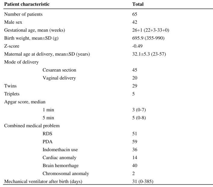
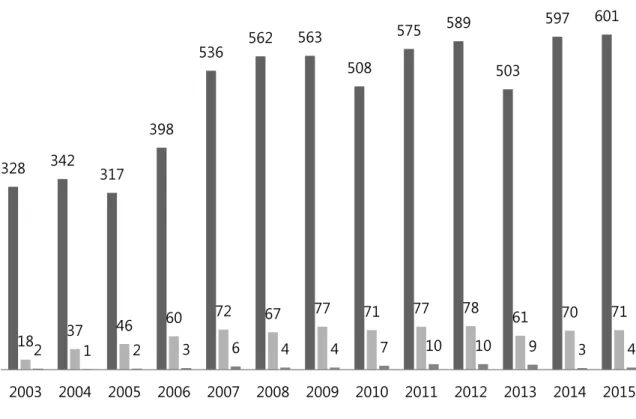
805 ELBW neonates who had admitted to neonatal intensive care unit (NICU) in Seoul National University Children’s Hospital between January 2003 and December 2015 were retrospectively reviewed. Identification of 65 ELBW neonates who underwent abdominal surgery for acute abdomen, including NEC, SIP, MRI, and MNRI was made from the medical records and electronic databases. (Figure 1)
NEC was defined as necrosis of the mucosal and submucosal layers of the bowel, while SIP was defined as focal punched-out intestinal perforation without mechanical obstruction or necrotic change. (7-10) MRI was defined as functional ileus characterized by impaired meconium excretion, small-sized colon extending to the distal ileum, with dilated proximal ileum filled with sticky meconium.(11,15) MNRI was defined as functional ileus and small- sized colon without meconium impaction. MNRI was not associated with Hirschsprung’s disease or intestinal atresia. (Figure 2)
Patients’ characteristic, combined medical problem, maternal age, mode of delivery, multiple pregnancy, surgical procedure, and follow-up data were investigated. Parenteral nutrition associated liver dysfunction (PNALD) was defined as a direct bilirubin level > 2 mg/dL with TPN use. Patients who had direct bilirubin level elevation before TPN use were excluded.
Outcomes of surgery was measured as post-operative complications and growth on the last clinical visit. Post-operative complications were divided into early and late complications by post-operative day 30. Growth was analyzed with age and height converted into Z-score on the last follow up in out-patient clinic.
Statistical analysis
Statistical analyses were performed using the SPSS 23.0 statistical software program (IBM
8
Corporation, Armonk, NY, USA) for Window. Descriptive data are reported using parameters such as frequency, mean, mode, and standard deviation. Factor difference between disease groups was analyzed with one-way analysis of variance. Tukey’s multiple comparison test was used to compare the difference between each pair of means with appropriate adjustment for the multiple testing. It was expressed as ‘a’, and ‘b’ for the statistically not different group in the tables. The cumulative survival rates and graphs were calculated using the Kaplan-Meier method and compared using a log-rank test. The hazard ratios and multivariate analysis were performed with Cox’s proportional hazards model. The other levels of significance was set at 0.05.
Definitions
Z-score is the number of standard deviations from the mean of the general population. The World Health Organization AnthroPlus program was used to calculate z-score of Bwt and height for postmenstrual age >40-week patients and PediTools (http://peditools.org/fenton 2013) for <40-week patients. Total parenteral nutrition (TPN)–related cholestasis was defined as a total bilirubin level > 5 mg/dL with TPN use. Patients with physiological cholestasis or an already elevated bilirubin level before TPN use were excluded. Enterostomy was defined as any type of stoma (including double barrel ileostomy or jejunostomy, loop ileostomy or jejunostomy). Follow-up body weight and height is the last body weight and height measurement at the latest outpatient clinic.
The Clavien-Dindo classification was defined as the therapy used to correct a specific complication is the basis of this classification in order to rank a complication in an objective and reproducible manner. Grade III is defined as patient’s post-operative condition requiring surgical, endoscopic or radiological intervention. Grade IV is defined as life threatening complication requiring ICU management. Grade IV is defined as death of a patient. (16) The
9
complication was defined as complication of the Clavien-Dindo classification above grade III.
Early and late post-operative complications were defined as complications before and after post-operative day 30.
Ethics statement
This study protocol was reviewed and approved by the institutional review board of Seoul national university (H-1502-113-651). Informed consent was waived by the board.
10
Result
Data
Of 805 ELBW patients, 65 (8.1%) received abdominal surgery. Patient characteristics are described in Table 1. There were 29 (44.6%) NEC, 18 (27.7%) SIP, 13 (20%) MRI, and 5 (7.7%) MNRI patients. Mean gestational age was 26+1 weeks (22+3 - 33+0) and mean birth weight was 695.9 g (355 - 990). Mean age at operation was 12.4 days (0 - 34 days) and mean body weight at operation was 710.7 g (490 - 970 g). There were 51 (68.2%) respiratory distress syndrome (RDS), 59 (90.8%) patent ductus arteriosus (PDA), 40 (61.5%) different brain hemorrhages associated, and 36 (55.4%) using indomethacin pre-operatively. Mean maternal age was 32.2 (24 - 57) years old. There were 5 (7.7%) maternal preeclampsa, 2 (3.1%) chorioamnionitis, 3 (4.6%) hypothyroidism, 2 (3.1%) thyrotoxicosis, and 1 (2.3%) case of long term usage of steroid. Infantogram and abdominal ultrasonography was taken for all patients pre-operatively. 51 (78.5%) showed free air in pre-operative infantogram. 8 (12.3%) had portal air, 17 (26.2%) showed intra-mural gas, and 13 (20.0%) showed ascites in ultrasonography. 45 underwent Cesarean section and 20 underwent vaginal delivery. Median Apgar score was 3 in 1 minute and 5 in 5 minute. Mean days of mechanical ventilator after birth was 31 days.
Operative characteristic are depicted in Table 2. Mean age at operation was 12.4 days. Mean body weight at operation was 710.7g. 22 cases underwent laparotomy before the year of 2010, and 43 underwent after 2010. Peritoneal drainage was inserted in 3 cases, but due to worsening prognosis all underwent following laparotomy. Laparotomy was performed in total of 65 cases.
In 65 laparotomy cases, ostoma formation of any fashion including loop, double barrel, or proximal loop ileostomy or jejunostomy was performed in 61 (93.8%). 4 underwent primary anastomosis without ostoma formation - 3 SIP patients underwent pirmary anastomosis without proximal ostoma formation, and 1 MRI patient underwent enterotomy, removal of the packed
11
meconium, and primary closure of the enterotomy site. All patients without ostoma encountered anastomosis leakage and underwent additional surgery for ostoma formation.
Segmental resection and double barrel ileostomy was the most common method of laparotomy in NEC patients followed by segmental resection with end ileostomy, proximal loop ileostomy (or jejunostomy) without bowel resection, and segmental resection and primary anastomosis with proximal loop ileostomy. 5 of 8 segmental resection with end ileostomy cases had underwent 2nd look operations. Loop ileostomy on perforation site, and segmental resection and double barrel ileostomy were two most common procedures for SIP, while loop ileostomy was most common for both MRI and MNRI. Mean post-operative duration of total parenteral nutrition (TPN) were 39.7 days, and 26 had PNALD. Mean hospital stay were 107.8 days.
Post-operative complications
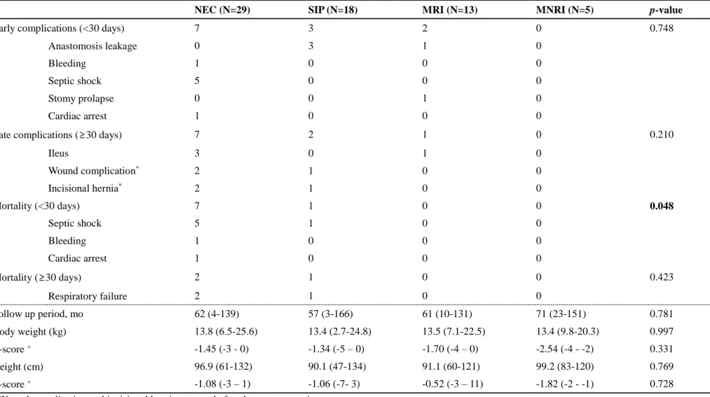
There were 24 (36.9%) post-operative complications before discharge. 14 cases were complications of extended Clavien‑Dindo classification over grade III, and the detail of the cases are presented in Table 3. There were 8 NEC, 4 SIP, and 2 MRI patients. Septic shock and anastomosis leakage were the most common type of complications, followed by 1 case of post- operative adhesive ileus, bleeding, ostoma prolapse, peri-stomal infection, respiratory failure, and 1 case of intra-operative cardiac arrest. The septic shock was defined as septic shock occurred peri-operatively. The 4 anastomosis leakage cases underwent additional surgery for ostoma formation. There were 7 (12.3%) 30-days mortalities, and 6 of them were NEC. Most common cause of death was sepsis.
The complications occurred before post-operative day 30 was defined as early complication, and complications occurred after post-operative day 30 was defined as late complications. The mortality was also divided into two categories of mortality occurred before and after post- operative day 30. Overall characteristics are described in Table 4. Most common case of early
12
complications was anastomosis leakage. Most common case of late complications was ileus, followed by wound complications and incisional hernia. Most common cause of mortality before post-operative day 30 was peri-operative sepsis, followed by culture proven sepsis, bleeding, and cardiac arrest. Mortality after post-operative day 30 were mainly due to respiratory failure.
Characteristics according to differential diagnosis
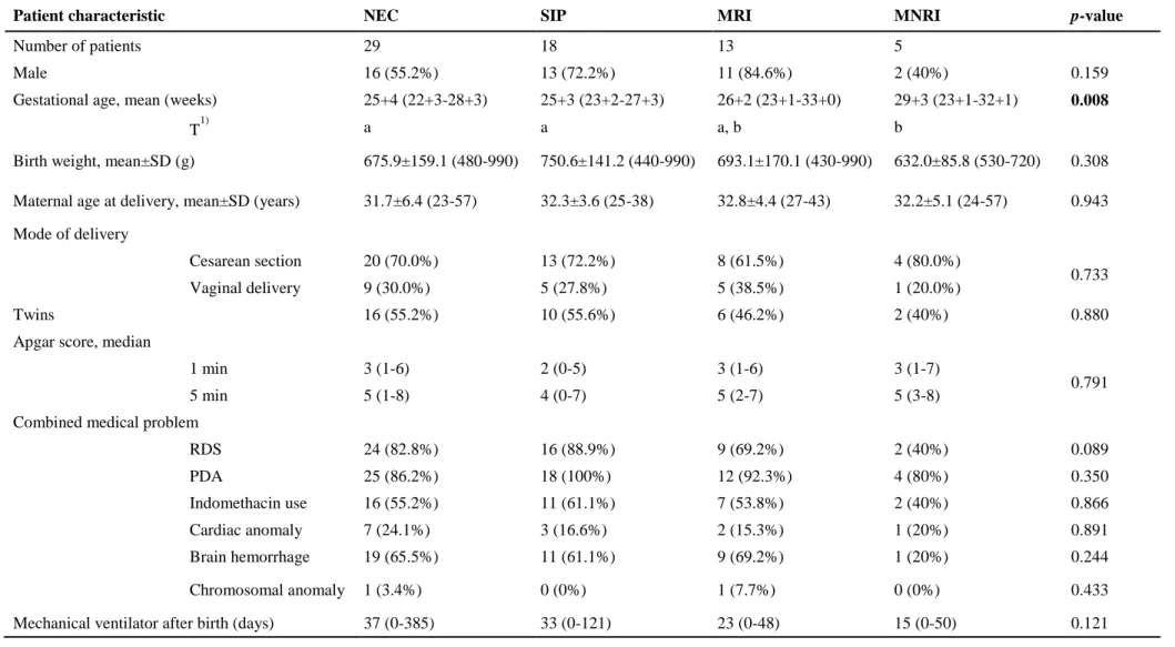
Patient characteristics were compared according to differential diagnosis was analyzed in Table 5. The comparison was analyzed with one-way analysis of variance method. The only significant difference between the diagnoses was mean gestational age. Patients diagnosed as MNRI had a significantly higher gestational age compared to patients diagnosed with NEC or SIP. Patients diagnosed with MRI did not show difference in gestational age compared to other diagnoses. Other factors such as incidence, gender, birth weight, maternal age at delivery, mode of delivery, whether they were twin or triplet, 1, and 5 minutes Apgar score, combined anomaly, and days of mechanical ventilator did not show any difference between the four differential diagnoses.
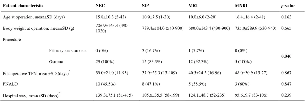
Operative characteristics were analyzed in Table 6. Mean age at operation, mean body weight at operation, post-operative duration of parenteral feeding, parenteral feeding related liver diseases, and mean hospital stay were not statistically different according to differential diagnosis. Only surgical procedure showed significant statistical difference. Primary anastomosis was performed only in SIP or MRI patients. All NEC or MNRI patients underwent ostoma formation.
Outcomes according to differential diagnosis
Outcomes of surgery was measured as post-operative complications and growth on the last clinical visit. The only significant factor different between the diseases was mortality before
13
post-operative day 30. Within seven 30-days mortality cases, six were NEC diagnosed patients followed by one SIP patient. Early mortality was significantly high in NEC compared to other differential diagnoses. The most common reason for early mortality in NEC patients was peri- operative sepsis, followed by culture proven sepsis, bleeding, and cardiac arrest.
Growth of the patient was analyzed in Z-score of body weight and height on the last follow up. Mean Z-score in body weight of last follow up was -1.52, and mean Z-score in height of last follow up was -1.16. (Table 4) There was no significant difference in follow up body weight and follow up height between the differential diagnoses.
Outcome according to surgical procedure
Three patients underwent peritoneal drainage for initial treatment. All three peritoneal drainage patients underwent following surgical laparotomy due to worsening prognosis. Four patients underwent primary anastomosis and sixty one patients underwent ostoma formation.
All four primary anastomosis cases resulted in leakage of the anastomosis site. Comparing two group of patients of initial surgical procedure with primary anastomosis and ostoma formation, there were significant difference in early complication and mean hospital stay. Early complication for anastomosis leakage and mean hospital stay was significantly higher in primary anastomosis group. Other factors of late complications, mortality and follow up growth did not showed significant difference.
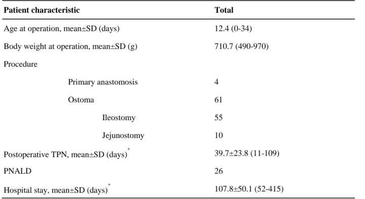
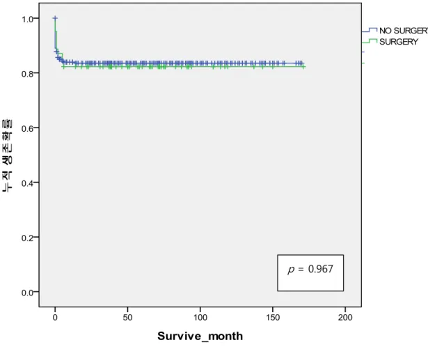
Survival outcome
Long-term survival rate was calculated in Figure 4. 5-year overall survival (OS) rate of the non-surgical and surgical group were 84% and 83% (p=0.967). (Figure 2-A) 5-year OS rate of NEC, SIP, MRI, and MNRI were 72%, 83%, 100%, and 100%. Survival outcomes between the four disease entities were not significantly different (p=0.109). (Figure 2-B) However, comparing survival outcomes of NEC and non-NEC group, NEC showed a significantly poor
14
survival outcome (p=0.033). (Figure 2-C) Multivariate analysis for survival
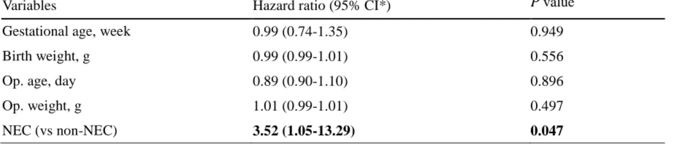
Logistic regression test for survival was performed with covariates of patient characteristics and follow up variables. Even after adjusting potentially confounding factors, NEC was still statistically significant factor for survival. (Hazards ratios = 3.521, 95% confidence interval (CI) 1.053-13.287, p=0.047). (Table 8)
15
Discussion
ELBW neonates were known to have high morbidity and mortality, however, recent improvements in neonatal ventilation and monitoring techniques made it possible for ELBW neonates to live. The incidence of ELBW in South Korea has grown over the years. (Figure 3) The treatment guidelines for ELBW neonates has been evolved over the recent years, but still much of its portions are controversial due to its rarity. There have been few reports about differential diagnosis which led to acute abdomen in ELBW neonates, and there have been reports discussing treatment modalities for acute abdomen, for example, peritoneal drainage or laparotomy. However, clinical characteristics and prognosis of acute abdomen according to differential diagnosis among ELBW neonates has not been established well and has been reported only for each disease categories. Our single institutional study provides data in support of the safety of laparotomy for ELBW neonates with acute abdomen with respect to complications, and overall survival according to differential diagnosis.
There are 4 different disease entities that lead to acute abdomen in ELBW, but are seldom emphasized. However, it is important to suspect and make a precise diagnosis since they have different prognosis, and different surgical procedure are recommended for each differential diagnosis. Making a right diagnosis may reduce post-operative complications and help patient to survive longer. SIP is defined as isolated single intestinal perforation that is typically found at terminal ileum. (7-9) Absences of traditional clinical signs of Bell’s criteria and histologic signs of typical NEC characterize SIP as a distinct entity. Some had reported that radiographic findings associated with NEC such as pneumatosis intestinalis and portal venous air are indicative factors to distinguish between SIP, but in the current series, it was statistically difficult to make differential diagnosis preoperatively. A report from the Pediatrix Medical Group (17) mentioned about clinical manifestation of the neonates with SIP have presented in
16
earlier age than those with NEC (median age 7 versus 15 days), but this was not adjusted in the current study. Kubota et al (11) first used the term MRI in their report defining bowel obstruction with meconium in neonates without cystic fibrosis. They suggested that inspissated meconium is the result of excessive water absorption in the hypoperistaltic bowel before birth, and reported 13 ELBW neonates with intestinal perforation secondary to MRI. MNRI is defined as ileus without a definite sticky meconium, and not associated with Hirschsprung’s disease or intestinal atresia. (20-22) It is often hypothesized that zonal hypoganglionosis may be the pathophysiology of the disease. Although the macroscopic findings of NEC, SIP, MRI, and MNRI are clearly different from each other, it is often difficult to make a definitive diagnosis without laparotomy. In this study, comparing characteristics of four differential diagnoses were performed with one-way analysis of variance method. In order to specify the difference Tukey’s multiple comparison test was used to compare the difference between each pair of means with appropriate adjustment for the multiple testing. It was expressed as ‘a’, and
‘b’ for the statistically not different group in the tables, and the mean gestational age was the only factor different among the differential diagnoses.
Abdominal surgery for ELBW was performed for infants on suspicion of bowel perforation diagnosed by pneumoperitoneum on infantogram, or infants with clinical deterioration despite medical treatment. In this series of case, all patients had serial examinations with infantogram and abdominal ultrasonography pre-operatively. 21 (47.7%) showed free air in pre-operative infantogram. 5 (11.4%) had portal air and 8 (18.2%) showed mural gas, and 9 (20.5%) showed ascites in ultrasonography. In most of the cases, loop type or double barrel type ostoma was performed. But primary closure was performed in three SIP patients, and one MRI patient, and all experienced additional surgery for proximal diversion due to re-perforation of the anastomosis. Whilst there are positive results about primary peritoneal drainage (13-14, 18-19),
17
the current study have adjusted primary peritoneal drainage in three SIP patients, which eventually turned out to receive laparotomies after a worsening progression of the disease. Thus, in our experience, for ELBW neonates, when abdominal perforation is suspected, laparotomy is recommended compared to peritoneal drainage.
5 years survival outcome for NEC in this study was 72%, similar or higher than the previous reports. (6, 23) 5 years survival for SIP was 83% which is also higher than the previous studies.
(7-9, 24) There is still no consensus on which surgical techniques may be most beneficial to the patient. A number of centers have stated that a primary anastomosis for NEC or SIP showed a decent outcome. (25) In the current study, we have performed intestinal resection and primary anastomosis in three selected cases, but all resulted in complication of leakage at the anastomosis site. Although SIP, MRI, and MNRI have focal intestinal perforation and limited inflammation around the perforation site, we recommend performing ostoma formation in ELBW neonates.
Recently, the survival rate of ELBW neonates is known to have increased due to development of perinatal health care in various studies. (1-3) In the study of Japanese Society of Pediatric Surgeons, mortality of intestinal perforation for ELBW neonates in the recent 15 years was 31.6%.(5) There is no Korean studies for the overall survival rate of ELBW neonates.
However, Korean Neonatal Network (KNN) database system has been launched recently and reported 69.6% of survival rate for VLBW neonates. (3, 26) In this study, the 5-years overall survival rate for ELBW neonates who have underwent abdominal surgery were 84%. The 5- years overall survival rate for ELBW neonates who did not receive abdominal surgery were 83%. As far as we are concerned, this is the first to report survival outcome for ELBW neonates with abdominal surgery in Korean data. Different from other previous studies, overall rate of surgery group did not show a significant difference compared to the non-surgical group. On
18
risk factor analysis, NEC was a single significant prognostic factor.
The limitation of this study was that it was a retrospectively reviewed study. The growth and development of the patients were not analyzed. However, with long-term, detailed single-center study, it may aid clinicians and surgeons in their medical decisions. This report was the first to explain abdominal surgical indication into four defined disease entities. As the ELBW neonates are increasing worldwide, this study may give a guideline to diagnosis and treatment of abdominal problems.
In conclusion, abdominal surgery for ELBW neonates is a feasible procedure with good survival outcome compared to non-surgical group. Ostoma formation is recommended as a procedure of choice in ELBW neonates. Diagnosis of NEC was a significant risk factor for survival, which most of the postoperative morbidity and mortality was encountered.
19
REFERENCES
1. Fitzgibbons SC, Ching Y, Yu D, et al. Mortality of necrotizing enterocolitis expressed by birth weight categories. J Pediatr Surg. 2009;44(6):1072-1076.
doi:10.1016/j.jpedsurg.2009.02.013
2. Patel RM, Kandefer S, Walsh MC, et al. Causes and Timing of Death in Extremely Premature Infants From 2000 Through 2011. Obstet Anesth Dig. 2015;35(4):213-214.
doi:10.1097/01.aoa.0000472734.42973.28
3. Shim JW, Jin HS, Bae CW. Changes in survival rate for very-low-birth-weight infants in Korea: Comparison with other countries. J Korean Med Sci. 2015;30:S25-S34.
doi:10.3346/jkms.2015.30.S1.S25
4. Stoll BJ, Hansen NI, Bell EF, et al. Neonatal outcomes of extremely preterm infants from the NICHD Neonatal Research Network. Pediatrics. 2010;126(3):443-456.
doi:10.1542/peds.2009-2959
5. Committee on Academic Survey and Advanced Medical Science (2004) Neonatal surgery in Japan—National survey on neonatal surgery 2003. J Jpn Soc Pediatr Surg 40:919–934
6. Neu J, Walker WA. Necrotizing enterocolitis. N Engl J Med. 2011;364:255-264.
doi:10.1056/NEJMra1005408
7. Mintz AC, Applebaum H. Focal gastrointestinal perforations not associated with necrotizing enterocolitis in very low birth weight neonates. J Pediatr Surg.
1993;28(6):857-860. doi:10.1016/0022-3468(93)90345-L
8. Aschner JL, Deluga KS, Metlay LA, Emmens RW, Hendricks-Munoz KD.
Spontaneous focal gastrointestinal perforation in very low birth weight infants. J Pediatr. 1988;113(2):364-367. doi:10.1016/S0022-3476(88)80285-3
20
9. Meyer CL, Payne NR, Roback SA. Spontaneous, isolated intestinal perforations in neonates with birth weight < 1,000 g not associated with necrotizing enterocolitis. J Pediatr Surg. 1991;26(6):714-717. doi:10.1016/0022-3468(91)90017-N
10. Kubota A, Imura K, Yagi M, et al. Functional ileus in neonates: Hirschsprung’s disease-allied disorders versus meconium-related ileus. Eur J Pediatr Surg.
1999;9(6):392-395. doi:10.1055/s-2008-1072290
11. Kubota A, Shiraishi J, Kawahara H, et al. Meconium-related ileus in extremely low- birthweight neonates: Etiological considerations from histology and radiology. Pediatr Int. 2011;53(6):887-891. doi:10.1111/j.1442-200X.2011.03381.x
12. Baird R, Puligandla PS, St Vil D, et al. The role of laparotomy for intestinal perforation in very low birth weight infants. J Pediatr Surg 2006; 41:1522.
13. Cass DL, Brandt ML, Patel DL, et al. Peritoneal drainage as definitive treatment for neonates with isolated intestinal perforation. J Pediatr Surg 2000; 35:1531.
14. Hall NJ, Curry J, Drake DP et al. Resection and primary anastomosis is a valid surgical option for infants with necrotizing enterocolitis who weigh less than 1000 g.
Arch Surg 2005;140:1149–1151
15. Kubota A, Yamanaka H, Okuyama H, et al. Focal intestinal perforation in extremely- low-birth-weight neonates: Etiological consideration from histological findings.
Pediatr Surg Int. 2007;23(10):997-1000. doi:10.1007/s00383-007-1984-9
16. Dindo, D., Demartines, N., Clavien, P., 2004. Classification of surgical complications:
a new proposal with evaluation in a cohort of 6336 patients and results of a survey.
Ann. Surg. 240, 205–213
17. Pumberger W, Mayr M, Kohlhauser C, Weninger M. Spontaneous localized intestinal perforation in very-low-birth-weight infants: A distinct clinical entity different from
21
necrotizing enterocolitis. J Am Coll Surg. 2002;195(6):796-803. doi:10.1016/S1072- 7515(02)01344-3
18. Hall NJ, Curry J, Drake DP, Spitz L, Kiely EM, Pierro A. Resection and primary anastomosis is a valid surgical option for infants with necrotizing enterocolitis who weigh less than 1000 g. Arch Surg. 2005;140(12):1149-1151.
doi:10.1001/archsurg.140.12.1149
19. Gfroerer S, Fiegel H, Schloesser RL, Rolle U. Primary laparotomy is effective and safe in the treatment of necrotizing enterocolitis. World J Surg. 2014;38(10):2730-2734.
doi:10.1007/s00268-014-2615-y
20. Rickham PP, Boeckman CR. Neonatal meconium obstruction in the absence of mucoviscidosis. Am. J. Surg. 1965; 109: 173–7.
21. Puri P. Variant Hirschsprung's disease. J Pediatr Surg 32 (1997) 149-157
22. Toyosaka A, Tomimoto Y, Nose K, Seki Y, Okamoto E. Immaturity of the myenteric plexus is the aetiology of meconium ileus without mucoviscidosis: A histopathologic study. Clin Auton Res. 1994;4(4):175-184. doi:10.1007/BF01826183
23. Zani A, Pierro A. Necrotizing enterocolitis: controversies and challenges.
F1000Research. 2015. doi:10.12688/f1000research.6888.1
24. Adderson EE, Pappin A, Pavia AT. Spontaneous intestinal perforation in premature infants: a distinct clinical entity associated with systemic candidiasis. J Pediatr Surg.
1998;33(10):1463-1467. doi:S0022-3468(98)90475-4
25. Blakely ML, Gupta H, Lally KP. Surgical Management of Necrotizing Enterocolitis and Isolated Intestinal Perforation in Premature Neonates. Semin Perinatol.
2008;32(2):122-126. doi:10.1053/j.semperi.2008.01.008
26. Chung SH, Bae CW. Improvement in the survival rates of very low birth weight
22
infants after the establishment of the Korean neonatal network: Comparison between the 2000s and 2010s. J Korean Med Sci. 2017;32(8):1228-1234.
doi:10.3346/jkms.2017.32.8.1228
Author Disclosure Statement: None of the authors of this study has any financial interest or conflict with any industries or parties.
23
Table Legend
1. Patient characteristics 2. Operative characteristics
3. Post-operative complications before discharge (Cases of Extended Clavien‑Dindo classification over Gr III)
4. Outcome of abdominal surgery
5. Demographics according to differential diagnosis
6. Operative characteristics according to differential diagnosis 7. Outcome of abdominal surgery according to differential diagnosis 8. Multivariate proportional hazards analysis
Figure Legend
1. Incidence of abdominal surgeries in ELBW neonates 2. Survival outcome
A. Survival rate (Surgery vs Non-Surgery) B. Survival rate (Between Disease Categories) C. Survival rate (NEC vs non-NEC)
3. Incidence of ELBW in Korea
24
Table 1. Patient Characteristics
Patient characteristic Total
Number of patients 65
Male sex 42
Gestational age, mean (weeks) 26+1 (22+3-33+0)
Birth weight, mean±SD (g) 695.9 (355-990)
Z-score -0.49
Maternal age at delivery, mean±SD (years) 32.1±5.3 (23-57) Mode of delivery
Cesarean section 45
Vaginal delivery 20
Twins 29
Triplets 5
Apgar score, median
1 min 3 (0-7)
5 min 5 (0-8)
Combined medical problem
RDS 51
PDA 59
Indomethacin use 36
Cardiac anomaly 14
Brain hemorrhage 40
Chromosomal anomaly 2
Mechanical ventilator after birth (days) 31 (0-385)
1) The same letters indicate non-significant difference between groups based on Tukey’s multiple comparison test.
2) PNALD; Parenteral nutrition associated liver dysfunction
* Analyzed without in-hospital mortality cases
+ Complication cases of Extended Clavien‑Dindo classification over Gr III before discharge
25
Table 2. Operative Characteristics
Patient characteristic Total
Age at operation, mean±SD (days) 12.4 (0-34) Body weight at operation, mean±SD (g) 710.7 (490-970) Procedure
Primary anastomosis 4
Ostoma 61
Ileostomy 55
Jejunostomy 10
Postoperative TPN, mean±SD (days)* 39.7±23.8 (11-109)
PNALD 26
Hospital stay, mean±SD (days)* 107.8±50.1 (52-415)
26
Table 3. Post-operative complications before discharge (Cases of Extended Clavien‑Dindo classification over Gr III)
No OP date GA Diagnosis Age at op Bwt at OP OP POD PostOP complication Treatment of
complication Outcome
1 2003-06-02 26+5 SIP 19 900 Primary closure 3 Anastomosis leakage Enterostomy stable
3 2004-09-09 33 MRI 2 900 Decompression and
closure 4 Anastomosis leakage Enterostomy stable
5 2006-04-08 25+3 SIP 1 600 Primary closure 9 Anastomosis leakage Enterostomy stable
9 2007-05-11 27+4 NEC 22 920 Enterostomy 36 Adhesive ileus Adhesiolysis stable
11 2007-07-10 27+2 NEC 28 970 Enterostomy 1 Bleeding Bleeding control expired
16 2008-10-04 25+3 MRI 7 520 Enterostomy 14 Ostoma prolapse Reduction stable
25 2010-08-27 25+1 NEC 24 570 Enterostomy 1 Sepsis 0 expired
29 2011-08-08 24 SIP 5 740 Primary closure 3 Anastomosis leakage Enterostomy stable
34 2011-10-30 23+1 NEC 10 580 Enterostomy 0 Cardiac arrest(intraop) 0 expired
37 2012-03-02 22+3 NEC 16 490 Enterostomy 14 Peristomal infection Enterostomy revision expired
46 2013-07-12 30+4 NEC 9 890 Enterostomy 28 Sepsis 0 expired
50 2013-09-07 27+1 NEC 15 700 Enterostomy 43 Respiratory failure 0 expired
58 2014-11-20 23+2 SIP 30 640 Enterostomy 22 Sepsis 0 expired
61 2015-12-24 26+3 NEC 9 1020 Enterostomy 1 Sepsis 0 expired
27
Table 4. Outcome of abdominal surgery
N=65 Early complications (<30 days)
Septic shock 5
Anastomosis leakage 4
Bleeding 1
Stomy prolapse 1
Cardiac arrest 1
Late complications (≥30 days)
Ileus 4
Wound complication 3
Incisional hernia 3
Mortality (<30 days)
Septic shock 5
Bleeding 1
Cardiac arrest 1
Mortality (≥30 days)
Respiratory failure 3
Follow up period, mo 62 (3-166)
Body weight (kg) 12.7 (2.7-25.6)
Z-score + -1.52 (-4.88 - 0.84)
Height (cm) 95.05 (47.3-134.1)
Z-score+ -1.16 (-6.82 – 1.52)
+ Z score measured on the last follow up visit to clinic
28
Table 5. Demographics according to differential diagnosis
Patient characteristic NEC SIP MRI MNRI p-value
Number of patients 29 18 13 5
Male 16 (55.2%) 13 (72.2%) 11 (84.6%) 2 (40%) 0.159
Gestational age, mean (weeks) 25+4 (22+3-28+3) 25+3 (23+2-27+3) 26+2 (23+1-33+0) 29+3 (23+1-32+1) 0.008
T1) a a a, b b
Birth weight, mean±SD (g) 675.9±159.1 (480-990) 750.6±141.2 (440-990) 693.1±170.1 (430-990) 632.0±85.8 (530-720) 0.308 Maternal age at delivery, mean±SD (years) 31.7±6.4 (23-57) 32.3±3.6 (25-38) 32.8±4.4 (27-43) 32.2±5.1 (24-57) 0.943 Mode of delivery
Cesarean section 20 (70.0%) 13 (72.2%) 8 (61.5%) 4 (80.0%)
0.733
Vaginal delivery 9 (30.0%) 5 (27.8%) 5 (38.5%) 1 (20.0%)
Twins 16 (55.2%) 10 (55.6%) 6 (46.2%) 2 (40%) 0.880
Apgar score, median
1 min 3 (1-6) 2 (0-5) 3 (1-6) 3 (1-7)
0.791
5 min 5 (1-8) 4 (0-7) 5 (2-7) 5 (3-8)
Combined medical problem
RDS 24 (82.8%) 16 (88.9%) 9 (69.2%) 2 (40%) 0.089
PDA 25 (86.2%) 18 (100%) 12 (92.3%) 4 (80%) 0.350
Indomethacin use 16 (55.2%) 11 (61.1%) 7 (53.8%) 2 (40%) 0.866
Cardiac anomaly 7 (24.1%) 3 (16.6%) 2 (15.3%) 1 (20%) 0.891
Brain hemorrhage 19 (65.5%) 11 (61.1%) 9 (69.2%) 1 (20%) 0.244
Chromosomal anomaly 1 (3.4%) 0 (0%) 1 (7.7%) 0 (0%) 0.433
Mechanical ventilator after birth (days) 37 (0-385) 33 (0-121) 23 (0-48) 15 (0-50) 0.121
29
Table 6. Operative Characteristics according to differential diagnosis
Patient characteristic NEC SIP MRI MNRI p-value
Age at operation, mean±SD (days) 15.8±10.3 (5-43) 10.9±7.5 (1-30) 10.0±6.0 (2-20) 16.4±16.4 (2-41) 0.163
Body weight at operation, mean±SD (g) 706.9±163.4 (490-
1020) 739.4±104.0 (540-900) 680.0±143.4 (430-900) 735.0±289.9 (530-940) 0.665
Procedure
Primary anastomosis 0 (0%) 3 (16.7%) 1 (7.7%) 0 (0%)
0.040
Ostoma 29 (100%) 15 (83.3%) 12 (92.3%) 5 (100%)
Postoperative TPN, mean±SD (days)* 39.0±21.0 (11-93) 37.9±25.3 (13-109) 40.5±24.2 (16-96) 48.0±30.9 (15-77) 0.867
PNALD 10 (45.5%) 8 (47.1%) 5 (38.5%) 3 (60%) 0.847
Hospital stay, mean±SD (days)* 139.3±75.1 (81-415) 105.6±35.5 (58-199) 124.1±48.7 (52-235) 95.6±9.7 (83-106) 0.239
30
Table 7. Outcome of abdominal surgery according to differential diagnosis
NEC (N=29) SIP (N=18) MRI (N=13) MNRI (N=5) p-value
Early complications (<30 days) 7 3 2 0 0.748
Anastomosis leakage 0 3 1 0
Bleeding 1 0 0 0
Septic shock 5 0 0 0
Stomy prolapse 0 0 1 0
Cardiac arrest 1 0 0 0
Late complications (≥30 days) 7 2 1 0 0.210
Ileus 3 0 1 0
Wound complication* 2 1 0 0
Incisional hernia* 2 1 0 0
Mortality (<30 days) 7 1 0 0 0.048
Septic shock 5 1 0 0
Bleeding 1 0 0 0
Cardiac arrest 1 0 0 0
Mortality (≥30 days) 2 1 0 0 0.423
Respiratory failure 2 1 0 0
Follow up period, mo 62 (4-139) 57 (3-166) 61 (10-131) 71 (23-151) 0.781
Body weight (kg) 13.8 (6.5-25.6) 13.4 (2.7-24.8) 13.5 (7.1-22.5) 13.4 (9.8-20.3) 0.997
Z-score + -1.45 (-3 - 0) -1.34 (-5 – 0) -1.70 (-4 – 0) -2.54 (-4 - -2) 0.331
Height (cm) 96.9 (61-132) 90.1 (47-134) 91.1 (60-121) 99.2 (83-120) 0.769
Z-score + -1.08 (-3 – 1) -1.06 (-7- 3) -0.52 (-3 – 11) -1.82 (-2 - -1) 0.728
* Wound complication and incisional hernia occurred after the ostoma repair
31
Table 8. Multivariate proportional hazards analysis
Variables Hazard ratio (95% CI*) P value
Gestational age, week 0.99 (0.74-1.35) 0.949
Birth weight, g 0.99 (0.99-1.01) 0.556
Op. age, day 0.89 (0.90-1.10) 0.896
Op. weight, g 1.01 (0.99-1.01) 0.497
NEC (vs non-NEC) 3.52 (1.05-13.29) 0.047
*CI denotes confidence interval.
32
Figure 1. Abdominal surgeries in ELBW neonates
328 342 317
398
536 562 563
508
575 589 503
597 601
18 37 46 60 72 67 77 71 77 78 61 70 71
2 1 2 3 6 4 4 7 10 10 9 3 4
2003 2004 2005 2006 2007 2008 2009 2010 2011 2012 2013 2014 2015 all admission <1kg <1kg (abdominal surgery)
33
Figure 2. Survival outcome
A. Survival rate (Surgery vs Non-Surgery)
p = 0.967
34
B. Survival rate (Between Disease Categories)
NEC SIP MRI MNRI
p = 0.109
35
C. Survival rate (NEC vs non-NEC)
NEC SIP MRI MNRI
p = 0.033
NEC non-NEC
36
Figure 3.