저작자표시-비영리-변경금지 2.0 대한민국 이용자는 아래의 조건을 따르는 경우에 한하여 자유롭게
l 이 저작물을 복제, 배포, 전송, 전시, 공연 및 방송할 수 있습니다. 다음과 같은 조건을 따라야 합니다:
l 귀하는, 이 저작물의 재이용이나 배포의 경우, 이 저작물에 적용된 이용허락조건 을 명확하게 나타내어야 합니다.
l 저작권자로부터 별도의 허가를 받으면 이러한 조건들은 적용되지 않습니다.
저작권법에 따른 이용자의 권리는 위의 내용에 의하여 영향을 받지 않습니다. 이것은 이용허락규약(Legal Code)을 이해하기 쉽게 요약한 것입니다.
Disclaimer
저작자표시. 귀하는 원저작자를 표시하여야 합니다.
비영리. 귀하는 이 저작물을 영리 목적으로 이용할 수 없습니다.
변경금지. 귀하는 이 저작물을 개작, 변형 또는 가공할 수 없습니다.
의학석사 학위논문
척수 상의하세포종의 임상적, 영상의학적, 병리학적 특성 및 수술적 치료에 대한 고찰
: 다기관 후향적 연구
Clinical, radiological and pathological characteristics of spinal cord
subependymoma
: multi-institutional retrospective study
2018 년 2 월
서울대학교 대학원 의학과 신경외과학전공
여 운 탁
의학석사 학위논문
척수 상의하세포종의 임상적, 영상의학적, 병리학적 특성 및 수술적 치료에 대한 고찰
: 다기관 후향적 연구
Clinical, radiological and pathological characteristics of spinal cord
subependymoma
: multi-institutional retrospective study
2018 년 2 월
서울대학교 대학원 의학과 신경외과학전공
여 운 탁
A thesis of the Master’s degree
Clinical, radiological and pathological characteristics of spinal cord
subependymoma
: multi-institutional retrospective study
February 2017
Department of Neurosurgery
Seoul National University College of Medicine
Woontak Yuh
척수 상의하세포종의 임상적, 영상의학적, 병리학적 특성 및 수술적 치료에 대한 고찰
: 다기관 후향적 연구
지도교수 강 현 승
이 논문을 의학석사 학위논문으로 제출함
2017
년 10월서울대학교 대학원 의학과 신경외과학전공
여 운 탁
여운탁의 석사학위논문을 인준함
2018
년 1월위 원 장 손 철 호 (인) 부 위 원 장 강 현 승 (인) 위 원 박 철 기 (인)
Clinical, radiological and pathological characteristics of spinal cord subependymoma
: multi-institutional retrospective study
Woontak Yuh Department of Neurosurgery The Graduate School Seoul National University
ABSTRACT
Objective: A spinal cord subependymoma is an uncommon, indolent, benign spinal cord tumor. It is radiologically similar to a spinal cord ependymoma, but surgical findings and outcomes differ. Gross total resection of the tumor is not always feasible. The present study was done to determine the clinical, radiological and pathological characteristics of spinal cord subependymomas.
Methods: We retrospectively reviewed the medical records of ten spinal cord subependymoma patients (M:F=4:6; median 38 years, range 21-77) from four institutions.
Results: The most common symptoms were sensory changes and/or pain in eight patients, followed by motor weakness in six. The median duration of symptoms was 9.5 months. Preoperative radiological diagnosis was
i
ependymoma in seven and astrocytoma in three. The tumors were located eccentrically in six and were not enhanced in six. Gross total resection of the tumor was achieved in five patients, whereas subtotal or partial resection was inevitable in the other five patients due to a poor dissection plane. Adjuvant radiotherapy was performed in two patients. Neurological deterioration occurred in two patients; transient weakness in one after subtotal resection and permanent weakness after gross total resection in the other. Recurrence or regrowth of the tumor was not observed during the median 31.5 months follow-up period (range, 8-89).
Conclusion: Spinal cord subependymoma should be considered when the tumor is located eccentrically and is not dissected easily from the spinal cord.
Considering the rather indolent nature of spinal cord subependymomas, subtotal removal without the risk of neurological deficit is another option.
Keyword: subependymoma; ependymoma; spinal cord; spinal cord neoplasm; surgery; outcome
Student Number: 2016-21942
ii
Contents
1. Abstract ……….. i
2. Contents ……….. iii
3. List of Tables ……….. iv
4. List of Figures ……….. v
5. Introduction ……….. 1
6. Materials and Methods ……….. 2
7. Results ……….. 4
8. Discussion ……….. 21
9. Conclusions ……….. 27
10. References ……….. 28
11. 국문 초록 ……….. 34
iii
List of Tables
Table 1. Modified McCormick Classification
……… 3
Table 2. Demographics, clinical presentations and clinical outcomes of 10 patients
……… 5
Table 3. Radiological findings of 10 patients
……… 7
Table 4. Summary of characteristics of SCSE in the previous and current studies
……… 22
iv
List of Figures
Figure 1. Preoperative magnetic resonance images of patient #9
……… 8
Figure 2. Intraoperative photograph of patient #9
……… 9
Figure 3. Histopathological features of patient #9
……… 11
Figure 4. Preoperative magnetic resonance images of the patient #3
……… 12
Figure 5. Preoperative magnetic resonance images of the patient #6
……… 14
Figure 6. Intraoperative photograph of patient #6
……… 16 Figure 7. Preoperative magnetic resonance images of the patient #8
……… 17 Figure 8. Intraoperative photograph of patient #8
……… 18
v
1
INTRODUCTION
Subependymoma, first described by Scheinker et al. 1 in 1945, is benign, non-invasive, slow-growing tumor corresponding histologically to WHO grade I 2,3. It accounts for 0.51% of all central nervous system tumors and 8.3% of all ependymal tumors. Subependymoma most often arises in the fourth (50-60%) and the lateral (30-40%) ventricles4. A spinal cord subependymoma (SCSE), first reported by Boykin et al. 5 in 1954,
represented 2-15.5% of all CNS subependymomas, 1.3% of all intramedullary tumors6. Since the first study by Boykin et al., 72 cases of SCSE have been reported4-45. Most of the studies were case reports, and the largest study was by Wu et al. 42 involving 13 cases from a single institution. In Korea, there was only one case report of SCSE by Jang et al.35 in 2009. Although SCSE is not extremely rare, it is not easily differentiable from a spinal cord
ependymoma with radiological findings. However, surgical findings and outcomes differ from those of an ependymoma, including a high risk of neurological deficit in the event of a poor dissection plane from the spinal cord with a low rate of recurrence. This study is the largest case series of SCSE in Korea and the second biggest study, second to the study by Wu et al.
We reviewed the clinical, radiological and pathological characteristics of ten cases of SCSE from the Korea Spinal Oncology Research Group (KSORG) database. Furthermore, we reviewed clinical, radiological and pathological characteristics of all previously reported 72 cases of SCSE and compared with characteristics of 10 SCSE patients of this study.
MATERIALS AND METHODS
The KSORG was established in 2009 by spine surgeons from the Seoul National University Hospital (SNUH), Seoul National University Bundang Hospital (SNUBH), Samsung Medical Center (SMC), and several affiliated hospitals sought to promote clinical research on the management of spinal and spinal cord tumors and to develop educational programs to improve neuro- oncological care46-48. The principal goal of the KSORG was to evaluate surgical treatments of spinal neoplasms in a prospective and retrospective multicenter clinical series. From April of 2000 to September of 2014, ten patients in total were pathologically confirmed as SCSE patients in four hospitals. During the same period, 88 cases of spinal cord ependymoma were surgically treated48. These medical records, including radiological images, were retrospectively reviewed.
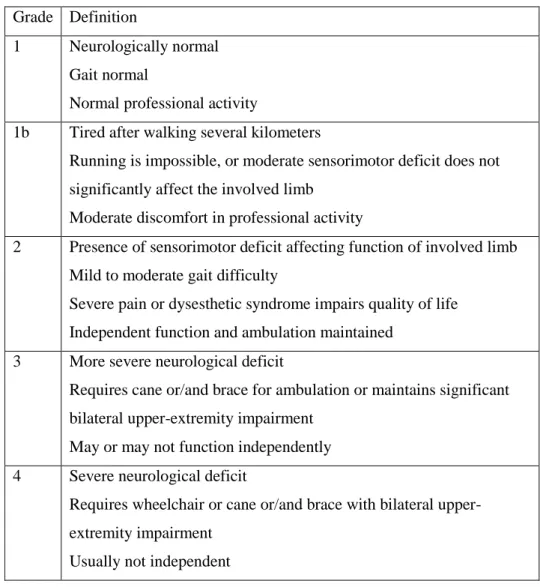
The characteristics and duration of symptoms, preoperative neurological function, intraoperative surgical findings, the extent of surgical resection, intraoperative electrophysiologic monitoring results, the use of an adjuvant treatment, the follow-up period, and postoperative complications were reviewed. The modified McCormick classification (MMC) (Table 1) 49 was applied to the assess neurological functions of the patients before surgery, at discharge, at one and six months after surgery and annually thereafter.
Magnetic resonance (MR) images were taken in all patients before and after surgery. The location and level of involvement of the tumor and the pattern of enhancement were evaluated in detail. In our study, all tumors were removed
3
with a conventional posterior approach by senior spine tumor surgeons, and microsurgical techniques did not differ from previously published techniques in the literature.
Table 1. Modified McCormick Classification
The extent of resection was classified as gross total resection (GTR), subtotal resection (STR) or partial resection (PR). GTR was defined if surgeons described a complete resection of the tumor and there was no Grade Definition
1 Neurologically normal Gait normal
Normal professional activity
1b Tired after walking several kilometers
Running is impossible, or moderate sensorimotor deficit does not significantly affect the involved limb
Moderate discomfort in professional activity
2 Presence of sensorimotor deficit affecting function of involved limb Mild to moderate gait difficulty
Severe pain or dysesthetic syndrome impairs quality of life Independent function and ambulation maintained
3 More severe neurological deficit
Requires cane or/and brace for ambulation or maintains significant bilateral upper-extremity impairment
May or may not function independently 4 Severe neurological deficit
Requires wheelchair or cane or/and brace with bilateral upper- extremity impairment
Usually not independent
evidence of the tumor in postoperative MR images. If a small piece of tumor was left in place according to the surgeon’s decision or an obvious retained fragment appeared in postoperative MR images (80-99% resection), the extent of resection was considered to be STR. In the same manner, cases involving less than 80% resection were defined as PR. Pathologically, after resection of the tumor, experienced neuropathologists in each institution reviewed both frozen sections and permanent specimens. The World Health Organization (WHO) classification system was used for histopathological diagnoses.
Immunohistochemistry and electron microscopic examinations were performed as needed.
Patient follow-up took place at an outpatient clinic postoperatively to evaluate their neurological statuses with MMC grades. Tumor recurrence and progression were defined both clinically and radiologically. Tumor recurrence was defined as regrowth of the tumor after GTR on follow-up MR images or any clinical aggravation. Tumor progression was defined as tumor regrowth after STR or PR on follow-up MR images or any clinical aggravation. The study was reviewed and approved by the institutional review board. (IRB No.
1306-106-500).
RESULTS
Patient demographics
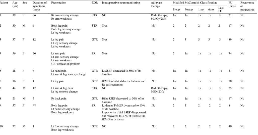
Table 2 summarizes the characteristics of all patients. Of the ten, four were
5
Patient Age (yrs)
Sex Duration of symptoms (mos)
Presentation EOR Intraoperative neuromonitoring Adjuvant therapy
Modified McCormick Classification FU (mos)
Recurrence or progression Preop Postop 1mo 6mo Last
FU
1 39 F 36 Rt arm sensory change
Rt arm weakness
STR NC Radiotherapy,
50.4Gy/28fx
1a 1a 1a 1a 1a 21 No
2 30 M 6 Both leg pain
Both leg sensory change Lt leg weakness
STR N/A No 2 2 2 2 2 17 No
3 37 F 12 Lt leg pain
Lt leg sensory change Lt leg weakness
GTR N/A No 2 3 3 3 3 89 No
4 56 F 36 Lt arm pain
Lt arm sensory change Lt arm weakness UR, defecation problem
PR N/A No 2 1a 1a 1a 1a 74 No
5 28 F 6 Lt hand pain
Lt arm & leg sensory change
GTR Lt SSEP decreased to 50% of its baseline
No 1a 1a 1a 1a 1a 41 No
6 36 F 1 Lt leg pain GTR fEMG in bilat abductor hallucis and
Rt gastrocnemius
No 1a 1a 1a 1a 1a 38 No
7 44 M 12 Lt arm & leg pain
Lt leg sensory change
STR NC Radiotherapy,
50Gy/20fx
1a 1a 1a 1a 1a 25 No
8 21 M 7 Rt back pain GTR Bilat SSEP decreased to 50% of its
baseline
No 1a 1a 1a 1a 1a 17 No
9 57 F 48 Both leg pain
Lt hand sensory change Both leg weakness
PR Lt thenar TcMEP decreased to 10%
of its baseline
Lt posterior tibial SSEP disappeared but recovered to 30% of its baseline fEMG in Lt thenar
No 2 3 2 2 2 8 No
10 77 M 1 Lt foot sensory change
Both leg weakness
GTR NC No 2 2 2 2 2 40 No
Table 2. Demographics, clinical presentations and clinical outcomes of 10 patients
Bilat,bilateral; EOR, extent of resection; F, female; fEMG, free-running electromyography; FU, follow-up; fx, fraction; GTR, gross total resection; Lt, left; M, male; mo, month; mos, months;
NC, no significant change; N/A, not available; Postop, postoperative; PR, partial resection; Preop, preoperative; Rt, right; SSEP, somatosensory evoked potential; STR, subtotal resection;
TcMEP, transcranial motor evoked potential; UR, urinary retention; yrs, years.
male and six were female, with ages at surgery ranging from 21 to 77 years (median, 38 years). The most common symptoms were sensory changes and pain in eight patients, followed by motor weakness in six, and bowel/bladder symptoms in one patient. The median duration of symptoms was 9.5 months (range, 1 to 48). The ratio of SCSE versus spinal cord ependymoma was 1:8.8 with the present database 46-48.
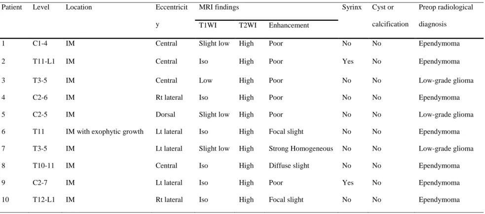
Radiological findings
Table 3 summarizes the radiological findings of all patients. The tumors were located at the cervical spinal cord in four patients, the thoracic spinal cord in four patients, and the thoracolumbar spinal cord in two. Magnetic resonance (MR) images of all patients showed iso- to low signal intensity on T1-weighted images (WI) and high signal intensity on T2-WI (Fig. 1). The pattern of contrast (gadolinium) enhancement was homogeneously strong in one patient, with partial uptake in three patients and scanty enhancement in six patients. The tumors
were located eccentrically in six patients. Syringomyelia existed in two patients, but intratumoral cystic changes, a hemosiderin cap, or calcification was not observed. The preoperative radiological diagnosis was ependymoma in seven patients and low-grade glioma in three patients, but a
subependymoma was not included in the list of differential diagnoses.
7
Patient Level Location Eccentricit
y
MRI findings Syrinx Cyst or
calcification
Preop radiological diagnosis
T1WI T2WI Enhancement
1 C1-4 IM Central Slight low High Poor No No Ependymoma
2 T11-L1 IM Central Iso High Poor Yes No Ependymoma
3 T3-5 IM Central Low High Poor No No Low-grade glioma
4 C2-6 IM Rt lateral Iso High Poor No No Ependymoma
5 C2-5 IM Dorsal Slight low High Poor No No Low-grade glioma
6 T11 IM with exophytic growth Lt lateral Iso High Focal slight No No Ependymoma
7 T3-5 IM Lt lateral Slight low High Strong Homogeneous No No Low-grade glioma
8 T10-11 IM Central Iso High Diffuse slight No No Ependymoma
9 C2-7 IM Lt lateral Iso High Poor Yes No Ependymoma
10 T12-L1 IM Rt lateral Iso High Focal slight No No Ependymoma
Table 3. Radiological findings of 10 patients
IM, intramedullary; Lt, left; Preop, preoperative; MRI, magnetic resonance imaging; Rt, right; T1WI, T1-weighted image; T2WI, T2-weighted image.
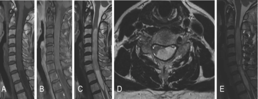
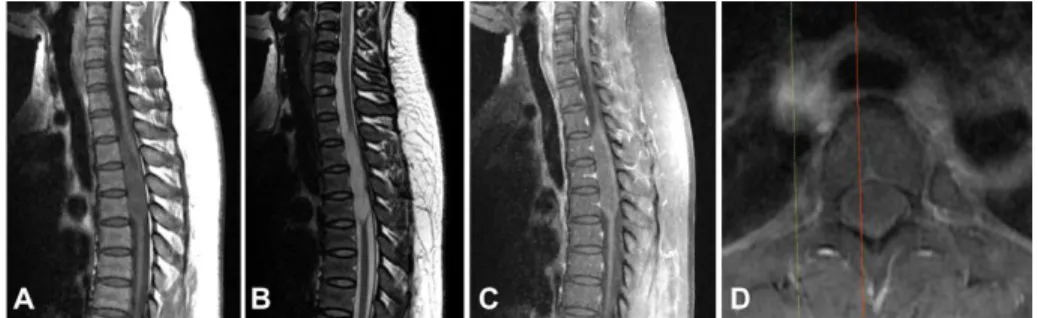
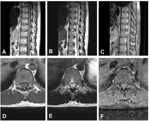
Fig 1. Preoperative magnetic resonance images of patient #9 showing an intramedullary mass located at the C2-7 level with associated hydrosyrinx extending to T3 level. A Sagittal T1-weighted image without contrast showing isointense lesion. B Sagittal T1-weighted image with contrast showing poor gadolinium enhancement. C Sagittal T2-weighted image exhibiting hyper-intense lesion. D Axial T2-weighted image showing the mass located at left and dorsal side of the spinal cord. e Postoperative T2- weighted image revealing partial removal state of the tumor.
Surgical findings
All operations were performed with the conventional posterior midline approach with laminectomy in a prone position. Tumors were grayish in color with a lobulated contour and mild vascularity (Fig 2). All tumors were intramedullary located, and additional exophytic growth was observed in one case. In five patients, tumors showed a well-demarcated margin from the spinal cord, and GTR was achieved. In contrast, in the others, the dissection
9
plane was not well developed, with STR performed in three patients and PR in two. Multimodal intraoperative neuro-monitoring (INM) with the transcranial motor-evoked potential (TcMEP), somatosensory evoked potential (SSEP) and free-running electromyography (fEMG) was performed in seven patients
50. Three patients showed a decrease of TcMEP or SSEP and two patients revealed fEMG events during tumor dissection (Table 2).
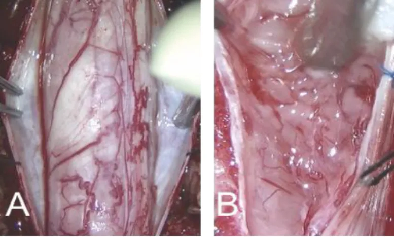
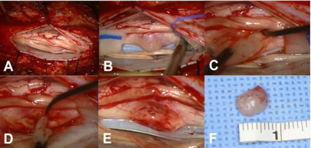
Fig 2. Intraoperative photograph of patient #9 A After durotomy an edematous spinal cord was exposed and rotated to right side. B After
myelotomy, the exposed tumor is gelatinous and light grayish with indistinct dissection plane.
Clinical outcome
Neurological deterioration occurred in two patients after surgery. In Case 9, the MMC grade was 3 after PR and returned to its preoperative state (MMC grade 2) one month after surgery. In Case 3, the MMC grade was 3 after GTR
without recovery to the preoperative state (MMC grade 2). No patient underwent a second surgery due to recurrence or progression of the residual tumor, and revision surgery was performed in one patient due to a surgical site infection. Adjuvant radiotherapy was performed for two patients with STR.
During the median of 31.5 months (range, 8 to 89) of follow-up, recurrence or progression of the tumor was not observed (Table 2).
Pathological findings
Intraoperative frozen sectioned examination was performed in nine out of ten patients. Interestingly, an intraoperative histological diagnosis was made correctly in only one patient, whereas eight patients were misdiagnosed as a low-grade glioma.
Histopathologically, these tumors were characterized by a lobular architecture and clusters of tumor cells, which resulted in an alternating cellular and acellular pattern (Figs. 3A and 3B). There were no high-grade features. Mitoses were scarce and necrosis was not present. However, a degenerative nuclear pleomorphism was found in the focal area (Fig. 3C). The vasculature was often prominent and perivascular hyalinization was noted.
Immunohistochemically, glial fibrillary acidic protein (GFAP) was diffusely and robustly positive in the tumor cells (Fig. 3D), but isocitrate
dehydrogenase 1 gene (IDH-1) and Olig2 were negative. The Ki-67 labeling index was less than 1% (Fig. 3E). An electron microscopic examination was performed in three patients.
11
Ultrathin sections showed loosely arranged round to oval tumor cells. The cells had some cytoplasmic processes filled with glial-type intermediate filaments. The perikaryal cytoplasm contained mitochondria and ribosomes.
Well-developed intracytoplasmic lumina with microvilli and cilli ranged from numerous to rare.
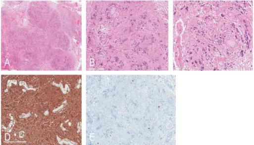
Fig 3. A Hematoxylin and eosin (H&E) staining of resected specimens
showing lobular architecture (H&E, x20), B and clustered nuclei (H&E, x100), C with focal degenerative nuclear enlargement or pleomorphism (H&E, x200).
D Immunostaining of specimens showing diffuse positive to GFAP (GFAP immunostaining, x150). E Ki-67 index was less than 1%.
Illustrative case
Case #3
A 37-year-old woman presented one year of left posterior thigh radiating pain and mild limping gait. Neurological examination revealed hypesthesia
below the level 5cm above nipples. MR images showed a centrally located mass, ranging from the T3 to T5 level, presenting a high signal intensity in T2-weighted images and iso-signal intensity in T1-weighted images without definite gadolinium enhancement. Associated syringomyelia or intratumoral calcification or cyst wasn’t showed. (Fig. 4)
Fig 4. Preoperative magnetic resonance images of the patient #3 showing an intramedullary mass located at the T3-5 level. A Sagittal T1-weighted image without contrast showing iso-intense lesion B Sagittal T2-weighed image showing hyperintense lesion. C Sagittal T1-weighed image with contrast showing poor gadolinium enhancement. D Axial T1-weighted image with contrast showing the mass located centrally.
The patient underwent T2-T5 laminectomy, midline durotomy and
arachnoid incision. The exposed spinal cord was swollen. Midline myelotomy was performed and extended to T3-T5 level. A grayish, gelatinous and moderately vascular tumor was exposed. Strong adhesion between the tumor and the spinal cord was observed especially at the lateral aspect of tumor at
13
was developed and GTR was achieved. The intraoperative histological diagnosis was a low-grade glioma.
A histopathological diagnosis was a subependymoma. A lobular architecture and clusters of tumor cells with an alternating cellular and acellular pattern were found without mitoses, vascular endothelial hyperplasia or necrosis. Immunohistochemically, GFAP, S-100 and Vimentin were positive. CD99 was weak dot-like positive and the Ki-67 labeling index was less than 1%. EM examination showed tumor cells with cord-like arrangement in loosely-organized background. The tumor nuclei are relatively round and electro-lucent. The cytoplasm has some short cytoplasmic processes containing intermediate filaments of glial type, and some polyribosomes, RERs and mitochondrias. Several cilia are found in the cytoplasm but microvilli formation are not identified.
Postoperatively, the patient presented grade 4/5 weakness of left lower limb and needed a cane for self-ambulation. Hypesthesia below the level 5cm above nipples was not changed. The patient presented residual urine more than 150cc, which needed clean intermittent catheterization (CIC) by herself.
Therefore, MMC grade was aggravated from grade 2 to grade 3 and didn’t recovered until the last follow up. Postoperative MR images revealed no residual tumor. She was followed up for 89 months without evidence of recurrence.
Case #6
A 36-year-old woman presented a one-month history of left leg pain.
Neurological examination revealed no other neurological symptom. MR images showed an eccentric intramedullary mass at T11 level, presenting a high signal intensity on T2 weighted images and iso signal intensity on T1- weighted images with focal slight gadolinium enhancement. Associated syringomyelia, intratumoral calcification or cyst wasn’t showed. (Fig. 5)
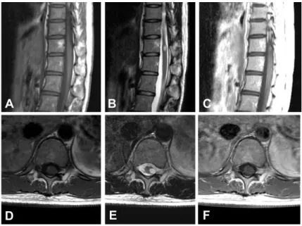
Fig 5. Preoperative magnetic resonance images of the patient #6 showing an intramedullary mass located at the T11 level. A Sagittal T1-weighted image without contrast showing iso-intense lesion B Sagittal T2-weighed image showing hyperintense lesion. C Sagittal T1-weighed image with contrast showing focal slight gadolinium enhancement. D Axial T1-weighted image without contrast. E Axial T2-weighted image showing the exophytic mass located at left lateral aspect of the spinal cord. F Axial T1-weighted image
15
The patient underwent T11-12 laminectomy, midline durotomy and arachnoid incision. With careful inspection among rootlets, the pinkish, glittering exophytic tumor was observed at the left lateral aspect of the spinal cord at T11 level (Fig. 6A, 6B). Incision was made at the tumor-cord interface and careful dissection was performed (Fig. 6C). At the deep portion of the tumor-cord interface, dissection plane became ambiguous and GTR was achieved (Fig 6D, 6E, 6F). Multimodal INM was done during the surgery.
Bilateral posterior tibialis SSEP was decreased to 30% of baseline voltage but recovered to 100% at the end of the surgery. Free running phasic EMG activities in both abductor halluces and right gastrocnemius were also observed during dissection. The intraoperative frozen sectioned histological diagnosis was a low-grade glioma.
A histopathological examination was consistent with subependymoma. A lobular architecture and clusters of tumor cells with an alternating cellular and acellular pattern were found. Increased cellularity was present without mitoses, nuclear polymorphism, vascular endothelial hyperplasia or necrosis.
Immunohistochemically, GFAP was positive and CD99 and epithelial membrane antigen (EMA) were focal dot-like positive. Ki-67 labeling index was positive in 0.69% (5/727) of tumor cells. EM examination showed loosely arranged round to oval tumor cells and the nuclei of tumor cells were heterochromatic. The cells had some cytoplasmic processes filled with glial type intermediate filaments. The perikaryal cytoplasm contained
mitochondrias, ribosomes and well developed intracytoplasmic lumen with numerous microvilli and cilia.
Postoperatively, the patient presented preoperative neurological status at neurological examination and there was no newly acquired symptoms or complication. Postoperative MR images revealed no residual tumor. She was followed up for 38 months without evidence of recurrence.
Fig 6. Intraoperative photograph of patient #6. A, B After durotomy the exophytic tumor was seen among the rootlets at the left lateral aspect of the spinal cord. C, D With careful dissection, tumor-cord interface was developed.
E, F Gross total resection was achieved and the tumor was pinkish and glittering.
Case #8
A 21-year-old man presented seven-month history of pain on lower back and right flank. MR images showed a centrally located intramedullary mass at T10-11 level, presenting a high signal intensity on T2 weighted images and iso signal intensity on T1-weighted images with diffuse slight gadolinium
17
enhancement. Associated syringomyelia, intratumoral calcification or cyst wasn’t showed (Fig. 7).
Fig 7. Preoperative magnetic resonance images of the patient #8 showing an intramedullary mass located at the T10-11 level. A, D Sagittal and axial T1- weighted image without contrast showing iso-intense lesion B, E Sagittal and axial T2-weighed image showing centrally located hyperintense lesion. C, F Sagittal and axial T1-weighed image with contrast showing diffuse slight gadolinium enhancement.
The patient underwent T9-11 laminectomy, midline durotomy and arachnoid incision. The exposed spinal cord was swollen (Fig 8A). Midline
myelotomy was performed and extended to T10-11 level and a grayish and friable tumor was exposed (Fig 8B). With careful dissection, interface
between the tumor and spinal cord was developed and GTR was achieved (Fig 8C). During the surgery, multimodal INM revealed bilateral SSEP was
decreased to 50% of baseline. The intraoperative frozen sectioned histological diagnosis was a low-grade glioma.
Fig 8. Intraoperative photograph of the patient #8. A After durotomy the swollen spinal cord was seen. B After midline myelotomy, grayish tumor was exposed. C, D, E With careful dissection, tumor-cord interface was developed and the tumor was gross totally resected. F The resected tumor was grayish and friable.
A histopathological examination revealed typical findings of a subepen- dymoma. A lobular architecture and clusters of tumor cells with focal psammomatous microcalcification and vascular endothelial hyperplasia were
19
found. Increased cellularity, nuclear polymorphism, mitoses or necrosis were not found.
Immunohistochemically, GFAP was positive and CD99 and EMA were negative. Ki-67 labeling index was positive in 3.82% (37/969) of tumor cells.
Phospho-Histon H3 (PHH3) mitotic count was 2/10 HPF. EM examination showed loosely arranged round to oval tumor cells. The cells had some cytoplasmic processes filled with glial type intermediate filaments. The perikaryal cytoplasm contained mitochondrias, ribosomes. Well developed intracytoplasmic lumina with numerous microvilli and cilia were rarely seen.
Postoperatively, the patient presented preoperative neurological status at neurological examination and there was no newly acquired symptoms or complication. Postoperative MR images revealed no residual tumor. He was followed up for 17 months without evidence of recurrence.
Case #9
A 56-year-old woman presented a four-year history of progressive bilateral leg numbness. She also revealed hypesthesia at the fourth and fifth fingers of her left hand, lasting for a year. Neurological examination revealed motor grade 4+/5 weakness in both lower extremities, which resulted in a mild gait disturbance. MR images showed an eccentrically located mass, producing a hyper-intense signal in T2-weighted and an iso-intense signal in T1-weighted images with focal weak gadolinium enhancement. The mass was extended to the left dorsal surface, ranging from the C3 to C7 level, and syringomyelia was combined below the mass (Fig. 1).
The patient underwent C3-C7 laminectomy, midline durotomy and
arachnoid incision. The exposed spinal cord was rotated to the right side (Fig.
2A). Midline myelotomy was performed and a gelatinous grayish tumor was exposed (Fig. 2B). The intraoperative histological diagnosis in this case was a low-grade glioma. The tumor was firm and strongly adherent to the normal spinal cord parenchyma, making dissection from the spinal cord difficult.
During the dissection, INM revealed repeated abnormal EMG at left thenar muscle and a reduction of the left thenar MEP amplitude to 10% of its baseline voltage. The left posterior tibial SSEP also disappeared. Thus, we stopped further dissection and the tumor was partially removed with a
cavitron ultrasonic aspirator (CUSA). The left posterior tibial SSEP amplitude recovered to 30% of its baseline voltage at the end of the surgery; however, the left thenar MEP amplitude did not recover.
A histopathological examination revealed typical findings of a
subependymoma. A lobular architecture and clusters of tumor cells with an alternating cellular and acellular pattern were found without mitoses or necrosis. Immunohistochemically, GFAP was diffusely positive and the Ki-67 labeling index was less than 1% (Fig. 3). Postoperatively, the patient
presented grade 3/5 weakness and hypesthesia of the bilateral lower limbs and left thenar muscle. At the postoperative one-month follow-up assessment, the patient recovered to her preoperative neurological status. She was followed up for eight months without evidence of regrowth.
21
DISCUSSION
Since the first report by Boykin et al. 5 in 1954, 72 cases of SCSE in total were reported in the literature before this study4-45. Most of the studies were case reports, and the largest study was by Wu et al. 42 involving 13 cases from a single institution. Table 4 summarizes the SCSE in the previously reported cases and current study4-6,28,35-45.
Because a SCSE is a benign and indolent WHO grade I tumor, GTR is not always necessary. In contrast, a spinal cord ependymoma is a WHO grade II tumor, and the extent of resection significantly affects the progression-free survival rate48. Thus, a preoperative differential diagnosis of SCSE especially from a spinal cord ependymoma is important when devising an optimal surgical plan. However, a preoperative differential diagnosis remains difficult due to the lack of specific clinical and imaging characteristics.
Preoperative clinical and radiological findings
In this study, the most common symptoms were sensory changes and pain followed by motor weakness and bladder symptoms. In the literature, instances of SCSE were most commonly located at the cervical spinal cord, followed by the thoracic spinal cord and the lumbar spinal cord (Table 4). In this study, SCSEs were most commonly located at the cervical and thoracic
Previous study4-6,28,35-45 (n=72) Current study (n=10) Overall (n=82)
Mean age (yrs) 43 (6-76, median 45) 42.5 (21-77, median 38) 43 (6-77, median 44) Sex
Male (%) 39 (54.2) 4 (40) 43
Female (%) 33 (45.8) 6 (60) 39
Mean duration of symptoms (mos) 48.1 (2-204, median 36) 16.5 (1-48, median 9.5) 43.5 (1-204, median 24) Level
C (%) 24 (33.3) 4 (40) 28 (34.1)
CT (%) 27 (37.5) 0 27 (32.9)
T (%) 11 (15.3) 4 (40) 15 (18.3)
TL (%) 8 (11.1) 2 (20) 10 (12.2)
L (%) 2 (2.8) 0 2 (2.4)
Gadolinium enhancement
Strong (%) 9 (24.3) 1 (10) 10 (21.3)
Slight (%) 8 (21.6) 3 (30) 11 (23.4)
No (%) 20 (54.1) 6 (60) 26 (55.3)
Eccentricity
Yes (%) 43 (86%) 6 (60) 49 (81.7)
No (%) 7 (14%) 4 (40) 11 (18.3)
EOR
GTR (%) 51 (73.9) 5 (50) 56 (70.9)
STR (%) 12 (17.4) 3 (30) 15 (19.0)
PR (%) 5 (7.2) 2 (20) 7 (8.9)
Biopsy (%) 1 (1.4) 0 1 (1.3)
Aggravation of symptoms
Permanent (%) 20 (35.1) 1 (10) 21 (31.3)
Transient (%) 12 (21.1) 1 (10) 13 (19.4)
No (%) 25 (43.9) 8 (80) 33 (49.3)
Radiotherapy
Preoperative 2 0 2
Postoperative 3 2 5
Recurrence 3 0 3
C, cervical; CT, cervicothoracic; T, thoracic; TL, thoracolumbar; L, lumbar; EOR, extent of Table 4. Summary of characteristics of SCSE in the previous and current studies
23
cord followed by the thoracolumbar cord. The location of the tumor was not discernable from cases of spinal cord ependymoma 48.
Common MR imaging findings of SCSEs were iso- to low signal intensity on T1-WI and high signal intensity on T2-WI, and these features were not different from those associated with a spinal cord ependymoma. However, the eccentric location, poor gadolinium enhancement, and rare intra-tumoral cysts and calcification may characterize SCSEs.
In the present study, the eccentric location in the axial MR images can serve as a clue for distinguishing a SCSE from an ependymoma, which was
observed in five out of ten cases. In previous reports, 86% of cases (43 cases/50 cases) showed an eccentric location. However, this finding may be indistinct if the tumor is large enough to occupy the entire spinal cord in the axial view of MR images, as shown in Figure 1. Eccentricity may be a clue only when the tumor is not large enough to occupy the entire spinal canal.
In this study, only one patient showed homogeneous strong contrast enhancement in the tumor, and such an enhancement was observed only in 24.3% of patients in previous reports. Although syringomyelia was combined in two patients in the present study, an intratumoral cyst or calcification was not found, in accordance with previous reports4-45
Intraoperative findings and surgical outcomes
Although preoperative radiological findings were similar, intraoperative
findings were not similar between cases of spinal cord ependymoma and SCSE. During surgery, the interface between the tumor and the spinal cord was clear only in half of the patients, whereas in the others, the dissection plane was not clearly developed. The tumors were completely removed only in 50% of cases, despite the fact that all surgical procedures were performed by senior spine surgeons who have more than ten years of experience with spinal cord tumors. The rate of GTR was much lower than the GTR rate of spinal cord ependymomas (72/88, 81.8%) during the same period by the same surgeons48. In the literature, the GTR rate of SCSEs was 73.9%, which is higher than that in the present study but nonetheless lower than the GTR rate of spinal cord ependymomas. There are two possible reasons for this: the poor dissection plane between the tumor and the spinal cord and the inaccurate intraoperative histological diagnosis as a low-grade glioma.
There was no recurred case in the present study during the median 31.5 months follow-up period (range, 8-89). Before this study, there have been three reported cases of recurrence after surgical resection of the SCSEs20,26,32. Follow-up periods of recurred cases were 7, 9 and 12 years, which is longer than that of all reported SCSEs (mean 45.7 months, median 39 months) 4-45. This result showed that long term follow-up is essential, though recurrence of SCSE is rare.
Pathological findings
The histopathogenesis of a subependymoma has yet to be revealed clearly.
25
An intracranial subependymoma is currently thought to derive from
subependymal glial precursor cells, which are bipotential cells with the ability to differentiate into either ependymal cells or an astrocyte 51. On the other hand, in SCSE, Krishnan et al.38 suggested subpial spinal white matter progenitor cells as a possible histogenesis for a better explanation of the predominant peripheral and exophytic location. This may explain the eccentric location of a SCSE.
Pathological features of subependymoma are distinct from features of spinal cord ependymoma and spinal cord astrocytoma. Histopathologically, subependymoma showed clusters of isomorphic tumor cells embedded in a dense fibrillary background3. In contrast to ependymoma, ependymal rosette or perivascular pseudorosette was only occasionally found. Immunohisto- chemically, GFAP was diffusely positive in all cases, which dertermines its glial origin. In contrast to ependymoma, subependymoma occasionally immunoreactive to EMA, which is attributed to poor development of the lumina in the rosettes of subependymoma. Ultrastructural investigation demonstrated both astrocytic and ependymal characteristics in
subependymoma. The former is evidenced by the processes containing intermediate filaments and the latter by microlumens, cilia, microvilli and intercellular junctions32.
Despite these distinct pathological features, however, differentiation with an intraoperative frozen section approach was challenging. In this study,
intraoperative histologic diagnoses by frozen section examination were performed in nine out of ten patients; however, eight patients were diagnosed
as having a low-grade glioma, and a correct diagnosis was made in only one case. Given that a subependymoma has both ependymal and glial components histologically, this high rate of misdiagnosis was attributed to the small number of specimens and a lack of suspicion. This problem can be overcome by sending a sufficient amount of specimens to a pathologist with appropriate suspicion by the clinician.
Treatment recommendation
Recommended treatments for SCSEs vary depending on the author.
Matsumoto at al. and Jallo et al.28,31 insisted that GTR should be conducted for a better clinical outcome, though GTR may not be possible in all cases. Wu et al. 42 and Jang et al. 35 showed that STR or PR was sufficient for a good clinical outcome when GTR is not feasible.
Because SCSEs are benign tumors and given that tumor progression is rare after STR or PR42, impellent GTR in the face of neurological damage cannot be recommended. The preservation of neurological function and the quality of life should be considered as the most crucial points. Thus, we propose STR without the risk of neurological deficit, if GTR is not feasible. Although adjuvant radiotherapy was performed in two patients after STR in the present study, it is not usually recommended.
27
CONCLUSION
A spinal cord subependymoma is a benign and indolent tumor. Clinical and radiological features are similar to those of an ependymoma, but a spinal cord subependymoma needs to be considered when the tumor is located
eccentrically and not easily dissected from the spinal cord. Impellent gross total removal may result in a neurological deficit. Considering the benign nature of subependymomas, subtotal removal without the risk of any neurological deficit may be a viable alternative option.
Limitation
Because SCSEs are uncommon spinal cord tumors, retrospective data from multiple institutes were utilized, but the small number of patients was the major limitation here. In the present study, although experienced specialists were involved in the radiological and pathological (intraoperative and permanent) diagnosis and surgeries, differences in experience must be considered. Most importantly, the follow-up period was not long enough to provide a treatment strategy. Nonetheless, we showed the importance of the preoperative suspicion of a SCSE and suggested STR or PR as an alternative option. Apparently, SCSEs are similar to spinal cord ependymomas, but the treatment strategy may differ, and this needs to be included in any differential diagnosis.
References
1. Scheinker IM. Subependymoma: a newly recognized tumor of subependymal derivation. J Neurosurg. 1945;2(3):232-240.
2. Im SH, Paek SH, Choi YL, Chi JG, Kim DG. Clinicopathological study of seven cases of symptomatic supratentorial subependymoma. J neurooncol. 2003;61(1):57-67.
3. Luis DN, Ohgaki H, Wiestler OD, Cavenee WK. World Health Organization Classification of Tumors of the Central Nervous System.
IARC; 2007.
4. Jain A, Amin AG, Jain P, et al. Subependymoma: clinical features and surgical outcomes. Neurol Res. 2012;34(7):677-684.
5. Boykin FC, Cowen D, Iannucci CA, WOLF A. Subependymal
glomerate astrocytomas. J Neuropathol Exp Neurol. 1954;13(1):30-49.
6. Wu Z, Iwanami A, Yasuda A, Mikami S, Toyama Y, Nakamura M.
Intramedullary cervicothoracic subependymoma: report of three cases and review of the literature. J Orthop Sci. 2014:1-8.
7. Slowik F, Pasztor E, Szöllösi B. Subependymal gliomas. Neurosurg Rev. 1979;2(2):79-86.
8. Pluchino F, Lodrini S, Lasio G, Allegranza A. Complete removal of holocord subependymoma. Acta Neurochir (Wien). 1984;73(3-4):243- 250.
9. Salcman M, Mayer R. Intramedullary Subependymoma of the Cervical Spinal Cord: Case Report. Neurosurgery. 1984;14(5):608.
10. Cervos-Navarro J, Artigas J, Perez-Canto A. Clinical and
29
Verh Dtsch Ges Pathol. 1986;70:376-379.
11. Lee KS, Angelo JN, McWhorter JM, Davis CH. Symptomatic subependymoma of the cervical spinal cord. Report of two cases. J Neurosurg. 1987;67(1):128-131.
12. Bardella L, Artico M, Nucci F. Intramedullary subependymoma of the cervical spinal cord. Surg Neurol. 1988;29:326-329.
13. Matsumura A, Hori A, Spoerri O. Spinal Subependymoma Presenting as an Extramedullary Tumor: Case Report. Neurosurgery.
1988;23(1):115.
14. Nagashima M, Isu T, Iwasaki Y. Intramedullary subependymoma of the cervical spinal cord. Case Report. Neurol Med Chir (Tokyo).
1988;28:303-308.
15. Herrmann H-D, Neuss M, Winkler D. Intramedullary Spinal Cord Tumors Resected with CO2 Laser Microsurgical Technique: Recent Experience in Fifteen Patients. Neurosurgery. 1988;22(3):518.
16. Guha A, Resch L, Tator CH. Subependymoma of the thoracolumbar cord. Case report. J Neurosurg. 1989;71(5 Pt 1):781-787.
17. Artico M, Bardella L, Ciappetta P, Raco A. Surgical treatment of subependymomas of the central nervous system. Acta Neurochir (Wien). 1989;98(1-2):25-31.
18. Vaquero J, Martinez R, Vegazo I, Pontn P. Subependymoma of the Cervical Spinal Cord. Neurosurgery. 1989;24(4):625.
19. Lach B, Russell N, Benoit B. Atypical Subependymoma of the Spinal Cord: Ultrastructural and Immunohistochemical Studies. Neurosurgery.
1990;27(2):319.
20. Bergman TA, Haines SJ. Subependymoma of the cervical spinal cord.
A case report of long-term survival. Minn Med. 1991;74(11):21-24.
21. Nakasu S, Nakasu Y, Saito A. Intramedullary Subependymoma with Neurofibromatosis. Report of Two Cases. Neurol Med Chir (Tokyo).
1992;32:275-280.
22. Pagni CA, Canavero S, Giordana MT, Mascalchi M, Arnetoli G.
Spinal intramedullary subependymomas: case report and review of the literature. Neurosurgery. 1992;30(1):115-117.
23. Salvati M, Raco A, Artico M, Artizzu S, Ciappetta P. Subependymoma of the spinal cord. Case report and review of the literature. Neurosurg Rev. 1992;15(1):65-69.
24. Casey AT, Marsh H, Wilkins P. Subependymoma of the thoracic cord:
potential pitfalls in diagnosis. Br J Neurosurg. 1993;7(3):319-322.
25. Roeder MB, Jinkins JR, Bazan C. Subependymoma of filum terminale:
MR appearance. J Comput Assist Tomogr. 1994;18(1):129-130.
26. Hoeffel C, Boukobza M, Polivka M, et al. MR manifestations of subependymomas. Am J Neuroradiol. 1995;16(10):2121-2129.
27. Tacconi L, Johnston FG, Thomas DGT. Subependymoma of the
cervical cord. Clinical Neurology and Neurosurgery. 1996;98(1):24-26.
28. Jallo GI, Zagzag D, Epstein F. Intramedullary subependymoma of the spinal cord. Neurosurgery. 1996.
29. Bret P, Bougeard R, Saint-Pierre G, Guyotat J, Ricci AC, Confavreux C. Intramedullary subependymoma of the cervical spinal cord. Review of the literature a propos of a case. Neurochirurgie. 1997;43(3):158- 163.
31
30. Dario A, Fachinetti P, Cerati M, Dorizzi A. Subependymoma of the spinal cord: case report and review of the literature. Journal of Clinical Neuroscience. 2001;8(1):48-50.
31. Matsumoto K, Nakagaki H. Intramedullary subependymoma occupying the right half of the thoracic spinal cord. Case report.
Neurol Med Chir (Tokyo). 2002;42(8):349-353.
32. Shimada S, Ishizawa K, Horiguchi H. Subependymoma of the spinal cord and review of the literature. Patholo Int. 2003;53:169-173.
33. Kremer P, Zoubaa S, Schramm P. Intramedullary subependymoma of the lower spinal cord. Br J Neurosurg. 2004;18(5):548-551.
34. Orakcioglu B, Schramm P, Kohlhof P, Aschoff A, Unterberg A, Halatsch M-E. Characteristics of thoracolumbar intramedullary subependymomas. J Neurosurg Spine. 2009;10(1):54-59.
35. Jang W-Y, Lee J-K, Lee J-H, et al. Intramedullary subependymoma of the thoracic spinal cord. Journal of Clinical Neuroscience.
2009;16(6):851-853.
36. Jabri HE, Dababo MA, Alkhani AM. Subependymoma of the spine.
Neurosciences (Riyadh). 2010;15(2):126-128.
37. Zenmyo M, Ishido Y, Terahara M, et al. Intramedullary subependymoma of the cervical spinal cord: a case report with immunohistochemical study. Int J Neurosci. 2010;120(10):676-679.
38. Krishnan SS, Panigrahi M, Pendyala S, Rao SI, Varma DR. Cervical Subependymoma: A rare case report with possible histogenesis. J Neurosci Rural Pract. 2012;3(3):366-369.
39. Iwasaki M, Hida K, Aoyama T, Houkin K. Thoracolumbar
intramedullary subependymoma with multiple cystic formation: a case
report and review. Eur Spine J. 2013;22 Suppl 3(3):S317-S320.
40. Dhandapani S, Anirudh S, Garg R, Vasishta RK, Vyas S.
Subependymoma causing conus-cauda syndrome: cured by total excision. Neurol India. 2013;61(6):675-677.
41. Takami T, Yamagata T, Ohata K. Posterolateral sulcus approach for spinal intramedullary tumor of lateral location: technical note. Neurol Med Chir (Tokyo). 2013;53:920-927
42. Wu L, Yang T, Deng X, et al. Surgical outcomes in spinal cord subependymomas: an institutional experience. J Neurooncol.
2014;116(1):99-106.
43. Cure LM, Hancock CR, Barrocas AM, Sternau LL, C Hirzel A.
Interesting case of subependymoma of the spinal cord. Spine J.
2014;14(5):e9-e12.
44. Tan LA, Kasliwal MK, Mhanna N, Fontes RBV, Traynelis VC.
Surgical resection of subependymoma of the cervical spinal cord.
Neurosurg Focus. 2014;37 Suppl 2(Suppl2):Video3.
45. Boström A, Lehe von M, Hartmann W, et al. Surgery for Spinal Cord Ependymomas: Outcome and Prognostic Factors. Neurosurgery.
2011;68(2):302-309.
46. Kim CH, Chung CK, Lee S-H, et al. Long-term recurrence rates after the removal of spinal meningiomas in relation to Simpson grades. Eur Spine J. November 2015:1-8.
47. Sohn S, Chung CK, Park S-H, Kim E-S, Kim K-J, Kim CH. The fate of spinal schwannomas following subtotal resection: a retrospective multicenter study by the Korea spinal oncology research group. J
33
48. Lee S-H, Chung CK, Kim CH, et al. Long-term outcomes of surgical resection with or without adjuvant radiation therapy for treatment of spinal ependymoma: a retrospective multicenter study by the Korea Spinal Oncology Research Group. Neuro-oncology. 2013;15(7):921- 929.
49. McCormick PC, Torres R, Post KD, Stein BM. Intramedullary ependymoma of the spinal cord. J Neurosurg. 1990;72(4):523-532.
50. Jin S-H, Chung CK, Kim CH, Choi YD, Kwak G, Kim BE.
Multimodal intraoperative monitoring during intramedullary spinal cord tumor surgery. Acta Neurochir (Wien). 2015;157:2149-2155.
51. Bi Z, Ren X, Zhang J, Jia W. Clinical, radiological, and pathological features in 43 cases of intracranial subependymoma. J Neurosurg.
2015;122(1):49-60.
국문 초록
척수 상의하세포종의 임상적, 영상의학적, 병리학적 특성 및 수술적 치료에 대한 고찰:
다기관 후향적 연구
서울대학교 대학원 의학과 신경외과학전공
여 운 탁
목적: 척수 상의하세포종은 비교적 드문 양성 척수 내 종양으로, 영상의학적 소견은 척수 내 상의세포종과 비슷하지만 수술적 치료 중 관찰되는 소견이나 치료 성적은 상이하다. 양성 종양으로 종양의 전절제를 시행하면 완치가 가능하나 항상 가능한 것은 아니다. 이 연구를 통해 척수 상의하세포종의 임상적, 영상의학적, 병리학적 특성을 파악하고 치료 지침을 제시하고자 하였다.
방법: 2000 년부터 2012 년 사이 4 개 병원에서 수술 후 척수 상의하세포종으로 진단받은 10 명의 환자의 의무기록을 후향적 방법으로 분석하였다.
결과: 가장 흔한 증상은 10 명 중 8 명의 환자가 호소한 감각 변화와 통증 이었고, 그 다음으로 6 명의 환자가 사지의
35
위약감을 호소하였다. 진단 전까지 증상이 지속된 기간의
중앙값은 9.5 개월 이었다. 수술 전 영상의학적 검사를 통한 감별 진단은 7 명의 환자는 척수 상의세포종 이었고 3 명의 환자는 척수 성상세포종이었다. 6 명의 환자에서 종양은 가측에 치우쳐 위치하고 있었다. 조영 증강 자기공명영상 검사에서 6 명의 환자는 조영 증강 되지 않았다. 종양의 전절제는 5 명의 환자에서 가능하였고, 나머지 5 명의 환자에서는 종양과 정상 척수 사이의 경계가 불분명하여 아전절제 또는 부분절제를 하여야 했다. 수술 후 추가 방사선 치료는 두명의 환자에서 시행하였다. 수술 직 후 두명의 환자에서 신경학적 증상 악화가 발생하였다. 그 중 부분절제를 시행한 환자는 한 달 뒤 수술 전의 신경학적 상태로 회복하였고, 전절제를 시행했던 환자는
회복되지 않았다. 수술 적 치료 후 중앙값 31.5 개월 (8-
89 개월) 동안 경과 관찰 하였고, 종양의 재발 및 재성장을 보인 환자는 없었다.
결론: 척수 상의하세포종은 수술 전 영상의학적 검사에서 종양이 척수 내에서 가측으로 위치하고, 수술 중 정상 척수와의 경계가 불분명할 때 의심해보아야 한다. 척수 상의하세포종은 전절제를 통해 완치를 기대할 수 있으나, 종양의 양성적인 경과를
고려했을 때 신경학적 증상의 악화를 유발하지 않고 부분절제를 시행하는 것 또한 치료 대안이 될 수 있다.