Case Report
원고 접수일 2012년 5월 17일, 원고 수정일 2012년 6월 13일, 게재 확정일 2012년 8월 27일
책임저자 김선종
(158-050) 서울시 양천구 안양천로 1071, 이화여자대학교 의학전문대학원 구강악안 면외과학교실
Tel: 02-2650-5041, Fax: 02-2650-5764, E-mail: [email protected]
*곽주희, 김지연 저자가 공동 제1저자로서 본 연구에 기여했습니다.
RECEIVED May 17, 2012, REVISED June 13, 2012, ACCEPTED August 27, 2012
Correspondence to Sun-Jong Kim
Department of Oral and Maxillofacial Surgery, Ewha Womans University School of Medicine
1071, Anyangcheon-ro, Yangcheon-gu, Seoul 158-710, Korea Tel: 82-2-2650-5041, Fax: 82-2-2650-5764, E-mail: [email protected]
*Juhee Kwak and Ji-Youn Kim equally contributed as a first author.
CC This is an open access article distributed under the terms of the Creative Commons Attribution Non-Commercial License (http://creativecommons.org/licenses/
by-nc/3.0) which permits unrestricted non-commercial use, distribution, and reproduction in any medium, provided the original work is properly cited.
체루비즘: 2예 보고
곽주희*ㆍ김지연*ㆍ김명래ㆍ김선종
이화여자대학교 의학전문대학원 목동병원 구강악안면외과
Abstract
Cherubism: Review of 2 Cases
Juhee Kwak*, Ji-Youn Kim*, Myung-Rae Kim, Sun-Jong Kim
Department of Oral and Maxillofacial Surgery, Ewha Womans University Mokdong Hospital, Ewha Womans University School of Medicine
Cherubism is a rare familial disease of childhood, characterized by proliferative lesion, which is within the maxilla and mandible.
In a typical case, painless symmetric expansile lesions develop in the jaws. It shows substitution of the bone by proliferating fibrous tissue exhibiting mature fibroblasts and a number of multinucleated giant cells within an intercellular matrix. Usually, the disease manifests in early childhood, and becomes more marked until puberty, at which time the bony lesions begin to regress. As such, conservative approaches to management are advisable. However, excision of tissue through enucleation or curettage appears to be necessary in more aggressive cases, to reduce the maxillofacial deformity after puberty and to ensure a successful outcome without the risk of progression, requiring additional resection. This report describes 2 cases of manifestation of cherubism of oral and maxillofacial region. We present diagnosis, radiological - histopathologic features, and treatment of cherubism.
Key words: Cherubism, Giant cells, Genes dominant, Maxilla, Mandible
서 론
체루비즘(cherubism)은 어린 연령대에서 호발하는 가족력과 연관된 드문 질환이다[1,2]. 임상적으로 상, 하악의 대칭적, 증식 성 병소를 유발하여 하안모가 팽창하는 특징을 가지고 있어[3], 르네상스 예술 작품에서 묘사된 아기 천사(cherub)의 모습처럼
보인다[4]. 체루비즘의 전형적인 증례는 무통성의 대칭적이며 다
방성 낭성 방사선투과상의 팽창성 병소가 하악각 혹은 하악 상행
지에 발생하는 것으로 관찰된다[2]. 대부분의 병소의 중심은 악골
의 후방부, 즉, 하악각-하악지 혹은 상악의 상악결절 부위에 존재
한다[3]. 이 병소는 병리학적으로 거대세포 육아종 유사 조직 소견
과 함께 세밀하고 과립성의 골과 다방성의 성긴 골소주를 가진
Fig. 1. The family tree. Child A (Case 1) and Child E (Case 2)
are diagnosed as cherubism clinically, radiologically and histopathologically. Their mothers (Mother A and Mother C) are sister. The Mother C remembered that Child C and D have also the bilateral chubby cheeks at childhood, suggestive of mild forms of cherubism.Fig. 2. (A, B) Pre-operative clinical photographs. The bilateral expansion of lower face seen in the photographs is the result of marked
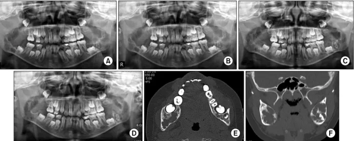
expansion of the mandible and maxilla. (C∼E) Pre-operative radiographs. The symmetric, expansile and multilocular radiolucent lesions are shown at panoramic view and computed tomography images. (F) Pre-operative bone scan image. There are shown high uptake at both posterior mandibular area, and also shown mild uptake at both maxillary posterior area. It may be shown the active bony expansile-remodeling lesion.체루비즘으로 진단받은 친인척 관계에 있는 두 증례를 고찰하였으 며, 각 증례에서 환자 및 가족들을 대상으로 개인 정보 제공 동의 하에 개별 면담을 통하여 정보를 제공받았다. 두 환아의 어머니는 자매관계로, 두 환아 외의 다른 남자 사촌 2명도 경도의 체루비즘 의 임상 소견을 보인다고 언급하였다(Fig. 1).
1. 증례 1
초진 내원 시 13세 남자 환아로, 양측 하안모의 종창을 주소로
내원하였다. 임상적으로 확연하게 진행된 양측성 하안모의 팽창
과 방사선학적으로 양측 상, 하악의 특징적인 다방성의 낭성 방사
선 투과상, 피질골판의 팽창과 비박화 및 다수의 미맹출치가 관찰
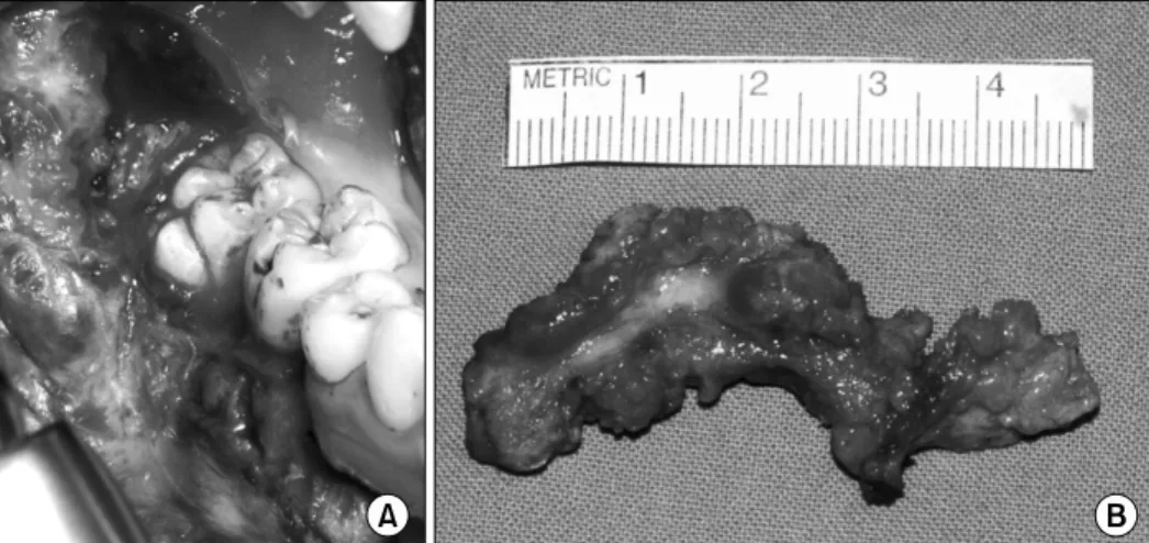
Fig. 3. (A) Intra-operative photograph.
There is shown a fibrous-osseous le- sion at mandibular body posterior- ramal area including alveolar bone of right lower first and second molar. (B) Resected fibrous-osseous lesion.
Fig. 4. (A) Post-operative 1 day panoramic view. (B) Post-operative 2 weeks panoramic view. (C) Post-operative 2 months panoramic
view. (D) Post-operative 5 years panoramic view. (E, F) Post-operative 5 years computed tomography images. Note signs of new bone formation.2 대구치를 포함한 하악체 후방부 및 하악지 부위의 외과적 종물 절제술을 시행하였다(Fig. 3). 조직병리학적 검사 결과, 상악의 병소는 낭성 조직을 동반한 섬유점액성 조직(fibromyxoid tis- sue) 소견이 관찰되었으며, 하악의 병소는 양성 섬유-골이형성 조직 소견을 보였다. 술 후 5년 동안의 경과 관찰 기간 동안, 주기적으로 촬영한 임상 사진에서 재발 소견을 보이지 않고, 정상 발육 양상을 보였으며, 파노라마 및 컴퓨터단층촬영 방사선 사진 에서도 병소 절제된 부위에서 신생골 형성 소견 보였으며, 재발 소견은 보이지 않았다(Fig. 4, 5).
2. 증례 2
초진 내원 시 8세 남자 환아로, 증례 1의 이종 사촌으로 양측 하안모의 종창이라는 동일한 주소로 내원하였다. 증례 1에 비해 임상적 증상의 정도는 약하나, 유사하게 하악 후방부의 양측성
팽창을 보였으며, 방사선학적으로 양측 하악체 후방부과 상행지
의 팽창성, 다방성 방사선 투과상 및 비후되어 있는 양측 상악결절
부의 방사선 투과상을 보였다(Fig. 6). 이에 대해 체루비즘
(Seward and Hankey grading system Grade 2)으로 진단하고
보존적으로 경과 관찰 및 하악 병소의 조직 절개 생검에 대한
외과적 수술을 계획하였다. 우측 하악체 후방부의 점막골막피판
거상 후 소파술로 병소부의 일부 조직을 채득하였다. 조직병리학
적 검사결과, 다수의 섬유아세포 및 다핵거대세포를 포함한 세포
와 혈관이 풍부한 섬유성 조직 및 작은 화생성 골 조직(small
foci of metaplastic bone tissue) 소견이 관찰되어 체루비즘으로
확진하였다. 이후 2년 동안 경과 관찰 중이며, 주기적으로 촬영한
임상적 방사선학적 소견상, 하악의 성장에 따라 병소의 크기가
더 증대되지는 않았으며, 유지되는 양상을 보였다(Fig. 7, 8).
Fig. 5. (A∼C) Post-operative 5 years follow-up clinical facial photographs. (D∼I) Post-operative 5 years follow-up clinical intraoral
photographs. There is no sign of recurrence.고 찰
체루비즘은 상염색체 우성으로 유전되는 가족성 질환이다 [1,2,8]. 여자에 비해 남자에서 더 일반적으로 이환되며, 이환되는 중증도 또한 더 심화되어 나타난다[9,10]. 2001년, Ueki 등[11]에 의해 체루비즘을 가진 12 가족에서 SH3-domain binding pro- tein 2 (SH3BP2) 의 이형성변형(heterozygous mutatain)이 보 고된 이래로 체루비즘 환자에서 이 유전자의 변형에 대해 계속 연구되었다. 대부분의 체루비즘 환자들은 면역반응 신호전달체계 에 관련된 adapter protein인 SH3BP2을 encoding하고 있는 이 유전자에 생식계열(germline) 유전자 변형이 있는 것으로 보 고되었으며, SH3BP2 에 유전자 변형이 있는 마우스 동물 모델에서 골연화증 및 팽창성 골용해성 병소가 발병되었음이 보고되었다[8].
질환의 유전자 침투도(penetrance)는 남자에서 100%, 여자에서 70∼50%로 매우 높으며, 더 정확한 평가를 위해 임상적 또는 방사선학적 진단 평가 기준이 적용된다[8-10]. 위 증례들에서도 이러한 유전적 변형의 연관성으로 어머니쪽 친인척인 두 명의 남자 환아에서 체루비즘이 임상적으로 발현되었음을 알 수 있다.
대부분의 어린 아이들의 얼굴이 통통하기 때문에 증상이 경미
한 경우에는 환아가 10대가 되기 전까지 진단되지 않은 경우가 많으며, 이런 경우, 방사선학적 소견에 의해 확진된다. 조직병리 학적 검사에서는 다수의 다핵거대세포가 공통적으로 관찰되는 소견을 보인다[2,5,6]. 풍부한 다핵거대세포를 포함한 광범위한 병소와 해당 질환 병소의 공격성과는 상관관계가 있다고 보고되어 있다[3]. 위 두 증례의 환아에서도 증식성 섬유성 결체조직으로 정상골조직이 치환된 조직 소견을 보였으며, 섬유성 결체 조직 내에 는 다수의 성숙된 섬유아세포 및 불균일하게 분포되어 있는 다수의 다핵거대세포들이 세포간질에 포함되어 있는 양상을 보였다.
체루비즘은 질환의 이환으로 인한 어린 연령대의 악골의 변형
으로 인해 영구적인 치열의 변형을 일으킬 수 있다[2]. 상악골이
이환되는 경우, 주변의 다른 안면골까지 병소가 퍼져서 이환될
수 있다. 병소는 보통 유아기(2∼5세)에 발현되며, 사춘기 때까지
점점 더 증식하는 양상 보이다가 이후 병변이 퇴행하여 소실되기
시작하며, 상악이 먼저 병소의 소실을 보인다. 반면 하악의 경우는
종종 20세까지 활동적으로 팽창-증식하기도 한다. 아직까지 체루
비즘의 치료에 대해서는 표준화된 바가 없다. 사춘기 이후 낭종과
같은 병소의 활동성이 정체되고 과립성 골조직으로 채워지고 골격
의 성장이 끝나기 때문에, 치료는 지연될 수 있다. 따라서,
Fig. 6. (A, B) Clinical photographs at first visit. The mild bilateral expansion of lower face is shown in the photographs. (C∼E) Pre-operative
panoramic view and computed tomography images. The bilateral, expansile and multilocular radiolucent lesions at both posterior mandible are shown. (F) Pre-operative bone scan image. There are shown mild uptake at both posterior mandibular and both maxillary tuberosities area than Case 1 patient. It may be shown that the activity of bony remodeling lesion is a little mild compared to Case 1 patient.Fig. 7. (A) Post-operative 2 days panoramic view after incisional bone biopsy. (B) Post-operative 2 weeks panoramic view. (C) Post-operative
3 months panoramic view. (D∼F) Post-operative 6 months panoramic view and computed tomography images. The lesions have been not shown enlargement.Seward와 Hankey의 체루비즘 등급에 따라 grade 1과 2에서는 2차적 변형이 존재하지 않는다면 외과적 치료는 꼭 필요하지 않을 수 있다[7]. 하지만, 공격적 성격이 강한 grade 3 이상의 병소의
경우, 사춘기 이후 발생 가능성이 있는 악골-안면부의 변형을
줄이고 더 진행되는 것을 막기 위해 외과적 적출술 및 소파술이
필요할 수 있다[2,3,7,12,13]. Dukart 등[14]은 체루비즘의 병소
Fig. 8. (A∼C) Clinical photographs at post-operative 6 months follow-up. The symmetric swelling of the mandibular angles has been
remained.가 급격히 커지는 시기 동안 외과적 소파술 및 recontouring을 시행하는 것은 술 후 향상된 외모뿐만 아니라 남아 있는 병소의 활동적 성장을 억제하며, 골의 재형성을 촉진한다고 언급하였다.
위 두 증례 중, 앞의 증례의 13세 환아의 경우, 상, 하악 전반적으 로 병소에 이환된 grade 3로, 외과적 적출술 후 경과 관찰 중이며, 술 후 신생골이 재형성되는 양상을 보였다. 뒤의 증례의 8세 환아 의 경우에는 절개 생검으로 확진 후 외과적 절제술을 시행하지 않고 경과 관찰을 시행하고 있다.
일반적으로 체루비즘의 예후는 양호하다[3]. 체루비즘은 성장 에 따라 병소 크기 증가를 스스로 제한하여 사춘기 이후에는 병변이 개선된다고 보고되었기 때문에, 우선 보존적 치료가 고려 되지만, 병소의 크기 및 공격성에 따라 수술이 필요할 수도 있다 [2,7]. 따라서 체루비즘의 임상적 치료 및 외과적 수술의 결정은 각 증례 병변의 공격성 및 각 환자의 기능적, 심미적 필요에 따라 결정되어야 할 것이다.
References