DOI:10.5125/jkaoms.2010.36.3.211
211 Abstract (J Korean Assoc Oral Maxillofac Surg 2010;36:211-3)
Ⅰ.
서 론
Glandular odontogenic cyst (GOC, 선양치성낭)은 1987년 Gardener가 보고한 이래 현재 문헌으로 보고된 증례가 111 case에 불과한 serre잔사로부터 유래한 희귀한 구강 내 낭
이다1. 이것은 중년, 하악에 호발하는 경향을 가지며, 임상
적 특징으로 피질골의 천공, 치근흡수의 국소적으로 공격 적인 양상을 특징으로 하며 환자는 보통 무통성의 종창을 우연히 발견하게 된다1-3. 크기는 0.5-12 cm으로 다양한 경 향을 나타내며. 재발률은 적출술, 소파술의 보존적 처치 시 64.3%, 부분절제술, 변연절제술 등의 광범위 절제술 시 0%
로 조사되어 치성각화낭, 치아모세포종과 같은 임상적 예 후를 보인다4. 따라서 재발에 대한 주기적인 경과 관찰이 필수적이다. 방사선학적으로는 다방성의 방사선투과성을
특징으로 한다1-3. 조직병리학적으로는 특징적으로 상피세 포구, 상피세포층 내의 점액세포, 위도관 구조의 세포배열 을 특징으로 한다1-4. 본 교실에서는 하악 우측에 발생된 GOC을 치험하여 그 증례를 공유하기 위하여 문헌 고찰과 함께 보고한다.
Ⅱ.
증례보고
30세 여자 환자가 2009년 5월 6일 하악 우측 구치부의 낭 종을 주소로 개인치과에서 의뢰되었다. 환자는 전신질환 은 없었으며 내원 수 일전부터 주소 부위의 불편감과 부종, 통증을 인지하고 있었다. 초진 내원 시 촬영한 파노라마사 진으로 명확한 경계를 가진 낭종이 #48 치아의 치관 주위 로 존재하는 것이 관찰되었고, 단방성의 양상을 의심하였 다.(Fig. 1) 임상검진으로 협측, 설측의 피질골 팽융은 관찰 되지 않았으며, 촉진 시 통증은 재현되지 않았다. #47 치아 는 타진 시 동통은 없었으며, 동요도는 관찰되지 않았다.
초진 내원 시, 치성각화낭 가진하에 하악 전산화단층촬영 을 시행하였다.
하악 전산화단층촬영영상으로 주위와 경계가 비교적 명 확한 골 내 낭종이 관찰되었다. 협측과 설측의 피질골판이 김 형 준
120-752 서울시 서대문구 성산로250 연세대학교 치과대학 구강악안면외과학교실 Hyung Jun Kim
Department of Oral and Maxillofacial Surgery College of Dentistry, Yonsei University
250 Seongsanno, Seodaemoon Gu, Seoul, 120-752, Korea TEL: 82-2-2228-3132 FAX: 82-2-2227-8022
E-mail: [email protected]
하악골에 발생한 선양치성낭의 치험례
권진일1∙김현우1∙한선희2,3∙남 웅1,3∙차인호1,3∙김형준1
연세대학교 치과대학 1구강악안면외과학교실, 2구강병리학교실, 3구강종양연구소
Glandular odontogenic cyst of mandible: case report
Jin-Il Kwon1, Hyun-Woo Kim1, Seon-Hee Han2,3,Woong Nam1,3, In-Ho Cha1,3, Hyung Jun Kim1
1Departments of Oral and Maxillofacial Surgery, 2Oral Pathology and 3Oral Cancer Research Institute, College of Dentistry, Yonsei University, Seoul, Korea
Glandular odontogenic cyst (GOC) is an intraoral cyst originated from serre remnants which has incidence of rare frequency. Only 111 cases have been reported since Gardener first introduced it in 1987. The clinical features are the following components: cortical bone thinning, locally aggressive root resorption, non-painful swelling. The following recurrences rate are 64.3% in conservative treatment, and 0% in wide excision for instance, seg- mental or marginal mandibulectomy. So, its prognosis is similar to that of odontogenic keratocyst and ameloblastoma. Therefore, periodic recall fol- low ups are essential to detect disease recurrence. Here, we will report the first case of GOC diagnosed in our department considering with references.
And we share this treatment experience because these aggessive lesions may be misjudged for simple dental cyst.
Key words: Glandular odontogenic cyst, Mandible
[원고접수일 2010.3.28 / 1차수정일 2010.4.19 / 2차수정일 2010.5.3 / 게재확정일 2010.5.26]
대구외지 2010;36:211-3
212
다소 얇아진 소견이 관찰되나 두드러진 골팽융의 소견은 관찰되지 않았다.(Fig. 2) 방사선판독 소견은 함치성낭과 감별진단이 필요한 치성각화낭으로 판독되었다.
동년 6월 12일 입원하여 전신마취하 구강 내 접근 시 이 미 골천공이 관찰되었다. 낭종적출술을 시행과 함께 병소 와 연결된 #48 치아를 발치 후 골공동과 발치와는 흡수성
콜라겐 충전재를 이용하여 충전하였다. 상방의 구강점막 은 1차 봉합 시행하였다.
병리조직학적 검사 결과는 GOC으로 환자분에게 설명한 후 주기적인 검진과 경과 관찰이 중요함을 설명한 후 술후 1개월, 3개월, 6개월 내원하여 재발 여부를 관찰 중이나 특 기할 소견은 관찰되지 않았다.(Fig. 4)
Fig. 1. Preoperative panoramic view showing well-defined cystic lesion on right ramus area with impacted tooth.
Fig. 2.Coronal and axial computed tomography scan show- ing large cystic lesion with cortical thinning.
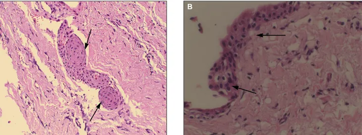
Fig. 3. A: Histologic section.(H&E staining, original magnification x200) This histologic section showing pseudo-glandular structure in connective tissue. B: Histologic section.(H&E staining, original magnification x400) This histologic section show- ing squamous epithelial lining, with a flat interface, and intraepithelial goblet cell.
Fig. 4. Postoperative 6-month panoramic view showing no evidence of recurrence on right ramus area.
A B
213
Ⅲ.
고 찰
GOC은 흔하지 않은 악골 내 치성 기원 낭종으로 Jones 등 은 2006년 GOC의 유병률을 0.2%라고 보고하였으며, Magnusson 등은 조직학적 기준을 모두 만족시키는 GOC는 0.012%라고 보고하였다2. 이 치성낭종의 기원은 초창기 논쟁 의 영역이 되었다. 특징적인 선구조를 가진 낭종상피의 소견 으로 인해 치성기원이 아닌 타액선기원으로 오인되었다1. (Figs. 3A, 3B)
그리하여 Gardner보다 1년 먼저 증례보고를 한 Padayachee 와 van Wyk는 sialodontogenic cyst라는 용어를 사용하였다1. 이 후 약 3년간 논쟁이 있었으나 1992년 Kramer 등5이 면역 조직학적으로 타액선 기원이 아님을 명확히 밝혀내어 논 쟁은 종결되었다.
임상적으로 전 연령대에 모두 고르게 나타날 수 있으나, 호발 연령대는 45.7세로 보고되고 있다. 남성이 여성에 비 하여 약 1.3배 더 호발하나 유의미한 정도는 아니며, 상악 (30%) 보다는 하악(70%)에 호발하며 전치부, 구치부에 나 타나는 정도는 거의 비슷한 정도로 보고되고 있다4,6. 환자 는 천천히 성장하는 무통성의 부종을 경험하게 된다1-3.
방사선학적으로 경계가 뚜렷한 평균 5 cm의 방사선투과 성을 특징으로 하며, 단방성(53.8%), 다방성(46.2%)으로 큰 차이는 보이지 않는다4.
전산화단층촬영영상 소견으로 피질골 천공(61%), 피질 골 희박화(24.4%)의 소견이 대부분 관찰된다4,6. 이것은 이 병소의 공격적인 생물학적 특성을 반영하는 결과이다. 이 것을 반영하는 것으로 또한 치근흡수와 치근이개(24%)가 관찰된다.
감별진단이 필요한 병소로는 치성각화낭, 함치성낭, 저 등도의 점액표피양 암종을 포함시켜야 한다. 부가적으로 조직학적인 명확한 감별진단이 어려울 경우, p53, Ki67 표 지자를 이용한 면역화학적 접근도 사용될 수 있다1.
재발률은 적출술, 소파술의 보존적 처치 시 64.3%, 부분 절제술, 변연절제술 등의 광범위 절제술 시 0%로 조사되어 치성각화낭, 법랑모세포종과 같은 임상적 예후를 보인다4,6.
따라서 광범위절제술을 시행하는 것이 요구되지만, 현실 적으로 공격적인 술식을 선택하기에는 구강악안면외과의 로서 많은 고민이 따른다. 아직 문헌으로 그 효율성이 검증 된 것은 없지만, 보존적인 낭종적출술 후 골공동을 기계적, 또는 화학적 방법으로 2차적으로 처리하는 것도 재발을 줄 이는 좋은 대안이 될 수 있을 것이다3,4.
이러한 고민의 연장선에서 Kaplan 등4은 GOC의 수술 시 아래의 순서도와 같은 원칙을 제창하였다.(Fig. 5) 즉 단방 성, 2치관 이하의 작은 병소의 경우 보존적 수술방법과 주 기적 관찰, 2치관 이상의 다방성 또는 단방성의 병소는 변 연절제술 등의 공격적 술식을 추천하였다. 따라서 외과의 의 판단에 따라 보존적인 수술방법을 택할 시 최소3년 이 상의 정기적인 재발의 검사는 필수적이라 할 수 있다4. 본 증례는 2치관 이하, 단방성의 증례로 환자는 주기적인 추 적관찰 중 이다.
References
1. Krishnamurthy A, Sherlin HJ, Ramalingam K, Natesan A, Premkumar P, Ramani P, et al. Glandular odontogenic cyst: re- port of two cases and review of literature. Head Neck Pathol 2009;3:153-8.
2. Macdonald-Jankowski DS. Glandular odontogenic cyst: system- atic review. Dentomaxillofac Radiol 2010;39:127-39.
3. Oliveira JX, Santos KC, Nunes FD, Hiraki KR, Sales MA, Cavalcanti MG, et al. Odontogenic glandular cyst: a case report.
J Oral Sci 2009;51:467-70.
4. Kaplan I, Anavi Y, Hirshberg A. Glandular odontogenic cyst: a challenge in diagnosis and treatment. Oral Dis 2008;14:575-81.
5. Kramer IR, Pindborg JJ, Shear M. The WHO histological typing of odontogenic tumours. A commentary on the second edition.
Cancer 1992;70:2988-94.
6. Kaplan I, Gal G, Anavi Y, Manor R, Calderon S. Glandular odontogenic cyst: treatment and recurrence. J Oral Maxillofac Surg 2005;63:435-41.
Fig. 5.2005 Kaplan’s treatment scheme of glandular odontogenic cyst 4. (GOC: glandular odontogenic cyst, FU: follow up)
Small Unilocular
Enucleation &
pathological evaluation
Dg: GOC
Recurrency
Retreat, major Prolonged FU
Large
multilocular or unilocular
Biopsy
Dg: GOC
Surgery (Major procedures)
Proximity to vital Structures
Marsupilalization
2ndphase surger
하악골에 발생한 선양치성낭의 치험례