대 한 류 마 티 스 학 회 지 V o l. 1 8 , N o . 2 , J u n e , 2 0 1 1
D O I:10.4078/jrd.2011.18.2.147
□ Clinical Im age □
147
<접수일:2010년 9월 13일, 수정일:2011년 6월 6일, 심사통과일:
2011년 6월 7일>
통신저자:전 재 범
서울 성동구 행당1동
한양대학교 의과대학 내과학교실 E-mail:[email protected]
Figure 1. Radiographies of the lumbar spine. The anteroposterior and lateral radiographs of the lumbar spine show abnormal contact of the apposing spinous processes and enlarged spinous processes that are flattened and sclerotic in their superior and inferior portions (arrows) of the whole lumbar spine.
허리 통증의 흔치 않은 원인; Baastrup’s Disease
김영삼1ㆍ전재범1ㆍ이승훈2ㆍ최윤영3
한양대학교 류마티스병원
1, 한양대학교병원 영상의학과
2, 핵의학과
3An Unusual Cause of Lower Back Pain; Baastrup’s Disease
Young-Sam Kim
1, Jae-Bum Jun
1, Seung-Hun Lee
2, Yun-Young Choi
3Department of Rheumatology, Hanyang University Hospital for Rheumatic Diseases
1,
Departments of Radiology
2and Nuclear Medicine
3, Hanyang University Medical Center, Seoul, Korea
증 례 환 자: 62세 여자
주 소: 간헐적인 허리 통증, 양측 무릎 통증 및 우측 세 번째 중수지 통증
병 력: 14년 전 좌측 무릎 활막제거술 시행 가족력: 특이 소견 없음
검사실 소견: RF (−), Anti-ccp Ab (−), HLA B27 realtime PCR (−), ANA skeleton (1 : 80), CRP 14.4 mg/dL, ESR 139 mm/h, Joint fluid in right knee {WBC 12,700/mm3 (neutrophil 74%, lymphocyte 26%), AFB stain: negative, KOH: negative, culture: no growth}, uric acid 4.0 mg/dL
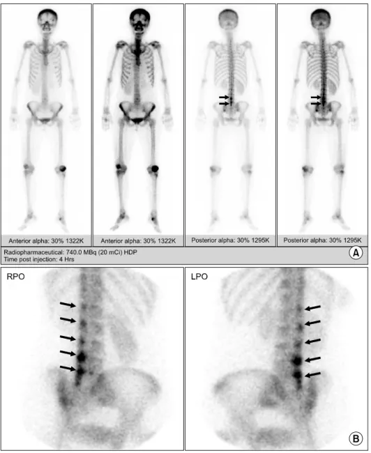
영상의학 소견: Sacroiliac joint series and pelvic X-ray: no remarkable finding, anteroposterior and lateral radiographs of the lumbar spine: 그림 1, computerized tomography (CT) of the lumbar spine: 그림 2, Whole body bone scan: 그림 3.
고 찰
Baastrup’s disease는 척추가 신전될 때 국소 부위의 허리 통증을 보이고 굴곡될 때 통증이 완화되는 임상증상을 보 이며 인접한 극상돌기들의 근접과 접촉을 특징으로 하는 질환으로 1933년 덴마크의 방사선과의사 Christian Baas- trup에 의해 소개되었다 (1). 이 질환은 인접한 극상돌기 사
이의 만성적인 접촉에 의해 발생되는 퇴행성 디스크 질환 이나 과다앞굽은증(hyperlordosis)을 가진 고령의 환자들에 서 종종 관찰된다 (2).
단순방사선사진 혹은 컴퓨터단층촬영술에서 극상돌기들 의 마주하는 표면들은 팽대(enlargement), 편평화(flatte- ning), 경화(sclerosis) 소견을 보이고 반응성 상아질화(reac- tive eburnation)가 되기 쉽다 (3). 자기공명영상은 뼈의 부 종과 극상돌기 사이의 윤활낭액의 존재를 평가하는데 도
148 김영삼 외
Figure 3. Focal increased uptake in the spinous process areas (arrows) of the lower L-spine (L4 and L5 level) on the posterior whole body bone scan image (A).
The right posterior oblique (RPO) and left posterior oblique (LPO) regional images show more clearly defined focal uptake lesions in the spinous process areas (arrows) and not only in the L4 and L5 levels, but also in the whole L-spine level (B). The focal uptake lesions on the bone scan images seem to match with the sclerotic lesions between the spinous processes, as detected on CT images, and the focal uptake lesions were possibly associated with active disease pro- cesses, including bony erosion and inflammatory changes, and these were most severe in the L4-5 level in this patient.
Figure 2. CT of the lumbar spine. The coronal and sagittal reformatted CT scans also show enlarged spinous processes of the whole lumbar spine with abnormal contact of apposing spinous processes (arrow).
움이 된다 (4). 조직학적으로 육아종성 반응과 혈관 주위 세포 침윤이 이러한 부착 건병증(insertion tendinopathy)의 특징으로 나타난다 (3).
치료로는 국소부위 마취제 주입방법과 관련된 극상돌기 들을 절제하는 방법이 있다 (5).
간헐적인 허리 통증을 호소하는 환자에서 흔치 않지만 드물지도 않는 Baastrup’s disease를 진단하였다. 이 질환이 허리 통증의 한가지 원인이 될 수 있다는 것을 알고 있어 야 되겠고 척추의 단순방사선사진에서 무시하기 쉽지만 극상돌기들을 관찰하는 습관을 가져야 되겠다.
간헐적인 허리 통증은 비마약성 진통제로 증상 조절하였 고 좌측 무릎관절염과 우측 세 번째 중수지 관절염은 미분 류성 관절염(undifferentiated arthritis)으로 진단하여 sulfasa- lazine을 투여하며 추적 관찰 중이다.
Synonyms: Arthrosis interspinosa, diarthrosis interspinosa,
허리 통증의 흔치 않은 원인; Baastrup’s Disease 149
kissing osteophytes, kissing spine, kissing spinous disease, os- teoarthrosis processus spinosi vertebrarum lumbalum, osteo- arthrosis interspinalis
참고문헌
1. Baastrup CI. On the spinous processes of the lumbar vertebrae and the soft tissues between them, and on pathological changes in that region. Acta Radiol 1933;14:52-5.
2. Meleger AL, Krivickas LS. Neck and back pain: muscu- loskeletal disorders. Neurol Clin 2007;25:419-38.
3. Resnick DL. Diagnosis of bone and joint disorder. 4th ed. p. 1411-4, Philadelphia, W.B. Saunders, 2002.
4. Bywaters EG, Evans S. The lumbar interspinous bursae and Baastrup's syndrome. An autopsy study. Rheumatol Int 1982;2:87-96.
5. Lin E. Baastrup's disease (kissing spine) demonstrated by FDG PET/CT. Skeletal Radiol 2008;37:173-5.