Pediatr Allergy Respir Dis(Korea) 2012;22:163-170
1)
접수 :2012년 월1 16 ,일 수정:2012년 월2 14 ,일 승인:2012년 월 일3 9
책임저자 김여향 대구광역시 중구 달성로: , 56계명대학교 의과대학 소아과학교실
Tel: 053)250-7524, Fax: 053)250-7783, E-mail: [email protected]
청소년 폐결핵의 임상적 및 영상학적 특징:
재활동성 결핵과의 관련성
계명대학교 의과대학 소아과학교실
1, 내과학교실
2, 영상의학과교실
3
경북대학교 의학전문대학원 소아과학교실
4
강석진1ㆍ김여향1ㆍ정치영2ㆍ이희정3ㆍ현명철4
Clinical Characteristics and Radiologic Patterns of
Adelescents with Pulmonary Tuberculosis:
Relevance to the Reactive Tuberculosis
Seok Jin Kang, MD
1, Yeo Hyang Kim, MD, PhD
1, Chi Young Jung, MD, PhD
2
Hee Jung Lee, MD, PhD
3, Myung Chul Hyun, MD, PhD
4
Departments of
1Pediatrics,
2Internal Medicine, and
3Radiology, Keimyung University School of Medicine, Daegu,
4
Department of Pediatrics, Kyungpook National University School of Medicine, Daegu, Korea
Purpose: To evaluate the clinical characteristics and radiologic patterns of adolescents with pul- monary tuberculosis (TB), and to assess whether they are related with primary TB or reactive TB.
Methods: Among the enrolled patients who were diagnosed with pulmonary TB from March 2000 to May 2011, 36 with plain radiography and/or chest computed tomography (CT) were reviewed.
We reviewed retrospectively their medical charts to collect clinical data and past history. Among these 36 patients, plain radiography of the 36 patients and chest CT of the 34 patients were retro- spectively evaluated.
Results: The patients consisted of 18 males and 18 females, and their median age was 14 years old. The most common clinical presentation was cough and fever. Half of them had chronic cough for more than two weeks. Ten patients had history of close contact with adult patients with active pulmonary TB: 7 patients with their parents, 2 patients with friends, 1 patient with their grandmother.
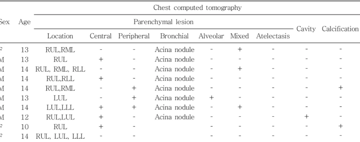
The most frequent pattern of plain radiography was pleural effusion (16/36). In the chest CT findings, all cases showed parenchymal lesions and lymphadenopathy. In addition, 91% of the cases showed acinar nodules. The pattern of pleural effusion revealed associated ipsilateral pleural lymph node and subpleural nodule. Rim enhancement and calcification of the lymph node demonstrated 9% (3/34) and 12% (4/34), respectively. Only two of them showed typical hilar lymphadenopathy in chest X ray and CT.
Conclusion: The radiologic findings of adolescents with pulmonary TB show patterns for rather reactive than primary TB. For diagnosis of adolescent pulmonary TB, chest CT is more helpful than that of plain radiography. [Pediatr Allergy Respir Dis(Korea) 2012;22:163-170]
Key Words : Adolescent, Computed tomography, Tuberculosis, pulmonary