INTRODUCTION
Despite recent advancements in the diagnosis and treat- ment of systemic lupus erythematosus (SLE), infection remains the most important cause of death in this disease (1). Patients with SLE appear to be predisposed to an increased risk for infection due to immunosuppressive therapies as well as the intrinsic immunological defects associated with the disease (2). We report here on a patient with SLE who recovered from cryptococcemia and cryptococcal lymphadenitis two years ago, and the patient went on to develop mesenteric cryptococ- cal lymphadenitis even in the absence of any immunosup- pressive treatment since the time of the original infection.
CASE REPORT
A 42-yr-old woman was admitted to Kangnam St. Mary’s Hospital suffering from high fever and diffuse abdominal pain for three weeks. She had been diagnosed with SLE with class V lupus nephritis fourteen years ago. Two years ago, she
had received corticosteroid and cyclophosphamide therapy for mesenteric vasculitis. After the treatment, she had fallen ill with cryptococcemia and retroperitoneal cryptococcal lym- phadenitis. She recovered after her administration with treat- ment using intravenous fluconazole. However, she stated that she had not taken any medication for SLE such as corticos- teroids and immunosuppressive agents.
On the physical examination, there was diffuse tenderness without rebound tenderness on her abdomen. There was nei- ther arthralgia nor pitting edema on the extremities. A com- plete blood count revealed a mildly low hemoglobin level of 10.6 g/dL, a slightly elevated WBC count (10,800/ L with a differential count of neutrophils 70.4% and lymphocytes 20.2%), and a normal platelet count of 394,000/ L. The erythrocyte sedimentation rate (ESR) was 102 mm/hr and C-reactive protein (CRP) was 6.43 mg/dL. Her renal function was slightly impaired (creatinine 1.45 mg/mL, proteinuria 6.9 g/day and creatinine clearance 35.4 mL/min). The serum electrolytes showed normal levels. ANA was positive (1:160 with homogeneous pattern), anti-dsDNA antibody was 13.91 IU/mL (normal range 0-7 IU/mL), and C3 and C4 levels were
Sang-Hyon Kim, Sung-Dong Kim, Hae-Rim Kim*, Chong-Hyeon Yoon, Sang-Heon Lee*, Ho-Youn Kim, Sung-Hwan Park
Division of Rheumatology, Department of Internal Medicine, Kangnam St. Mary’s Hospital, College of Medicine, The Catholic University of Korea, and Konkuk University*, Seoul, Korea
Address for correspondence Sung-Hwan Park, M.D.
Division of Rheumatology, Department of Internal Medicine, Kangnam St. Mary’s Hospital, The Catholic University of Korea, 505 Banpo-dong, Seocho-gu 137-040, Korea
Tel : +82.2-590-1427, Fax : +82.2-599-3589 E-mail : [email protected]
*This work was supported by grant (R11-2002-098-01- 001-0) from the Korea Science & Engineering Founda- tion through the Rheumatism Research Center at Catho- lic University of Korea.
1059 J Korean Med Sci 2005; 20: 1059-61
ISSN 1011-8934
Copyright � The Korean Academy of Medical Sciences
Intraabdominal Cryptococcal Lymphadenitis in a Patient with Systemic Lupus Erythematosus
Cryptococcal infection is a rare, yet well recognized complication of systemic lupus erythematosus (SLE). We present a case of mesenteric and retroperitoneal crypto- coccal lymphadenitis resulting in the obstruction of the stomach and proximal duo- denum in a patient suffering from SLE, while recently she did not receive any immu- nosuppressive treatment. A 42-yr-old woman was admitted due to high fever and diffuse abdominal pain for three weeks. Abdominal computed tomography (CT) scan showed multiple conglomerated lymphadenopathies in the retroperitoneum and the mesentery resulting in luminal narrowing of the third portion of the duode- num. Cryptococcal lymphadenitis was proven by needle biopsy and she was treat- ed with intravenous liposomal amphotericin B, followed by oral fluconazole. After fourteen-month antifungal therapies, the clinical symptoms and follow-up images improved. This case emphasize that the intrinsic immunological defects of SLE may be directly responsible for the predisposition to fungal infections.
Key Words : Mesenteric Lymphadenitis; Cryptococcus neoformans; Lupus Erythematosus, Systemic
Received : 13 August 2004 Accepted : 6 November 2004
1060 S.-H. Kim, S.-D. Kim, H.-R. Kim, et al.
in the normal range. Cryptococcal antigen was negative and there was no microorganism on the blood and stool cultures.
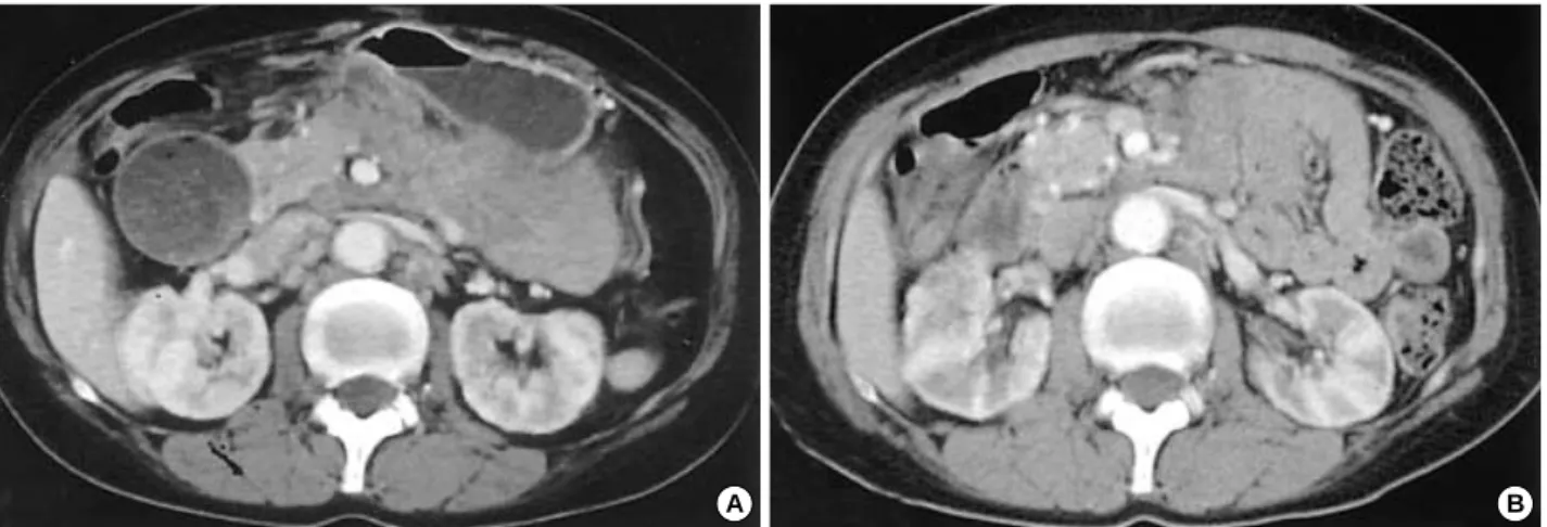
Abdominal computed tomography (CT) scan showed mul- tiple conglomerated soft tissue density lesions noted in the mesenteric root, aortocaval space and left paraaortic space and smooth luminal narrowing was noted at the third por- tion of the duodenum: this resulted in the marked dilatation of the stomach and proximal duodenum (Fig. 1A). Under sonographic guidance, needle biopsy was performed at the conglomerated lymphadenopathies. Histopathologic find- ing showed chronic granulomatous inflammation with focal necrosis and the presence of fungal organisms, which were consistent with cryptococci (Fig. 2).
She was treated with intravenous amphotericin B 0.5 mg/
kg/day, but after two weeks the serum creatinine was elevated up to 2 mg/dL, so amphotericin B was replaced with liposo-
mal amphotericin B 1 mg/kg/day for eight weeks. This was followed by oral fluconazole 400 mg/day. Eight weeks after the liposomal amphotericin B administration, a follow up abdominal CT scan was performed. It showed marked decrease in the sizes and extents of the multiple conglomerated lym- phadenopathies in the mesentery, and resolution of the pre- vious noted obstruction of the duodenal third portion (Fig.
1B). The ESR and CRP were decreased to 38 mm/hr and 0.47 mg/dL. Oral fluconazole (200 mg/day) was maintained for twelve months and there was no sign of remained infec- tion in clinical findings and last CT. For lupus nephritis, she was managed by angiotensin receptor blocker without any immunosuppressive agents due to the infection, and there was no change in renal function and proteinuria.
Fig. 1.Abdominal computed tomography (CT) scan shows the multiple conglomerated soft tissue density lesions noted at the mesenteric root, aortocaval space and left paraaortic space; the CT scan also shows the smooth luminal narrowing noted at the third portion of the duodenum, resulting in marked dilatation of the stomach and proximal duodenum (A). Eight weeks later, the follow up abdominal CT scan shows that there is a marked decrease in the sizes and extents of multiple conglomerated lymphadenopathies in the mesenteric root, aor- tocaval and left paraaortic space, and the previous noted obstruction of the duodenal third portion is resolved (B).
A B
Fig. 2.Under sonographic guidance, percutaneous needle biopsy was performed on the mesenteric conglomerated lymph nodes, and it shows chronic granulomatous inflammation with focal necrosis and a fungal organism: this is consistent with the cryptococcal infection noted on silver staining (A, ×400) and hematoxylin and eosin staining (B, ×100).
A B
Intraabdominal Cryptococcal Lymphadenitis in SLE 1061
DISCUSSION
Cryptococcosis is an infection caused by a yeast-like fungus Cryptococcus neoformans. C. neoformans is an ubiquitous encap- sulated yeast that can be isolated from soil and avian habitats, and most infections are thought to be acquired by inhalation of fungus into the lungs (3). Pulmonary infection has a ten- dency toward spontaneous resolution and this condition is usually asymptomatic. The organisms spread through the blood system and it mainly causes meningoencephalitis, but it can also affect other sites (4). Cryptococcosis due to C. neo- formans is a common complication of late infection concur- rent with HIV (5). Patients who have undergone solid-organ transplantation or glucocorticoid therapy are at increased risk for infections with C. neoformans (3).
This case is unique in that intraabdominal cryptococcal lymphadenitis occurred in the absence of immunosuppres- sive treatment, although the initial infection developed after immunosuppressive treatment. It is supposed that the intrin- sic immune defects related to the SLE could have been respon- sible for cryptococcal lymphadenitis. Most cryptococcal infec- tions present as meningoencephatlitis, followed by pulmonary and skin infection. Intraabdominal lymphatic cryptococco- sis is very rare. There is only 2 reports of intraabdominal cryp- tococcal lymphadenitis in a patient with AIDS and omental cryptococcoma (6, 7).
The host defense mechanisms protecting the body against cryptococcal infections are complex and not completely under- stood. Cell-mediated immunity (CMI) is the crucial compo- nent of the immune system for resistance against cryptococ- cosis (8). This is supported by evidence that the most of the serious cryptococcal infections usually occur in individuals with defective CMI such as patients with acquired immun- odeficiency syndrome (AIDS), corticosteroid treatment, retic- uloendothelial malignancies or post-organ transplantation (9, 10).
Mok et al. have reported on a female SLE patient who de- veloped cryptococcal meningitis concurrently with active SLE and in the absence of immunosuppressive treatment or other predisposing conditions (11). They suggested that her low complement level, low natural killer cell count and the prob- able defective CD4 positive T cell and macrophage functions resulting from her SLE disease activity might have been res- ponsible for her susceptibility to cryptococcal meningitis.
A lot of immunological abnormalities that are present may either be the cause or the effect of SLE activity. These SLE related abnormalities include lymphopenia, defective CD4
positive T cell proliferation and interleukin 2 production to antigenic and mitogenic stimulation, reduced cytotoxicity of CD8 positive T cells, the impaired antigen-presenting func- tion of monocytes and macrophages, hypocomplementemia, defective opsonization and the impaired neutrophil chemo- taxis and phagocytosis (11, 12).
This case highlights that the importance of the intrinsic immunological defects of SLE predisposition to opportunistic fungal infection. To our knowledge, this is the first case report of mesenteric cryptococcal lymphadenitis occurred in a SLE patient without any immunosuppressive drugs.
REFERENCES
1. Abu-Shakra M, Urowitz MB, Gladman DD, Gough J. Mortality stud- ies in systemic lupus erythematosus. Results from a single center. II.
Predictor variables for mortality. J Rheumatol 1995; 22: 1265-70.
2. Kang IS, Park SH. Infectious complications in SLE after immuno- suppressive therapies. Curr Opin Rheumatol 2003; 15: 528-34.
3. Bennet JE. Cryptococcosis. In: Braunwald E, Fauci AS, Kasper DL, Hauser SL, Longo DL, James JL, editors, Harrison’s principles of internal medicine. 15th ed. New York: McGraw-Hill, 2001; 1174-5.
4. Lewis JL, Rabinovich S. The wide spectrum of cryptococcal infec- tions. Am J Med 1972; 53: 315-22.
5. Eng RH, Bishburg E, Smith SM, Kapila R. Cryptococcal infections in patients with acquired immune deficiency syndrome. Am J Med 1986; 81: 19-23.
6. Scalfano FP Jr, Prichard JG, Lamki N, Athey PA, Graces RC. Abdomi- nal cryptococcoma in AIDS: a case report. J Comput Tomogr 1988;
12: 237-9.
7. Chong PY, Panabokke RG, Chew KH. Omental cryptococcoma. An unusual presentation of cryptococcosis. Arch Pathol Lab Med 1986;
110: 239-41.
8. Mitchell TG, Perfect JR. Cryptococcosis in the era of AIDS: 100 years after the discovery of Cryptococcus neoformans. Clin Microbiol Rev 1995; 8: 515-48.
9. Perfect JR, Durack DT, Gallis HA. Cryptococcemia. Medicine (Bal- timore) 1983; 62: 98-109.
10. Miller GP. The immunology of cryptococcal disease. Semin Respir Infec 1986; 1: 45-52.
11. Mok CC, Lau CS, Yuen KY. Cryptococcal meningitis presenting concurrently with systemic lupus erythematosus. Clin Exp Rheuma- tol 1998; 16: 169-71.
12. Tsokos GC, Kovacs B, Sfikakis PP, Theocharis S, Vogelgesang S, Via CS. Defective antigen-presenting cell function in patients with systemic lupus erythematosus. Arthritis Rheum 1996; 39: 600-9.