INTRODUCTION
Calcifications within primary tumors can be seen in thy- roid, breast, and ovarian neoplasms. They are also described within benign lesions of the gastrointestinal tract, such as leiomyomas and angiomas, or in metastases from colonic carcinoma. However, calcifications within primary gastric cancer are a rare finding (1, 2). Moreover, gastric uptake of bone seeking agents in a patient with advanced gastric can- cer is an even rare entity.
Tc-99m 3,3-diphosphono-1,2-propanedicarboxylic acid (DPD) bone scintigraphy is used primarily for skeletal imag- ing to detect abnormalities of the bone. However, a variety of soft tissue abnormalities can also be detected on bone scin- tigraphy through accumulation of bone seeking agents. Neo- plastic, hormonal, inflammatory, ischemic, traumatic, excre- tory, and artifactual entities demonstrate abnormal soft-tis- sue uptake of Tc-99m DPD (3).
Recently, we experienced a case of bone scintigraphy that demonstrated diffusely increased gastric uptake of Tc-99m DPD in a patient with advanced gastric cancer. We report this case along with a review of the literatures.
CASE REPORT
A 69 yr-old male patient was admitted to our hospital due to epigastric discomfort for 2 months duration. Epigastric discomfort was exacerbated after meals. An upper GI endos- copy revealed a huge ulcerative creator on the posterior wall of the prepyloric area with creamy exudates and a biopsy diag- nosed the lesion to be adenocarcinoma. Non-contrast enhanced computed tomography (CT) of the abdomen showed a mass involving the lower body and antrum of the stomach with diffuse and circumferential wall thickening, punctate calci- fications within the gastric walls, and several lymphadeno- pathies along the retropancreatic area (Fig. 1). Laboratory stud- ies revealed normochromic and normocytic anemia (hemo- globin of 10.3 g/dL, normal; 12-16 g/dL), but other labora- tory findings including serum calcium (normal; 8.4-10.2 mg/
dL) and phosphorus (normal; 2.5-4.5 mg/dL) concentrations were normal.
To evaluate the extent of the disease, 3 hr after injection of 20 mCi (740 MBq) of Tc-99m DPD, bone scintigraphy and single photon emission computed tomography (SPECT) were performed using a dual-head gamma camera (Vertex; ADAC laboratories, Milpitas, CA, U.S.A.). Bone scintigraphy showed gastric uptake but no evidence of bone metastasis (Fig. 2).
Dae-Weung Kim, Hwan-Jeong Jeong*, Soon-Ah Park, Chang-Guhn Kim
Department of Nuclear Medicine, Wonkwang University School of Medicine, Iksan; Department of Nuclear Medicine and Research Institute of Clinical Medicine*, Chonbuk National University Medical School, Jeonju, Korea
Address for correspondence Chang-Guhn Kim, M.D.
Department of Nuclear Medicine, Wonkwang University School of Medicine, 344-2 Sinnyong-dong, Iksan 570-711, Korea
Tel : +82.63-850-1364, Fax : +82.63-852-1310 E-mail : [email protected]
*This paper was supported by Wonkwang University in 2004.
153 J Korean Med Sci 2007; 22: 153-5
ISSN 1011-8934
Copyright � The Korean Academy of Medical Sciences
Gastric Accumulation of Bone Seeking Agent in a Patient with Advanced Gastric Cancer
Soft tissue uptake of Tc-99m labeled bone seeking agents, such as Tc-99m 3,3- diphosphono-1,2-propanedicarboxylic acid (DPD), is commonly seen in clinical prac- tice, even though bone scintigraphy is mainly used to detect bone disease. How- ever, gastric uptake of bone agents in patients with gastric cancer is very rare. And it has been reported that calcified gastric adenocarcinoma appears in only about 5%
of all gastric cancer. We report a rare case of bone scintigraphy, single photon emis- sion computed tomography and computed tomography fusion images that demon- strated diffuse gastric uptake of Tc-99m DPD in a patient with advanced gastric cancer.
Key Words : Radionuclide Imaging; Tomography, Emission-Computed, Single-Photon; Stomach Neoplasms
Received : 29 August 2005 Accepted : 14 November 2005
154 D.-W. Kim, H.-J. Jeong, S.-A. Park, et al.
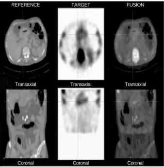
The gastric uptake was thought to be calcified tumoral up- take. To confirm this, fusion images were constructed by com- bining morphologic CT and functional SPECT images on a computer workstation. The SPECT and CT fusion images show the intense Tc-99m DPD uptake to be located in the anterior and posterior wall of the gastric antrum (Fig. 3).
DISCUSSION
The first report of calcified gastric cancer was by Gruber in 1913 (4), and subsequently several other cases were reported (5, 6). When histology was considered, most of these tumors were proved to be mucinous adenocarcinomas. It has been
reported that mucinous gastric adenocarcinoma compromise about 5% of all gastric cancers (7). To explain the presence of calcifications, ‘‘dystrophic’’ and ‘‘metastatic’’ mechanisms have been proposed (8). Dystrophic calcifications develop in necrotic tissues, probably because denatured proteins prefer- entially bind phosphate ions, which react with calcium ions to form a precipitate of calcium phosphate. In these cases plasma levels of calcium are within normal range. Metastat- ic calcifications are simple mineral deposits in undamaged normal tissues caused by persistent hypercalcemia. In our case, normal level of serum calcium and phosphorus concen- trations suggests the presence of dystrophic calcification.
Tc-99m DPD bone scintigraphy is used primarily for skele- tal imaging to detect abnormalities of the bone. However, bone scintigraphy also delineates a wide spectrum of non- osseous disorders. Neoplastic, hormonal, inflammatory, ische- mic, traumatic, excretory, and artifactual entities demonstrate abnormal soft-tissue uptake of Tc-99m DPD. Mechanisms leading to increased extraosseous Tc-99m DPD uptake include extracellular fluid expansion, enhanced regional vascularity and permeability, and elevated tissue calcium concentration.
Bone scintigraphy can show gastric uptake in various con- ditions, including artifact due to Tc-99m-pertechnetate in radiopharmaceutical preparation, hypercalcemia due to me- tastatic calcinosis, dialysis patients and gastric carcinoma. In our case, because the thyroid was not visible, visualization of the stomach on the bone scintigraphy cannot be attribut- ed to a faulty preparation containing free Tc-99m-pertech- netate (9).
Fig. 1.Non-contrast enhanced CT of the upper abdomen shows diffuse thickening of gastric antral wall with punctate calcifica- tions (arrows).
Fig. 2.Bone scintigraphy shows increased uptake of tracer in the gastric wall (arrow) but no evidence of bone metastasis.
A B
Fig. 3.The single photon emission computed tomography (SPE- CT) and CT fusion images show that intense Tc-99m DPD uptake is located in the anterior and posterior wall of the gastric antrum.
However, the extent of tracer uptake is diffuse and broader than the gross calcifications seen on CT images.
TARGET FUSION
REFERENCE
Transaxial Transaxial Transaxial
Coronal Coronal
Coronal
Gastric Uptake of Bone Agent in a Gastric Cancer Patient 155
There are a few cases showing gastric uptake of tracer in advanced gastric cancer patients (10). However, the fact that the extent of tracer uptake is more diffuse and broader than that of gross calcifications seen on CT images makes our case unique. This may suggest the presence of micro-calcifications in the gastric wall which is not seen on CT images but is detected in bone scintigraphy.
In summary, we describe a rare case showing gastric uptake of Tc-99m DPD in an advanced gastric cancer patient. The extent of tracer uptake in the gastric walls was more diffuse and broader than that of gross calcifications seen on CT ima- ges. Calcified gastric cancer is rare, but it was possible to detect even micro-calcifications with Tc-99m DPD bone scintig- raphy. As exemplified by this case, recognition of extra-skele- tal Tc-99m DPD uptake may enhance the diagnostic value of bone scintigraphy.
REFERENCES
1. Rotondo A, Grassi R, Smaltino F, Greco A, Leung AW, Allison DJ.
Calcified gastric cancer: report of a case and review of the literature.
Br J Radiol 1986; 59: 405-7.
2. Balestreri L, Canzonieri V, Morassut S. Calcified gastric cancer-CT
findings before and after chemotherapy. Case report and discussion of the pathogenesis of this type of calcification. Clin Imaging 1997;
21: 122-5.
3. Peller PJ, Ho VB, Kransdorf MJ. Extraosseous Tc-99m MDP uptake:
a pathophysiologic approach. Radiographics 1993; 13: 715-34.
4. Gruber GB. Knochebildung in einem magen karzinom. Z Beitr Path Anat 1913; 55: 368-70.
5. Kunieda K, Okuhira M, Nakano T, Nakatani S, Tateiwa J, Hiramat- su A, Mizuno T, Shiozaki Y, Sameshima Y. Diffuse calcification in gastric cancer. J Int Med Res 1990; 18: 506-14.
6. De Carvalho JC, Francischetti EA, De Barros Filho GA, Cerda JJ.
Calcified mucinous adenocarcinoma of the stomach. Am J Gastroen- terol 1978; 69: 481-4.
7. Adachi Y, Mori M, Kido A, Shimono R, Maehara Y, Sugimachi K.
A clinicopathologic study of mucinous gastric carcinoma. Cancer 1992; 69: 866-71.
8. Ashley DJB. Evan’s Histological Appearances of Tumors. Edinburgh;
Churchill Livingstone, 1990; 307-8.
9. Delcourt E, Baudoux M, Neve P. Tc-99m-MDP bone scanning detec- tion of gastric calcification. Clin Nucl Med 1980; 5: 546-7.
10. Watanabe N, Haida M, Mikuni I, Shimizu M, Nakajima A, Seto H.
Bone imaging in advanced gastric cancer. Clin Nucl Med 1998; 23:
384.