pISSN : 1229-5418
Implantology 2018; 22(2): 88-98
https://doi.org/10.32542/implantology.20180008
Received: May 31, 2018 Revised: June 27, 2018 Accepted: June 29, 2018
Copyright © 2018. The Korean Academy of Oral &
Maxillofacial Implantology
This is an Open Access article distributed under the terms of the Creative Commons Attribution Non-Commercial License (http://creativecommons.
org/licenses/by-nc/4.0/) which permits unrestricted non-commercial use, distribution, and reproduction in any medium, provided the original work is properly cited.
OPEN ACCESS
Purpose: The purpose of this study was to analyze the mean residual alveolar bone height according to various measuring points of male edentulous patients. And to compare the residual alveolar bone height differences observed in panoramic and computed tomography images for analyzing the predictable distortion trends in panoramic radiography.
Materials and Methods: The study used 40 images of the maxilla and mandible, excluding computed tomography and panoramic images. Based on the anatomical indices, the measurement values of each image were obtained by setting 7 measuring points of the maxilla and 9 measuring points of the mandible. The significant difference was statistically analyzed by paired t test comparing the measurement values observed on computed tomography and panoramic radiography ( p<0.05).
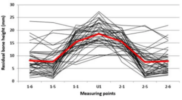
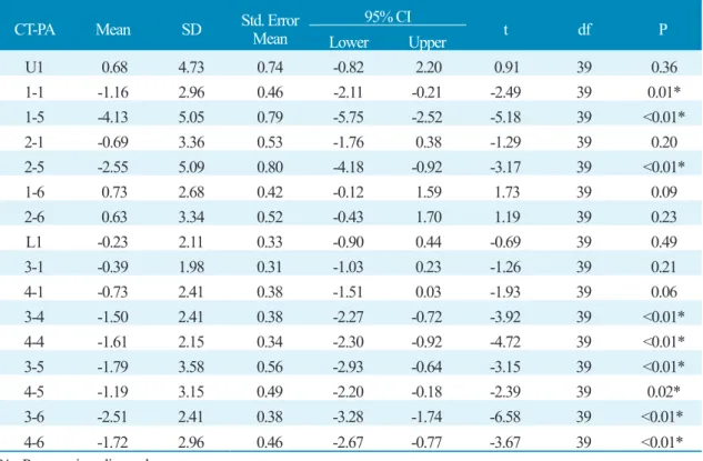
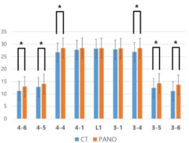
Results: The mean residual alveolar bone height was more than 15.5 mm in the maxillary anterior part, 8.2 mm in posterior part, 27.7 mm in the mandibular anterior part, and the mandibular posterior part was 11.1 mm. The result of paired t-test comparing the computed tomography and panoramic images showed that the maxillary right central incisor position, maxillary left second premolar, maxillary right second premolar, mandibular left first premolar, mandibular right first premolar, mandibular left second premolar, mandibular right second premolar, mandibular left first molar, mandibular right first molar positions of residual alveolar bone height were significantly different between two images.( p<0.05).
Conclusion : It is considered that the reliability of the panoramic radiography is low because there is a significant difference in the residual alveolar bone height observed on computed tomography and panoramic radiography images.
Keywords: Computed tomography, Panoramic radiography, Edentulism, Residual bone height
Abstract
한국인 남성 무치악환자에서 파노라마 방사선사진상의 치조골 높이 왜곡에 대한 후향적 연구
백주현, 송영균*
단국대학교 치과대학 치과보철학교실
*Corresponding author: Young-Gyun Song, [email protected]
Retrospective Study on Distortion of Alveolar Bone Height in Panoramic Radiographs of Edentulous Korean Male Patients
Joo-Hyun Baik, Young-Gyun Song*
Department of Prosthodontics, College of Dentistry, Dankook University, Cheonan, Korea
Ⅰ. 서론
임플란트와 연조직에서 일차 지지를 얻는 보철은 환자의 심미, 기능, 안정, 발음 등에 관한 요구를 만 족시켜야 한다. 임플란트 수술 시 보철치료의 필요에 맞는 임플란트의 직경, 수, 위치를 정해야 하며 잔 존 치조골의 상태가 중요한 요인으로 작용한다
1.
임플란트의 성공에 영향을 미치는 주요 요인은 정확한 수술과 보철 치료계획이며, 치료 계획 중 방사 선학적 평가는 필수적인 요소이다
2,3. 잔존 치조골의 상태를 방사선학적 평가하는 방법에는 측방 두부 규격방사선사진(lateral cephalometric radiography)과 파노라마 방사선사진(panoramic radiography) 같은 다양한 방법이 있다
4-8. 이중 잔존치조골 높이 측정 방법에는 파노라마 촬영술과 전산화 단층 촬영 술(computed tomography/CT)이 있으며 파노라마 촬영술은 가장 보편적 방법이지만, 이차원 이미지 영 상으로 협설 방향의 정보가 부족하고 수평적, 수직적 영상의 확대가 나타날 수 있다
9-11. 또한 다양한 확 대율을 가진 영상으로 해부학적 구조물의 왜곡이 나타날 수 있으며 촬영 중 환자의 머리 위치에 따라 길 이가 달라지는 등 기준을 잡기 어려운 한계가 있다
12,13. 전산화 단층 촬영술은 3차원 영상으로 수직적, 수 평적 위치 확인이 가능하며 파노라마 촬영에 비해 정확한 해부학적 구조물의 관찰이 가능하나 높은 비 용과 방사선 노출량으로 인한 단점이 있다. 임플란트 수술 시 전산화 단층 촬영술 뿐만 아니라 기본 방 사선사진을 포함한 검사가 모두 실시되어야 하는데 파노라마 촬영술은 무치악과 유치악 환자 모두에서 가장 보편적으로 사용되는 방법이다
14,15. 임플란트 수술 시 주의해야 할 해부학적 구조물은 상악동, 이 공, 하 치조 신경관 등 다양하며, 특히 신경혈관계는 임플란트 수술 중 침범시에 위험을 초래할 수 있으 므로 수술 전 우선적으로 고려해야한다
16.
이번 연구의 목적은 한국인 남성 전부 무치악 환자의 다양한 위치에 따른 평균 잔존 치조골 높이를 파 노라마 촬영술과 전산화 단층 촬영술 영상에서 비교분석하여, 파노라마 촬영술에서 예측할 수 있는 왜 곡경향을 분석함으로써, 파노라마 영상만을 통해 임플란트 수술 시 주의해야 할 해부학적 구조물을 파 악하고자 하는 것이다.
Ⅱ. 연구재료 및 방법
1. 연구대상
2010년 6월부터 2016년 9월까지 대전보훈병원치과 전산화 단층 촬영술(computed tomography/CT)
을 시행한 환자 중 상악과 하악 완전 무치악 환자 100명 중 같은 기간 파노라마 촬영술(panoramic radi-
ography)을 시행한 환자 상악 46명 하악 43명을 대상으로 하였으며, 단국대학교 임상시험심사위원회
에서 승인받았다(DKU2017-06-002). 잔존 골에 영향을 주는 갑상선질환, 부갑상선기능 항진증, 당뇨,
만성 신장질환, 골다공증 등의 과거병력을 가진 환자는 제외했다. 전산화 단층 촬영술과 파노라마 촬영
술 영상 모두에서 다음과 같은 해부학적 기준에 적합한 영상을 선정하였다.
상악골의 해부학적 지표
① 비중격, 전비극, 비구개공 명확함
② 이상구 하연 명확함
③ 상악동 하연 명확함
④ 수술 및 골절 과거병력 없음
하악골의 해부학적 지표
① 이공 명확함
② 하치조신경관 상연 명확함
③ 수술 및 골절 과거병력 없음
상기 기준에 부합하지 않는 영상은 제외되었고 상악 40명, 하악 40명 환자의 영상이 분석에 사용되었 다. 전산화 단층 촬영기기로는 Lightspeed VCT 64channel (GE Healthcare, Waukesha, WI, USA)가 사 용되었으며 파노라마 촬영기기로는 PAX-500 (VATECH, Hwasung, Korea)이 사용되었다.
2. 연구방법
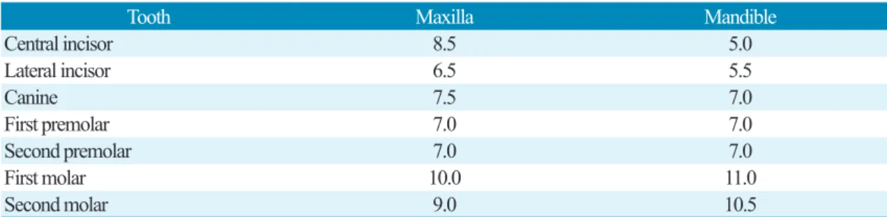
상악과 하악의 잔존 치조골 높이 측정을 위한 참고 위치는 상, 하악 치아의 평균 치관부 직경을 이용 정중선으로부터의 수평적 거리로 정했다(Table 1)
17.
상악골에서 사용한 해부학적 지표는 전비극, 이상구 하연, 상악동 하연으로 정중선을 기준으로 수평 이동한 위치에서 평행선을 그은 7개의 기준선을 정해 각 위치의 해부학적 지표로부터 잔존치조제정까 지 거리를 측정하였다.
Table 1. The mean width of all the natural crown (unit : mm)
Joo-Hyun Baik et al. : Retrospective Study on Distortion of Alveolar bone Height in Panoramic Radiographs of Edentulous Korean Male Patients.
Implantology 2018
Tooth Maxilla Mandible
Central incisor 8.5 5.0
Lateral incisor 6.5 5.5
Canine 7.5 7.0
First premolar 7.0 7.0
Second premolar 7.0 7.0
First molar 10.0 11.0
Second molar 9.0 10.5
1) 상악 영상 계측점
① U1 : 정중선, 전비극에서 잔존치조제정까지 거리
② 중절치 1-1, 2-1 : 정중선 좌(2-1), 우(1-1) 4.25 mm 평행선의 이상구 하연에서 잔존치조제정까 지 거리
③ 제 2 소구치 1-5, 2-5 : 정중선 좌(2-5), 우(1-5) 33 mm 평행선의 상악동 하연에서 잔존치조제정 까지 거리
④ 제 1 대구치 1-6, 2-6 : 정중선 좌(2-6), 우(1-6) 41.5 mm 평행선의 상악동 하연에서 잔존 치조제 정까지 거리
2) 하악 영상 계측점
① L1 : 정중선, 하악골 하연에서 잔존치조제정까지 거리
② 중절치 3-1, 4-1 : 정중선 좌(3-1), 우(4-1) 2.5 mm 평행선의 하악골 하연에서 잔존치조제정까지 거리
③ 제 1 소구치 3-4, 4-4 : 3-5, 4-5 근심 7 mm 평행선의 하악골 하연에서 잔존치조제정까지 거리 (파노라마: 하악체 하연과 하악각 의 접선의 수직으로 이공 관통선 하치조 신경관 상연에서 잔존 치조제정까지 거리)
④ 제 2 소구치 3-5, 4-5 : 이공 위치, 하 치조 신경관 상연에서 잔존치조제정까지 거리
⑤ 제 1 대구치 3-6, 4-6 : 3-5, 4-5 원심 9 mm 평행선의 하 치조 신경관 상연에서 잔존치조제정까지 거리
파노라마 영상의 경우, 평균 확대율을 적용하였고, 각 계측점 간 거리 측정은 최장거리를 기준으로 측 정 하였으며, 각 영상의 계측점 거리 측정은 같은 조사자가 측정, 기록하였다.
3. 통계 분석