13
Copyrights © 2014 The Korean Society of Radiology
INTRODUCTION
Cherubism is a rare hereditary disease of non-neoplastic ori- gin that occurs during childhood, leading to the characteristic bony enlargement of the maxilla and mandible (1). The condi- tion was initially thought to be hereditary, but a few sporadic cases have been described (2). We describe here the characteris- tic computed tomography (CT) features of this uncommon condition in a 6-year-old Korean boy.
CASE REPORT
A 6-year-old boy presented with progressive nasal obstruc- tion and painless swelling of both cheeks which occurred over 4 months. He visited a dental clinic one year previously due to left gingival pain with swelling, and was diagnosed with secondary dental caries. On clinical examination, he showed bilateral sym- metrical swelling of the cheeks with bony consistency. There was no local tenderness on palpation. He had no specific medi- cal or family history.
The panoramic view showed expansile, multilocular, osteolytic
lesions involving the maxilla as well as the body and rami of the mandible (Fig. 1). Bilateral condylar extension was absent. CT images clearly revealed the presence of variably expansile, multi- locular cystic lesions in the maxilla and mandible. The lesions showed expansile osseous remodeling with cortical thinning of the orbital walls and the nasal cavity, causing severe narrowing of the nasal airway (Fig. 2). There was no cortical breakdown, peri- osteal reaction, or associated soft-tissue mass. In the soft-tissue window setting, the lesions were shown to be filled with a low at- tenuation material containing multifocal sclerotic foci.
The patient was diagnosed with cherubism based on the char- acteristic clinical and radiologic findings. As the symptoms as- sociated with airway obstruction were not severe, conservative management and regular follow-up were recommended.
DISCUSSION
The term ‘cherubism’ was first used in 1933 by Jones to de- scribe clinical manifestations of full round cheeks, which resem- bled those of baby angels (cherubs) in Renaissance art (1, 3).
The condition was initially thought to be hereditary with auto-
Case Report
pISSN 1738-2637 / eISSN 2288-2928 J Korean Soc Radiol 2014;70(1):13-15 http://dx.doi.org/10.3348/jksr.2014.70.1.13
Received August 13, 2013; Accepted September 17, 2013 Corresponding author: Jinna Kim, MD
Department of Radiology and Research Institute of Radiological Science, Severance Hospital, Yonsei University College of Medicine, 50 Yonsei-ro, Seodaemun-gu, Seoul 120-752, Korea.
Tel. 82-2-2228-2392 Fax. 82-2-393-3035 E-mail: [email protected]
This is an Open Access article distributed under the terms of the Creative Commons Attribution Non-Commercial License (http://creativecommons.org/licenses/by-nc/3.0) which permits unrestricted non-commercial use, distri- bution, and reproduction in any medium, provided the original work is properly cited.
Cherubism is a rare hereditary disease that affects the jaws in children. This condi- tion shows distinctive computed tomography (CT) imaging features of multilocular, expansile, cystic lesions limited to the maxilla and mandible bilaterally, which can play a key role in the diagnosis of cherubism. We report here a case of sporadic cherubism with characteristic radiologic findings in a 6-year-old Korean boy.
Index terms Cherubism
Computed Tomography Pediatrics
CT Findings of Sporadic Cherubism in a 6-Year-Old Boy
1 6세 남아에서의 산발성 천사얼굴증 컴퓨터단층촬영 소견1Beomseok Sohn, MD
1, Jinna Kim, MD
1, Na-Young Shin, MD
1, Chang-Hoon Kim, MD
21Department of Radiology and Research Institute of Radiological Science, 2Department of Otorhinolaryngology, Severance Hospital, Yonsei University College of Medicine, Seoul, Korea
CT Findings of Sporadic Cherubism in a 6-Year-Old Boy
14
J Korean Soc Radiol 2014;70(1):13-15 jksronline.orgpurposes of symptom relief have been considered as the treat- ments of choice (7).
The imaging features of cherubism are very distinctive on CT, which clearly depict the limited extent of the disease in the max- illa and mandible. The most representative imaging features are bilateral, well-defined, multilocular cystic lesions of the mandible with expansile remodeling of the bone and thinning of the cor- tex. The usual involvement of the disease begins at the angle of the mandible and extends into the ramus and body (6). Sparing of the mandibular condyles was considered before as one of the typical characteristics of the condition, but a few recent reports have described condylar involvement (3). Approximately 60-70%
of lesions may show multifocal irregular patchy sclerosis within osteolytic lesions, which are thought to replace multilocular areas of diminished densities (6). Mandibular involvement is typically bilateral, but unilateral involvement has also been documented (8). Maxillary involvement is less common than mandibular in- volvement and shows a less extensive pattern. Maxillary lesions are always accompanied with mandibular involvement.
There have been a few studies on the MR imaging findings of cherubism (3), but they have been nonspecific and provided little help in evaluating this condition. In addition, several papers have reported that radiologic abnormalities could be observed during the screening of family members of patients with cherubism, al- though they showed no apparent clinical manifestations (9).
The radiologic differential diagnoses of cherubism are cranio- facial fibrous dysplasia, giant cell lesions (including central giant cell granulomas and giant cell tumors), and brown tumors of hyperparathyroidism (3, 5). However, based on the clinical and somal dominant inheritance. However, there have been a few
sporadic forms of the disease without any definite familial histo- ry (2), and recent studies have identified the gene responsible for cherubism mapped to chromosome 4p16.3 (4).
Cherubism usually manifests before the age of five years with painless progressive swelling of the cheeks, causing upward turn- ing of the eyes (“upward-to-heaven-looking eyes”) with exposure of the sclera inferior to the iris or protosis (1, 5). The clinical pre- sentation is variable, and depends on the severity of the disease.
Severe forms of the disease show not only massive deformity of the jaws but also respiratory difficulty with maxilla involvement.
Dental problems such as incomplete or non-developed teeth, root resorption, and displacement or loss of teeth are also frequently present (6). Because the lesions usually progress until puberty and show spontaneous involution later, conservative management is preferred until puberty. However, surgical intervention may be in- dicated in patients with serious cosmetic or functional problems.
If surgical intervention is needed, curettage or shaving for the
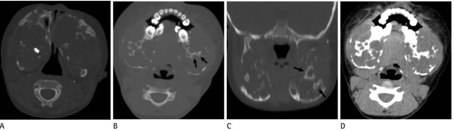
Fig. 2. Cherubism in a 6-year-old boy.
A-C. Axial and coronal CT images reveal expansile, multilocular cystic lesions containing internal sclerotic matrix (arrows) in the maxilla and mandible. Obstruction of the nasal lumen and remodeling of the bony orbit by the expansile maxilla is noted.
D. At soft-tissue window setting, these cystic areas contain low attenuation material.
A B C D
Fig. 1. A panoramic view shows expansile remodeling of the maxilla and mandible replaced by multilocular osteolytic lesions bilaterally.
Sparing of both mandibular condyles (arrows) is also noted.
Beomseok Sohn, et al
15
jksronline.org J Korean Soc Radiol 2014;70(1):13-15
REFERENCES
1. Jones WA. Familial multilocular cystic disease of the jaws.
Am J Cancer 1933;17:946
2. Grünebaum M, Tiqva P. Non familial cherubism: report of two cases. J Oral Surg 1973;31:632-635
3. Beaman FD, Bancroft LW, Peterson JJ, Kransdorf MJ, Mur- phey MD, Menke DM. Imaging characteristics of cherubism.
AJR Am J Roentgenol 2004;182:1051-1054
4. Mangion J, Rahman N, Edkins S, Barfoot R, Nguyen T, Sig- urdsson A, et al. The gene for cherubism maps to chromo- some 4p16.3. Am J Hum Genet 1999;65:151-157
5. Cornelius EA, McClendon JL. Cherubism--hereditary fibrous dysplasia of the jaws. Roentgenographic features. Am J Roentgenol Radium Ther Nucl Med 1969;106:136-143 6. Meng XM, Yu SF, Yu GY. Clinicopathologic study of 24 cases
of cherubism. Int J Oral Maxillofac Surg 2005;34:350-356 7. Hamner JE 3rd, Ketcham AS. Cherubism: an analysis of
treatment. Cancer 1969;23:1133-1143
8. Jones WA, Gerrie J, Pritchard J. Cherubism--familial fi- brous dysplasia of the jaws. J Bone Joint Surg Br 1950;32- B:334-347
9. Faircloth WJ Jr, Edwards RC, Farhood VW. Cherubism in- volving a mother and daughter: case reports and review of the literature. J Oral Maxillofac Surg 1991;49:535-542 10. Gorlin RJ, Goltz RW. Multiple nevoid basal-cell epithelioma,
jaw cysts and bifid rib. A syndrome. N Engl J Med 1960;
262:908-912 radiologic manifestations, cherubism can be quite easily distin-
guished from other conditions involving facial bones. Various conditions such as fibrous dysplasia, giant cell lesions, and brown tumors are not usually familial, present at later ages, do not show the typical facial features of swollen cheeks and upward turning of the eyes, and do not involute during puberty. These conditions rarely show the symmetrical involvement seen in cherubism. In addition, a few odontogenic lesions including am- eloblastomas and keratocystic odontogenic tumors should be included in the differential diagnosis. Among these, basal cell nevus syndrome presenting as multiple keratocystic odontogen- ic tumors in the posterior body of the mandible can mimic cherubism on imaging, but this syndrome usually also causes skin lesions or rib abnormalities without the characteristic facial swelling (10).
Cherubism is a very rare disease of the jaw in pediatric pa- tients and shows the characteristic imaging findings of multiloc- ular, expansile, radiolucent lesions involving the mandible and/
or maxilla bilaterally. This condition can be diagnosed based on its clinical and radiologic findings, and, thus, it is important for radiologists and clinicians to recognize these typical manifesta- tions when evaluating children with facial and dental problems.
CT imaging plays an important role in diagnosing as well as as- sessing the extent of the lesions, may prevent unnecessary inva- sive procedures such as biopsies or surgeries, and can aid in family screening.
6세 남아에서의 산발성 천사얼굴증 컴퓨터단층촬영 소견1
손범석
1· 김진아
1· 신나영
1· 김창훈
2천사얼굴증은 드문 유전성 질환으로서 소아의 하악을 침범하는 질환이다. 이 질환은 컴퓨터단층촬영에서 다발성, 확장성 의 낭성 병변이 상악과 하악을 양측으로 침범하는 특이한 소견을 보이며, 이는 천사얼굴증을 진단할 수 있는 중요한 소견 이다. 우리는 한국 6세 남아에서 발견된 특징적인 영상학적 소견을 보이는 산발성 천사얼굴증 증례를 보고하고자 한다.
연세대학교 의과대학 세브란스병원 1영상의학교실, 2이비인후과학교실