서 론
급성백혈병에서의 세포유전학적 검사는 질환의 진단, 치료방 침의 결정 및 예후판정에 매우 중요한 정보를 제공한다[1-6]. 통 상적인 염색체 핵형 분석은 염색체 이상을 규명하는 기본적인 방법이지만, 분열 중인 세포에서만 분석이 가능하고, 표지 염색
371 DOI 10.3343/kjlm.2009.29.5.371
371 371 371 Received :April 24, 2009 Manuscript No :KJLM09-055 Revision received :September 5, 2009
Accepted :September 10, 2009
Corresponding author :Sun-Hee Kim, M.D.
Department of Laboratory Medicine & Genetics, Samsung Medical Center, Sungkyunkwan University School of Medicine, 50 Irwon-dong, Gangnam-gu, Seoul 135-710, Korea
Tel : +82-2-3410-2704, Fax : +82-2-3410-2719 E-mail : [email protected]
급성백혈병의 유전적 이상 검출을 위한 형광제자리부합 프로필 검사의 임상적 유용성
Clinical Utility of Fluorescence in-situ Hybridization Profile Test in Detecting Genetic Aberrations in Acute Leukemia
Suk Ran Kim, M.D., Hee-Jin Kim, M.D., and Sun-Hee Kim, M.D.
Department of Laboratory Medicine & Genetics, Samsung Medical Center, Sungkyunkwan University School of Medicine, Seoul, Korea
김석란∙김희진∙김선희
성균관의대 삼성서울병원 진단검사의학과
Background : Cytogenetic abnormalities are one of the most reliable prognostic factors in acute leukemia. Combination of conventional chromosome analysis (CCA) and FISH provides higher sensitivity in detecting these genetic abnormalities, and it is effective to apply several FISH probes as a profile test. The objective of this study was to investigate the utility of FISH profile analyses in the initial diagnosis of acute leukemia.
Methods : Two hundred and forty one de novo acute leukemia patients diagnosed from January, 2002 to November, 2007 were included. For acute lymphoblastic leukemia profile test, FISH probes for BCR/ABL, TEL/AML1, MLL gene rearrangement and CDKN2A deletion were used. For acute myeloid leukemia profile test, probes for AML1/ETO, MLL and CBFβgene rearrangement were used. The results of CCA and FISH profile tests were collected, and the positive rates were com- pared.
Results : ALL FISH profile tests revealed additional genetic aberrations not detected by chromo- some analysis in 48.6% (67/138) of cases, including those with normal karyotypes or no mitotic cells (37%, 51/138). Among these 51 cases, TEL/AML1 abnormalities were detected in 44.3%, followed by the abnormal CDKN2A signal (24.6%) and hyperdiploidy (18.0%). AML FISH profile tests revealed additional genetic abnormalities in 7.8% (8/103) of cases.
Conclusions : FISH analysis as a profile test detected additional genetic aberrations in a signifi- cant proportion of acute leukemia, and was effective especially in detecting cryptic translocations, submicroscopic deletions and complex karyotypes. Our study supports the need to incorporate FISH profile test at initial work up in acute leukemia. (Korean J Lab Med 2009;29:371-8)
Key Words : Acute leukemia, Cytogenetic analysis, Fluorescence in-situ hybridization
핵형 분석의 기술적 한계점을 보완하기 위해 형광제자리부합법 (FISH)을 이용하는 것이 유용하다[8, 9]. FISH 검사는 민감도 와 해상도가 높고, 분열 중이 아닌 세포에서도 검사가 가능하며, 혈액뿐 아니라 골수 및 림프절, 조직에도 시행할 수 있어 백혈병 을 비롯한 많은 종양의 진단에 이용된다. 급성 백혈병은 그 계열 (lineage) 및 아형(subtype)에 따라 흔히 일어나거나 예후와 밀 접한 관계가 있는 특징적인 유전적 이상들이 존재하므로, 이러 한 변이들을 밝히기 위한 몇 개의 FISH 탐색자(probe)들의 조 합을 프로필(profile)화하여 검사하는 것이 효율적이다[10].
본 연구에서는 이러한 FISH 프로필 검사의 임상적 유용성을 알아보기 위해, 급성 백혈병의 초진 시 시행한 세포유전학적 검 사결과들을 후향적으로 분석하여 FISH 프로필 검사 적용 시의 검출률을 확인하고자 하였다.
대상 및 방법
1.연구 대상 및 진단기준
2002년 1월부터 2007년 11월까지, 급성 백혈병이 의심되어 골수 염색체 검사 및 FISH 프로필 검사가 의뢰된 환자들을 대 상으로 하였다. 검사는 환자의 치료 시작 전 골수 검체로 시행 되었다. 백혈병의 재발이나, 환자가 이전에 다른 혈액종양으로 치료받은 적이 있었던 경우는 제외하고, 치료효과와 예후를 확 인하기 위해 1년 이상 본원에서 추적 검사를 받았던 경우만을 포함시켰다. 이로써 총 241명의 환자들이 연구에 포함되었고, 급성 림프모구백혈병(ALL) FISH 프로필 검사를 시행한 환자가 138명, 급성 골수성백혈병(AML) FISH 프로필 검사를 시행한 환자가 103명이었다. 급성백혈병의 진단과 분류는 WHO 진단 기준에 근거하였다[11].
Sodium heparin 용기에 채취된 골수를 직접법, 24시간 단기 배양법 및 methotrexate 처리법을 선택적으로 활용하여 배양하 였다. 배양 후에는 세포를 수확, 고정한 뒤 GTG-분염법(GTG- banding, G-bands by Trypsin using Giemsa)을 시행하여 현미경으로 관찰하였다. 핵형 분석 시 총 20개의 세포분열 중기 세포(metaphase cell)를 계수하였으며, 결과의 표기는 Interna- tional System for Human Cytogenetic Nomenclature (ISCN) 1995 [12] 또는 2005 [13] 기준에 따랐다.
3.
형광제자리부합법(
FISH)
ALL FISH 프로필 검사는BCR/ABL, TEL/AML1및MLL 재배열(rearrangement), 그리고CDKN2A결손을 검출하는 탐 색자들로 구성되어 있으며 AML FISH 프로필 검사에는AML1/
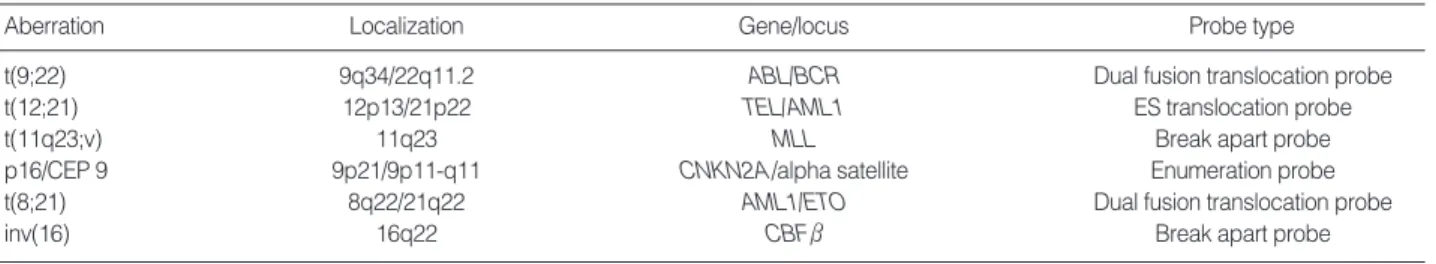
ETO, MLL과CBFβ유전자 재배열 검사가 포함되었다(Table 1).
FISH 프로필 항목의 구성은 세포유전학적 이상의 동반빈도가 높은 것을 우선으로 선택하였으며, AML의 경우 2001 WHO 진 단기준[11]에 따른“AML with recurrent genetic abnormal- ities”진단을 위해 필요한 항목으로 구성하였다. 이 조합은 본 연구에 포함된 환자들 모두에게 동일하게 시행되었다. 검사의 시행과정은 다음과 같다. 부합(hybridization) 영역이 표시된 Probe-on plus slide (Fisher Scientific, Pittsburgh, PA, USA)에 세포 부유액을 떨어뜨린다. 37℃로 가온한 2×Stan- dard saline citrate (SSC)/0.1% Nonyl phenoxylpolyetho- xylethanol-40 (NP-40)에 30분간 담근 후 실온에서 70%, 85%, 100% 에탄올 순으로 탈수, 건조시켰다. 탐색자 0.5 μL를 부합 완충액 3.5 μL, 3차 증류수 1 μL와 혼합한 후 원침하여 전 처리가 끝나면 HYBrite denaturation/hybridization system (Vysis, Abbott Molecular Inc., Downers Grove, USA)에 올 려놓은 슬라이드에 이를 떨군 후 70℃에서 3분간 변성시키고
All aberration was detected by dual color set and Abbott Vysis.
Abbreviations: ES, extra signal; CEP 9, chromosome 9 enumeration probe.
Aberration Localization Gene/locus Probe type
t(9;22) 9q34/22q11.2 ABL/BCR Dual fusion translocation probe
t(12;21) 12p13/21p22 TEL/AML1 ES translocation probe
t(11q23;v) 11q23 MLL Break apart probe
p16/CEP 9 9p21/9p11-q11 CNKN2A/alpha satellite Enumeration probe
t(8;21) 8q22/21q22 AML1/ETO Dual fusion translocation probe
inv(16) 16q22 CBFβ Break apart probe
Table 1. List of the probes used for fluorescence in-situ hybridization analysis in this study
다시 37℃에서 20시간 동안 부합되도록 하였다. 부합과정 후 SSC/NP-40을 이용하여 세척한 뒤 슬라이드를 건조시키는데, 이때 4,6-diamidino-2-phenylindole (DAPI)로 대조 염색을 시행하고 형광현미경으로 관찰하였다. 최소 200개 이상의 간기 세포(interphase cell)를 관찰하였다.
4.
연구 결과의 분석
연구결과는 분석을 위해 형광제자리부합 검사결과를 기준으 로 I, II군으로 크게 분류하였는데, I군은 형광제자리부합 검사 결과가 정상이거나 염색체 검사결과에 합당한 경우이고, II군은 형광제자리부합 검사로 추가 유전적 변이를 검출한 군으로 하였 다. 이것을 다시 5개의 카테고리(category)로 분류하여 표시하 였다. 카테고리 1은 염색체 검사상 정상이거나 분열세포가 없으 면서 FISH 검사가 정상이었던 경우, 카테고리 2는 두 검사방법 으로 발견한 유전적 이상에 차이가 없었던 경우, 그리고 카테고 리 3은 염색체 검사로만 이상을 발견한 경우로 하였고, 이들은 I군에 해당하는 것으로 분류하였다. II군 환자들은 FISH 검사를 통해 추가로 주요한 유전적 변이가 발견된 경우(카테고리 4)와 염색체 검사상 정상 핵형, 혹은 분열세포가 없었던 경우(카테고 리 5)로 세분하였다. 대상환자들의 치료효과, 생존여부 등의 임상 소견은 전자의무기록 조회를 통해 정리하였다. 완전 관해(com- plete remission, CR)는 치료 후 골수검사상 골수모세포(blast) 가 5% 이하이면서 잔존 질병(residual disease)이 없는 상태로 정의하였다. Event-free survival은 진단 시점부터 질병의 재 발, 치료 실패 또는 사망 등의 event가 나타난 시점까지로 하였 다. 통계처리는 MedCalc (version 9.4.2.0, MedCalc Software,
Mariakerke, Belgium)를 이용하였다. 범주형 자료(categorical data)의 비교는 Fisher’s exact test를 사용하고, 생존율은 log- rank test로 비교하였다.
P
value는 0.05를 기준으로 하였다.결 과
1.
급성 림프모구백혈병 형광제자리부합 프로필(
FISH, ALL profile)
ALL FISH 프로필 검사를 시행한 138명의 환자들의 임상적 분류는 Table 2와 같다. 18세 미만 환자들이 100명으로 72.5%
를 차지하였고, 환자들의 나이의 중앙값(median)은 15세(범위 0-62세)였다. Table 3은 환자들의 염색체 검사와 FISH 프로필 검사의 양성건수를 비교한 표이다. 검사결과들은 연구방법에서 기술한 바와 같이 분류하였다. 138명의 환자에서 염색체 검사상
“분열 중인 세포를 찾을 수 없음(no mitotic cells)’’으로 보고한 경우가 18명, 정상 핵형을 보였으나 중기세포가 20개 이하였던 경우는 30명으로, 34.8% (48/138)의 환자에서 염색체 검사결과 가 불충분하다고 할 수 있었다. 이에 반해 FISH 검사로 유전적 변이를 검출한 제II군에 해당하는 경우는 총 67명(67/138, 48.6
%)이었고, 이 중 16명은 카테고리 4로 분류되었고, 51명(51/138, 37.0%)이 카테고리 5에 해당하였다.
카테고리 4에 해당하는 경우는 염색체 검사상 정상 핵형을 보 인 것은 아니었으나 염색체 검사로 검출하지 못한 이상이 FISH 프로필 검사를 통해 발견된 경우들이었다. 추가로 발견된 세포 유전학적 이상으로는 16명의 환자들에게서 발견된 총 18건의 이상 중 잠재성
CDKN2A
결손이 8건(8/18, 44.4%)으로 가장 Table 2. Characteristics of acute leukemia cases where fluorescence in-situ hybridization profile test were appliedParameter/
immunophenotype
B-cell type (109)*
T-cell type (14)*
Biphenotype/
mixed lineage (15)*
Parameter/
classification
AML with recurrent genetic abnormalities (29)*
AML, not otherwise specified (65)*
Others (AML with MD,
etc.) (9)*
Age (yr)
Children (age<18)
<1 3 0 0
1-10 61 2 5
>10 24 3 2
Adult
<65 21 9 8
Gender
Male 53 9 8
Female 56 5 7
Age (yr)
Children (age<18) 14 10 (M7:4) 1
Adult
<55 9 36 6
>55 6 19 2
Gender
Male 16 39 7
Female 13 26 2
ALL FISH profile test were applied AML FISH profile test were applied
*( ) Number of patients with the specific immunophenotypic classification.
Abbreviations: AML with MD, acute myeloid leukemia with multilineage dysplasia; M7, acute megakaryoblastic leuekemia.
많았고, 4건의
TEL/AML1
재배열을 포함하는TEL/AML1
이 상이 6건(6/18, 33.3%)이었다.Table 4는 카테고리 5에 해당하는 51명의 환자들의 FISH 검 사 결과를 정리한 것으로서, precursor B-cell ALL 환자 39명, precursor T-cell ALL 환자 5명, 그리고 acute leukemia of ambiguous lineage 환자 7명의 결과가 포함되었다. 관찰된 이상은 모두 61건이었는데, 2가지 이상의 항목에서 양성을 보 인 환자가 8명이었고, 한 환자는
MLL
break-apart signal,CDKN2A
결손,TEL
유전자 결손을 동시에 보였다.TEL/AML1
이상이 가장 흔하여, 27건(27/61, 44.3%)이 발견되었다. 변이 형태로는
TEL/AML1
재배열뿐 아니라 각 유전자의 결손이나 중복, 증폭이 발견되었다.TEL/AML1
이상은 소아 precursor B-cell ALL 환자에서 특징적으로 나타났는데,TEL/AML1
이 상 양성을 보인 소아 환자들은 모두 21명이었고, 대부분 10세 이 하(19/21, 90.5%)였다. 잠재성(cryptic)CDKN2A
유전자 결손 은 14명에서 발견되었다. 이 중 다른 이상 없이 단독CDKN2A
결손을 보인 환자들은 7명이었는데, 모두 항암화학요법 후 완전 관해에 도달하였고, 추적기간 동안 재발이나 치료실패는 관찰되 지 않았다. 과배수성(hyperdiploidy)이 발견되었던 경우는 모두 11명(18%, 11/61)으로, 이 중 10명인 소아환자들은 모두 항암화 학요법 후 완전관해에 도달하였고, 재발이나 치료실패는 관찰 되지 않았다.2.
급성 골수성백혈병 형광제자리부합 프로필(
FISH, AML profile)
AML FISH 프로필 검사를 시행한 103명의 환자들의 진단명 및 성별과 나이에 따른 분류는 Table 2와 같다. 진단 시 나이의 중앙값은 40세(범위 0-79세)였다. Table 3의 하단에 AML의 아형별로 염색체 검사와 FISH 프로필 검사의 양성 수를 비교하 였는데, 이 중 FISH 검사를 통해서만 유전적 이상이 발견된 환
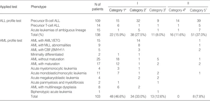
Applied test Phenotype N of
patients
I
Category 1* Category 2� Category 3�
II
Category 4� Category 5‖
ALL profile test Precursor B-cell ALL 109 15 32 9 14 39
Precursor T-cell ALL 14 6 1 1 1 5
Acute leukemias of ambiguous lineage 15 1 5 1 1 7
Total (%) 138 22 (15.9%) 38 (27.5%) 11 (8.0%) 16 (11.6%) 51 (37.0%)
AML profile test AML with AML1/ETO 15 14 1
AML with MLL abnormalities 9 8 1
AML with CBFβ/MYH11 5 3 2
Minimally differentiated 2 1 1
AML without maturation 25 18 1 5 1
AML with maturation 17 12 1 2 2
Acute myelomonocytic leukemia 4 3 1
Acute monoblastic/monocytic leukemia 11 7 1 2 1
Acute megakaryoblastic leukemia 4 2 2
Acute panmyelosis and myelofibrosis 2 1 1
AML with multilineage dysplasia 8 6 2
Biphenotypic acute leukemia 1 1
Total 103 48 (46.6%) 34 (33.0%) 13 (12.6%) 0 8 (7.8%)
Categories: 1*, CCA: normal or ‘no mitotic cells’/FISH profile test: normal; 2�, Compatible results of cytogenetic abnormalities between the two detec- tion methods; 3�, CCA: abnormal / FISH profile test: normal; 4�, Additional cytogenetic abnormalities were detected only by FISH profile test; 5‖, CCA:
normal or ‘no mitotic cells’/FISH profile test: abnormal.
Analyzed gene
N of abnormal cases (%)
Rear- range-
ment
Deletion Amplifi- cation
Extra copies related to aneuploidy
CDKN2A(p16) 15 (24.6) 14 1
TEL/AML1 27 (44.3) 13 10(TEL), 3 (AML1) 7(AML1) 1(AML1)
BCR/ABL 5 (8.2) 4 1(ABL)
MLL 3 (4.9) 3
Hyperdiploidy 11 (18.0)
Total 61* (100)
Table 4. Frequencies of the abnormalities identified by ALL FISH profile in 51 acute leukemia cases with normal karyotypes or without cytogenetic results (category 5)
*Number of patients harboring 2 abnormalities, 8; number of patients harboring 3 abnormalities, 1.
자들(제II군)은 8명으로 전체의 7.8% (8/103)에 해당하였다. 제 II군 환자 8명 중 4명은 최종진단명이“AML with recurrent genetic abnormalities”군으로 바뀐 경우였으며, 이들을 Table 5에 정리하였다. 다른 4명은“AML not otherwise categorized”
에 포함되었다. 카테고리 4에 해당하는 환자는 한 명도 없었다.
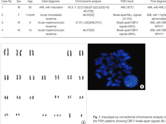
Table 5에서, t(8;21)(q22;q22)이 관찰되었던 첫 번째 환자는 염색체 검사상 45,X,-Y,add(2)(q32),del(8)(q22)[14]/46XY[6]
로 보고하고 AML with maturation으로 진단하였다가
AML1/
ETO
FISH 검사를 통해 복합 균형 전위(balanced complex translocation)을 가진 것을 파악할 수 있었다. 두 번째 환자는 염색체 검사상 정상 핵형을 보였으나, FISH 검사상MLL
break- apart signal이 35%의 세포에서 관찰되었다. Case 3 환자의 경우 염색체 검사상 trisomy 22만 발견되어 acute myelomo- nocytic leukemia로 진단되었던 환자인데, FISH 검사를 통해CBF
βcryptic break-apart signal이 관찰되어 최종 진단명을 수정하였다(Fig. 1). 제II군 환자 중“AML not otherwise cat-egorized”에 포함된 환자 4명은 염색체 검사상 정상 핵형을 보 이거나“no mitotic cells”로 보고된 경우로, FISH 프로필 검사 상
CBF
β,MLL
혹은ETO
triple signal이 관찰되어 이를 추적 검사에 이용할 수 있었다.AML1/ETO
이상은 총 15명(15/102, 14.7%)의 AML 환자에 서 발견되었다. 이 중 14명은 전위를 보였고, 9명은 성염색체 결 실과 같은 다른 이상을 동반하였다. 환자 15명 중 7명이 치료에 반응하지 않거나 재발되었으며, 염색체 검사상 정상 핵형을 보 인 AML 환자들에 비해 event-free survival에 유의한 차이는 없었다(P
=0.94).MLL
재배열은 9명(9/102, 8.8%)에서 관찰되 었고, 본 연구에 포함된 영아 AML 환자 5명 중 4명이MLL
이 상을 보였다.고 찰
본 연구에서, 20개의 중기세포가 모두 정상 핵형을 보였던 급
Case No. Sex Age Initial diagnosis Chromosome analysis FISH result Final diagnosis
1 M 55 AML with maturation 45,X,-Y, t(2;21;8)(q37;q22;q22)[14]/ AML1/ETO AML with AML1/ETO
46,XY[6]
2 F 1 month Acute monoblastic 46,XX[20] Break-apart MLL signals AML with 11q23(MLL)
leukemia (31.5%) abnormalities
3 M 6 Acute myelomonocytic 47,XY,+22[3]/46,XY[1] Break-apart CBFβ AML with CBFβ/
leukemia signals (83%) MYH11
4 M 15 Acute myelomonocytic 46,XY[20] Break-apart CBFβ AML with CBFβ/
leukemia signals (89%) MYH11
Table 5. Summary of the results of conventional chromosome analysis and FISH profile test of 4 cases of ‘‘AML with recurrent genet- ic abnormalities’’ and category 5
Fig. 1. Karyotype by conventional chromosome analysis (A) and the FISH patterns showing CBFβbreak-apart signals (B) shown in case 1.
A
B
용되었다.
ALL FISH 프로필 검사로 48.6%의 환자에서 염색체 검사로 는 발견하지 못한 유전적 이상을 발견할 수 있었다. 이것은 ALL 에서 흔히 잠재성 이상이 나타나기 때문이며[14, 15], 이 중
TEL/
AML1
이상은 특히 통상적인 염색체 검사로 진단하기 힘들어 FISH 검사를 이용하는 것이 중요하다[16].TEL/AML1
재배열 은 소아 precursor B-cell ALL에서 흔히 동반되는데, 본 연구 의 소아 ALL 환자 93명 중 25명(26.9%)에서 발견되었다. 이 변 이는 예후가 좋은 군으로 알려져 있어[17, 18], FISH 검사를 적 용하여 검출률을 높이는 것이 임상적으로 큰 의의가 있다 하겠다.염색체 검사상 정상 핵형을 보였거나 분열 세포가 없는 환자 에서 AML FISH 프로필 검사를 적용하여 유전적 이상을 발견 한 경우는 전체의 7.8%였다. 이 수치는 ALL에서보다는 빈도가 적지만, AML의 유전적 변이도 잠재적으로 일어나는 경우가 있 으며 이를 발견하는 것이 진단적으로 중요함을 보여준다[19].
AML1/ETO
재배열은 통상 t(8;21) 형태로 나타나는데 비해, t(2;8;21)(p37;q22;q22)을 보였던 환자의 경우 이 드문 복합 전 위를[20] 염색체 검사로 판독하기 어려워 FISH 검사가 매우 유 용하였다.AML1/ETO
변이를 가진 경우 보통 양호한 예후를 보이는 것으로 알려져 있으나[21, 22], 본 연구에서는 정상 핵형 을 보인 환자들에 비해 event-free survival에 있어 유의한 차 이를 보이지 않았고, 15명의 환자 중 7명이 치료에 실패하거나 재발하였다. inv(16)(p13q22)로 대표되는CBF
β유전자 재배열 은 비교적 흔하며, 결과에서 본 환자의 경우와 같이 trisomy 22와 동반되는 경향이 있으므로[23] FISH 검사로 확인하는 것 이 중요하겠다.MLL
의 구조적 이상은 AML의 5-8%, ALL의 7-10%에서 관찰되는 것으로 알려져 있는데[24], 본 연구에서는 AML 환자의 8.8%, ALL 환자의 6.5%에서MLL
재배열이 발 견되었다.MLL
재배열이 있었던 18명의 환자들 중 FISH 검사 로만 이를 발견한 경우는 22.2% (4/18)로, 보고된 바와 같이[25]FISH 검사에 의한
MLL
전위의 검출이 염색체 검사보다 민감 한 방법임을 확인할 수 있었다.MLL
유전자는 25개 이상의 다 양한 상대유전자와 재배열을 일으킬 수 있으므로[26], 이상 전 사체(transcript)를 검출하는 RT-PCR 방법으로는 그 변이를 찾기 어려울 수 있어 특히 FISH 검사가 유용하다.급성 백혈병의 유전적 변이를 검출하는데 있어 FISH 검사는 필수적인 항목이라고 할 수 있다. 질병의 정확한 진단과 예후 판 정을 위해 이는 초진 시 반드시 염색체 검사와 함께 적용되어야 할 것이다. 또한 각 FISH 검사항목의 양성률 및 유용성을 검증
요 약
배경 : 급성백혈병에서 유전적 변이는 가장 중요한 예후인자 중 하나이다. 이를 검출하는 방법으로써 통상적인 염색체 검사 에 형광제자리부합 검사를 추가하는 것이 검출의 민감도를 높일 수 있으며, 이때 몇 가지 탐색자들을 프로필화하여 검사하는 것 이 효율적이다. 이에 본 연구는 급성백혈병 환자의 진단 시 시행 된 형광제자리부합 프로필 검사의 유용성에 대해 알아보고자 하였다.
방법 : 2002년 1월부터 2007년 11월까지 삼성서울병원에서 새로 진단된 241명의 급성백혈병 환자를 대상으로 하였다. 급성 림프모구백혈병 프로필에는
BCR/ABL
,TEL/AML1
,MLL
재배 열 및CDKN2A
유전자 이상을 검사하는 탐색자들이 포함되었으 며 급성 골수성백혈병 프로필로는AML1/ETO
,MLL
및CBF
β 유전자 이상 검출용 탐색자들을 이용하였다. 환자들의 초진 시의 염색체 검사와 형광제자리부합 프로필 검사 결과를 비교하고, 각각의 검사를 통해 유전적 이상을 밝혀낸 빈도를 조사하였다.결과 : 급성 림프모구백혈병 형광제자리부합 프로필 검사를 시행한 환자들 중 48.6% (67/138)에서 염색체검사로는 발견하 지 못한 유전적 이상을 발견할 수 있었으며 이 중 37.0% (51/138) 는 염색체 검사상 정상이거나 분열 중인 세포를 확인할 수 없어 핵형 분석이 불가능한 경우였다. 이들 51명의 환자에서 발견된 유전적 이상으로는
TEL/AML1
이상이 44.3%,CDKN2A
이상 이 24.6%를 차지하였고 18%에서 과배수성을 보였다. 급성 골 수성백혈병 형광제자리부합 프로필 검사를 통해서만 유전학적 이상을 발견할 수 있었던 환자들은 7.8% (8/103)이었다.결론 : 본 연구에서, 급성백혈병의 진단 시 형광제자리부합 프로필 검사를 통해 추가 유전적 변이의 검출률이 유의하게 높 아짐을 확인하였고, 형광제자리부합 프로필 검사는 잠재적 유 전 이상, 복잡 핵형 및 드문 형태의 전위를 검출, 판독하는데 특 히 유용하였다. 따라서, 급성백혈병의 진단 시에는 초진 시에 형 광제자리부합 프로필 검사를 염색체 검사와 함께 시행해야 할 것으로 사료되었다.
참고문헌
1. Micallef-Eynaud PD, Eden OB, Grace E, Ellis PM. Cytogenetic ab- normalities in childhood acute lymphoblastic leukemia. Pediatr
Hematol Oncol 1993;10:25-30.
2. Faderl S, Kantarjian HM, Talpaz M, Estrov Z. Clinical significance of cytogenetic abnormalities in adult acute lymphoblastic leukemia.
Blood 1998;91:3995-4019.
3. Grimwade D, Walker H, Oliver F, Wheatley K, Harrison C, Harri- son G, et al. The importance of diagnostic cytogenetics on outcome in AML: analysis of 1,612 patients entered into the MRC AML 10 trial. The Medical Research Council Adult and Children’s Leukaemia Working Parties. Blood 1998;92:2322-33.
4. Slovak ML, Kopecky KJ, Cassileth PA, Harrington DH, Theil KS, Mohamed A, et al. Karyotypic analysis predicts outcome of prere- mission and postremission therapy in adult acute myeloid leukemia:
a Southwest Oncology Group/Eastern Cooperative Oncology Group Study. Blood 2000;96:4075-83.
5. Grimwade D, Walker H, Harrison G, Oliver F, Chatters S, Harrison CJ, et al. The predictive value of hierarchical cytogenetic classifica- tion in older adults with acute myeloid leukemia (AML): analysis of 1065 patients entered into the United Kingdom Medical Research Council AML11 trial. Blood 2001;98:1312-20.
6. Ribera JM, Ortega JJ, Oriol A, Granada I, Hernandez-Rivas JM, Par- ody R, et al. Prognostic value of karyotypic analysis in children and adults with high-risk acute lymphoblastic leukemia included in the PETHEMA ALL-93 trial. Haematologica 2002;87:154-66.
7. McGrattan P, Campbell S, Cuthbert R, Jones FG, McMullin MF, Humphreys M. Integration of conventional cytogenetics, compara- tive genomic hybridisation and interphase fluorescence in situ hyb- ridisation for the detection of genomic rearrangements in acute leu- kaemia. J Clin Pathol 2008;61:903-8.
8. Cox MC, Panetta P, Venditti A, Del Poeta G, Franchi A, Buccisano F, et al. Comparison between conventional banding analysis and FISH screening with an AML-specific set of probes in 260 patients.
Hematol J 2003;4:263-70.
9. Wolff DJ, Bagg A, Cooley LD, Dewald GW, Hirsch BA, Jacky PB, et al. Guidance for fluorescence in situ hybridization testing in hema- tologic disorders. J Mol Diagn 2007;9:134-43.
10. Sreekantaiah C. FISH panels for hematologic malignancies. Cyto- genet Genome Res 2007;118:284-96.
11. Jaffe ES, Harris NL, et al., eds. World Health Organization classifi- cation of tumours: pathology and genetics of tumours of haemato- poietic and lymphoid tissues. Lyon: IARC Press, 2001.
12. Mitelman F, ed. An international system for human cytogenetic
nomenclature. Basel: S Karger, 1995.
13. Shaffer LG and Tommerup N, eds. An international system for human cytogenetic nomenclature. Basel: S Karger, 2005.
14. Andreasson P, Hoglund M, Bekassy AN, Garwicz S, Heldrup J, Mitelman F, et al. Cytogenetic and FISH studies of a single center consecutive series of 152 childhood acute lymphoblastic leukemias.
Eur J Haematol 2000;65:40-51.
15. Perez-Vera P, Salas C, Montero-Ruiz O, Frias S, Dehesa G, Jarquin B, et al. Analysis of gene rearrangements using a fluorescence in situ hybridization method in Mexican patients with acute lymphoblas- tic leukemia: experience at a single institution. Cancer Genet Cyto- genet 2008;184:94-8.
16. Alvarez Y, Gaitan S, Perez A, Bastida P, Ortega JJ, Dastugue N, et al. ETV6/RUNX1 rearrangement in childhood B-precursor acute lymphoblastic leukemia with normal karyotypes or without cyto- genetic results. Cancer Genet Cytogenet 2004;152:77-80.
17. Loh ML, Silverman LB, Young ML, Neuberg D, Golub TR, Sallan SE, et al. Incidence of TEL/AML1 fusion in children with relapsed acute lymphoblastic leukemia. Blood 1998;92:4792-7.
18. Kempski H, Chalker J, Chessells J, Sturt N, Brickell P, Webb J, et al.
An investigation of the t(12;21) rearrangement in children with B- precursor acute lymphoblastic leukaemia using cytogenetic and molecular methods. Br J Haematol 1999;105:684-9.
19. Cuneo A, Bigoni R, Cavazzini F, Bardi A, Roberti MG, Agostini P, et al. Incidence and significance of cryptic chromosome aberrations detected by fluorescence in situ hybridization in acute myeloid leu- kemia with normal karyotype. Leukemia 2002;16:1745-51.
20. Zhang J, Liu Z, Shao H, Ma Y, Tong H, Wang Y. Laboratory study of a complex translocation t(2;8;21) (p12;q22;q22) in a patient with acute myelogenous leukemia. Leuk Lymphoma 2008;49:1925-8.
21. Miyamoto T, Nagafuji K, Akashi K, Harada M, Kyo T, Akashi T, et al. Persistence of multipotent progenitors expressing AML1/ETO transcripts in long-term remission patients with t(8;21) acute myel- ogenous leukemia. Blood 1996;87:4789-96.
22. Cho EK, Bang SM, Ahn JY, Yoo SM, Park PW, Seo YH, et al. Prog- nostic value of AML 1/ETO fusion transcripts in patients with acute myelogenous leukemia. Korean J Intern Med 2003;18:13-20.
23. Litmanovich D, Zamir-Brill R, Jeison M, Gershoni-Baruch R. Is inver- sion 16 a prerequisite and id trisomy 22 invariably associated with inversion 16 in AML-M4eo? Cancer Genet Cytogenet 2000;121:106.
24. Thirman MJ, Gill HJ, Burnett RC, Mbangkollo D, McCabe NR, Ko-
slocations. N Engl J Med 1993;329:909-14.
25. Kim HJ, Cho HI, Kim EC, Ko EK, See CJ, Park SY, et al. A study on 289 consecutive Korean patients with acute leukaemias revealed fluorescence in situ hybridization detects the MLL translocation
26. Meyer C, Schneider B, Jakob S, Strehl S, Attarbaschi A, Schnittger S, et al. The MLL recombinome of acute leukemias. Leukemia 2006;
20:777-84.