© The Korean Society of Plant Pathology
http://dx.doi.org/10.5423/RPD.2013.19.4.313First Report of Freesia sneak virus in Freesia spp. in Korea
Ju-Yeon Yoon
1, Youn-Jung Choi
2, Gug-Seoun Choi
3and Seung-Kook Choi
3*
1
Department of Horticulture and Landscape, Seoul Women’s University, Seoul 139-774, Korea
2
Department of Floral Science, National Institute of Horticultural and Herbal Science, Rural Development Administration, Suwon 441-440, Korea
3
Department of Horticultural Environment, National Institute of Horticultural and Herbal Science, Rural Development Administration, Suwon 441-440, Korea
(Received on September 30, 2013; Revised on October 29, 2013; Accepted on October 30, 2013)
In March, 2013, twenty symptomatic freesia plants (10 plants of cultivar Shiny Lemon and 10 plants of cultivar Shiny Gold), with striking virus-like symptoms were collected in Cheongju, Korea. The plants showed chlorotic, coalescing, interveinal, whitish, necrotic, mosaic, mottling or dark brown-to-purple necrotic spots on leaves. Freesia crude sap was directly analyzed by transmission electron microscopy, which potyvirus particles as well as long virus-like particles were detected. Total RNA extracts were analyzed for the infection of Freesia sneak virus (FreSV) by reverse transcription (RT)-PCR with primers specific to FreSV coat protein (CP) gene based on the sequences of FreSV isolates (GenBank No. GU071089, FJ807730 and DQ885455), showing 9 of 20 plants were infected. All 1305bp RT-PCR products were cloned and sequenced.
Comparisons of nucleotide and deduced amino acid sequences using BLAST and bioinformatics tools resulted in 99 to 100% sequence identity with FreSV isolates FOV, Virginia, and Italy, confirming FreSV in 9 symptomatic freesia plants. Of 9 determined cDNAs of FreSV isolates, sequences of 5 cDNA clones were identical (GenBank No. AB811437) and sequences of 4 cDNA clones were identical (GenBank No. AB811792).
To our knowledge, this is the first report of FreSV from Freesia spp. in Korea.
Keywords : Coat protein, Freesia, Freesia sneak virus, Identification, Olphiovirus, RT-PCR
Freesia is a small genus of southern African Iridaceae (subfamily Ixioideae), which has been familiar to horticulturists and valued by them for the beauty and fragrance of the flowers. Freesia was first grown in Europe in the mid-18
thcentury and become one of the most popular plants in horticulture in the last half of the 19
thcentury (Wang, 2006). Freesia flowers have an intense scent which is sometimes likened to citrus.
Freesias generally produce a very sweet, rich scent, although it can become more peppery in certain soil types, and the smell can be very pervasive. Appealing shapers of freesias make them suitable line flowering for any arrangement, and their wide range of color increases their versatility resulting that freesias are excellent cut flowers (Ao et al., 2013). The flowers are popular used for weddings and make fragrant additions to bouquets and body flowers. Freesias are known to be infected by Bean yellow mosaic virus (BYMV),
Freesia mosaic virus (FreMV), Cucumber mosaic virus, Tobacco rattle virus and ophioviruses (Brunt, 1995; Choi et al., 2010; Choi et al., 2013; Derks et al., 1987;
Kumar et al., 2009; Vaira et al., 2006). Among the viruses, the genus Ophiovirus consists of Citrus psorosis virus (CPsV), Ranunculus white mottle virus, Tulip mild mottle mosaic virus, Mirafiori lettuce bigvein virus (MiLBVV), Lettuce ring necrosis virus and Freesia sneak virus (FreSV) that is provisionally named (García et al., 1994; Martín et al., 2005; Roggero et al., 2000;
Sasaya et al., 2008; Sasaya and Koganezawa, 2006;
Vaira et al., 2003; Vaira and Milne, 2008; van der Wilk et al., 2002). FreSV has been identified as a new species, based on amino acid identity of inter-species coat protein (CP) sequences (Vaira et al., 2003; Vaira and Milne, 2008). The identity of CP amino acid sequences between ophioviruses ranges from 30 to 70%
(Navarro et al., 2005). Morphology of FreSV particles is indistinguishable from that characteristic of other ophiovirus particles (Vaira et al., 2006; Navarro et al., 2005), being naked filamentous nucleocapsids about 3 nm in diameter forming circularized structures of
*Corresponding author
Phone)+82-31-290-6236, Fax) +82-31-290-6259 Email) [email protected]
Note Open Access
different lengths. However, it is not clearly shown the fine morphology of FreSV particles under electronic microscopy due to difficulty of FreSV purification, so it remains to be determined the precise size and shape of FreSV particles. Although the complete genome sequence of FreSV has not been determined yet, FreSV shares somewhat common features with the members of the genus Ophiovirus that contain segmented negative- stranded RNA genomes.
Virus particles contain nearly equimolar amounts of RNA molecules of both polarities, which use negative and possibly ambisense coding strategies for replication and infection in plants (van der Wilk et al., 2002).
Based on the genome organization of MiLBVV, RNA1 is negative-sense and encodes two putative proteins with a molecular mass of approximately 263 and 25 KDa. The 263 KDa protein is a RNA-dependent RNA polymerase (RdRp) that contains the five conserved motifs of RdRp core module (Naum-Onganía et al., 2003). RNA2 is proposed to be ambisense and to encode two putative proteins with a molecular mass of approximately 10 and 55 KDa in the 5'-proximal region of the virion-sense strand and the virion-complementary sense-strand, respectively. A recent study clearly demon- strated that the 55 KDa was the movement protein using complementation assays with movement-deficient Tomato mosaic virus (Hiraguri et al., 2013). RNA3 is negative- sense and encodes CP (approximately the 49 KDa protein) which is a major component of thin filamentous particles. RNA4 is negative-sense and comprises two overlapping open reading frames in different reading frames. To date, presence of FreSV RNA4 molecule has not been proven experimentally. Natural transmission of olphioviruses is mediated by soil-borne zoospores of the fungus Olpidium brassicae or obligate root-inhabiting fungus Olpidium virulentus (Sasaya and Koganezawa, 2006; Vaira et al., 2011). Although FreSV was demon- strated to be soil-transmitted, the putative FreSV-trans- missible fungus was not identified. FreSV has been reported to infect two ornamental bulb plants, Freesia sp. (Iridaceae) and Lachenalia sp. (Hyacinthaceae) in Europe, South African and USA, respectively (Vaira et al., 2006; 2007; Verbeck et al., 2004). A few FreSV isolates were frequently detected from freesia plants showing necrotic or a variety of symptoms in leaves, the correlation between symptom inductions and FreSV infection has clearly not been validated yet. Since bulbous freesia cultivars are propagated vegetatively, it is highly possible that a large number of bulbs are infected by FreSV or other freesia-infecting viruses and/or mix infection. In this study, a few FreSV isolates were identified from freesia cultivars showing different symptoms and
the CP sequences of two FreSV Korean isolates were compared with those of other FreSV isolates reported previously.
In March 2013, twenty symptomatic freesia plants (10 plants of cultivar ‘Shiny Lemon’ and 10 plants of cultivar ‘Shiny Gold’), with virus-like symptoms similar to freesia leaf necrosis disease were collected by Dept.
Floral Science from a cut-flower nursery in Cheongju and the samples were forwarded for diagnostic analysis to the Virology Unit. To identify freesia-infecting viruses from the diseased chrysanthemum plant, total RNA was extracted from 1g of frozen (−80
oC) leaves of the infected freesia plants using Plant RNeasy
®Mini kit (Qiagen, USA), according to the manufacturer’s instruc- tions. One-step RT-PCR analysis was carried out to detect three major viruses in freesia plants, using FreMV CP- specific primers (FreMV-CP-For, 5'-GCAAACCAGCGC ACCAGAGCAACTTG-3'; FreMV-CP-Rev, 5'-TTACAT GTGACGTACACCCAACAG-3'), BYMV CP-specific primers (BYMV-CP-For, 5'-TCTGACCAAGAACAA CTCAATGCA-3'; BYMV-CP-Rev, 5'-TCTGACCAAGAA CAACTCAATGCA-3'), FreSV CP-specific primers (FreSV- CP-Forward, 5'-ATGTCTGGAAAATACTCTGTCAAG-3';
FreSV-CP-Reverse, 5'-TTAGATAGTGAATCCATAAGCT GCT-3'), based on the sequence of FreSV isolates from Freesia spp. (GenBank No. GU071089, FJ807730, and DQ885455). The thermo-cycling conditions were as follows: 60 min at 50
oC for reverse transcription (RT), 5 min at 95
oC (1 cycle), 94
oC, 30 s, 55
oC, 30 s and 72
oC, 1min (40 cycles), and a final extension at 72
oC for 7 min. RT-PCR product was analyzed in 1.2%
agarose gel and visualized after soaking in ethidium bromide solution. Then, RT-PCR product synthesized from the diseased freesia plants was directly cloned into the pCR4-TOPO
®vector (Invitrogen, USA) to determine sequences of the entire insert cDNA, using BigDye terminator cycle sequencing kit in both directions, according to manufacturer’s instructions (Applied Biosystems, USA). Both Nucleotide and amino acid sequence identities were calculated using the Jukes and Cantor index and genetic diversity at synonymous positions was estimated using the Hasegawa-Kishino- Yano model in the MEGA 5.0 Software (Tamura et al., 2011). Basically, phylogenetic tree analysis with FreSV CP sequences was constructed using Neighbor-Joining (NJ) with Jukes and Cantor index or Hasegawa-Kishino- Yano model in the MEGA 5.0 software, DNAMAN, and DNASTAR software. Bootstrap analyses with 1000 replicates were performed to evaluate the significance of the interior branches.
Freesia plants (cvs. Shiny Lemon and Shiny Gold)
showed chlorotic, coalescing, interveinal, whitish, necrotic,
mosaic, mottling or dark brown-to-purple necrotic spot symptoms on leaves (Fig. 1). Subsequently, freesia crude sap was analyzed by transmission electron microscopy, resulting that potyvirus particles were clearly observed from 7 of 20 plants (Fig. 2). Unusual virus-like particles that range approximately from 1000 nm to 3000 nm in length were observed from 8 of 20 plants (Fig. 2).
Morphology of virus-like particles is quite different from that of ophioviruses (García et al., 1994; Roggero et al., 2000; Vaira et al., 2011), though the long virus- like particles seem to be FreSV. In spite of purifying the virus-like particles several times, we failed to identify whether the unusual virus-like particles are a member of ophioviruses or authentic FreSV. It is noteworthy that the correlation between dark brown-to-purple necrotic spot symptoms and FreSV infection remains to be determined. In particular, it is interesting to determine whether the virus-like particles are responsible for the expression of different symptoms on freesia leaves.
BYMV, one of freesia-infecting viruses was demonstrated to cause leaf-yellowing in combination with corm
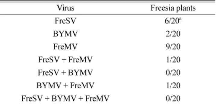
necrosis (Derks et al., 1987). Since virus examination of freesia cultivars in 2011 showed clearly that BYMV was the most common virus in freesia plants in Korea (Choi et al., 2013), total RNA extracts of 20 freesia samples were analyzed for the detection of BYMV by RT-PCR with primers specific to BYMV CP gene (Choi et al., 2013). The diagnostic 822-bp fragment was amplified from 2 of 20 plants. Sequencing of the clones containing 822-bp cDNA confirmed that the viruses were identified as BYMV. Subsequently, RT-PCR analysis for FreMV that is one of causal viruses in freesia showed the diagnostic 833-bp fragment from 6 of 20 plants (Choi et al., 2010). Cloned sequence from these plants was identified as FreMV (Table 1). Similarly, the CP genes of FreSV using the same total RNA samples were amplified by RT-PCR and cloned from 9 of 20 symptomatic freesia plants using primers specific to FreSV CP gene. Table 1 summarizes the results obtained from RT-PCR analyses of the three freesia infecting viruses. Neither double infection (FreSV + BYMV) nor triple infection (FreSV + BYMV + FreMV) was detected from the freesia samples tested (Table 1).
Results of virus identification in this study are different from the results surveyed previously (Choi et al., 2013).
For instance, BYMV isolates were all detected from a total of 50 freesia cultivars examined, but 2 BYMV isolates were detected from 20 freesia samples in this study (Table 1). It remains to be determined whether this difference is due to fewer samples than before or distribution of viruses in freesia cultivars has been currently changed.
To determine the variability of FreSV CP genes in our samples, RT-PCR fragments of FreSV isolates from 9 freesia plants were cloned and sequenced. All cDNA inserts in the clones were 1305 bp long and the CP gene encodes a putative product of approximately 48.5 kDa.
The resulting nucleotide and deduced amino acid Fig. 1. Symptoms on leaves of freesia plants. Chlorotic,
coalescing, interveinal, whitish, necrotic, mosaic, mottling or dark brown-to-purple necrotic spot symptoms are shown on freesia leaves used in this study.
Fig. 2. Electron micrographs of potyvirus and olphiovirus-like particles negatively stained in uranyl acetate from a crude extract of freesia leaves. Potyvirus particles that are approximately 780 nm in length are indicated by blue arrows and ophiovirus-like particles that are 1000−3000 nm in length are shown by red arrows. Bars indicate 500 nm long.
Table 1. Identification of freesia viruses using RT-PCR analysis in freesia plants
Virus Freesia plants
FreSV 6/20
aBYMV 2/20
FreMV 9/20
FreSV + FreMV 1/20
FreSV + BYMV 0/20
BYMV + FreMV 1/20
FreSV + BYMV + FreMV 0/20
aNumber of plants that show positive reactions/number of plants assayed.