First isolation report and molecular phylogenetic characteristics of Raphidiopsis raciborskii in South Korea
10
0
0
전체 글
(2) Isolation of Unrecorded Harmful Algae R. raciborskii in Korea ∙. 계로 분포 영역을 확장하였다(Padisák, 1997; Antunes et al.,. 351. 명의 탈수 및 설사 환자가 발생하였던 것으로 보고된 바 있다.. 2015). 현재는 대부분의 대륙 및 중국, 일본을 포함한 아시아. R. raciborskii은 국외에서 유입된 외래종으로, 일본이나 중. 등 여러 국가에서도 관찰되고 있는 것으로 보고되고 있으며,. 국에서 관찰되는 동일 종들은 조류 독소를 생산할 수 있는 것. 1935년과 1995년에 각각 일본과 중국에서 분포가 관찰된 바. 으로 알려져 있다(Zarenezhad et al., 2012). 또한, 지구온난화. 있다(Wilk-Woźniak et al., 2016). 이렇듯 가까운 주변국가에. 와 같은 환경변화에 의해 국내 수계에서도 발생하는 것으로. 서 비교적 이른 시기에 발견되었다는 점에서 국내에도 최초. 알려져 있으며, 국외 사례와 같이 조류 독소 등으로 인한 피해. 현미경 관찰을 통해 분포가 보고된 2017년보다 훨씬 이전에. 를 유발할 가능성이 있다. 특히, 2012년 수행된 낙동강 수계 내. 전파되었을 것으로 추정된다(경기도보건환경연구원 물환경. 남조류 유전자 분석 연구 결과, 비록 현미경을 통해 확인되지. 연구부, 2019).. 는 않았지만, 낙동강 수계 내에서 Raphidiopsis (Cylindrosper-. R. raciborskii는 자연 환경에서 안정적으로 유지되는 사상. mopsis) 종이 소수이지만 유전자 단계에서의 검출되었던 사. 체(trichome)의 말단세포(apical cell), 이형세포(heterocyst),. 례가 보고된 바 있다(국립환경과학원 낙동강물환경연구소.. 휴면포자(akinete) 등의 형태적 특징을 바탕으로 동정이 가능. 2013). 본 연구를 통해 동정 된 R. raciborskii GIHE 2018는. 한 것으로 알려져 있다(Komárek and Kling, 1991; Saker and. 2017년 인천 남동구 소재의 해오름 공원 내 저수지의 과산화. Neilan, 2001). 하지만 R. raciborskii은 환경변화에 따라 형태. 수소를 이용한 조류 제거 사업 제거효율 평가 과정에서 최초. 가 변할 수 있으며, 형태가 다른 경우에도 분자유전학적으로. 현미경 관찰을 통해 발견된 남조류이며, 같은 해 경기도보건. 매우 유사해서 종의 구분이 어려운 경우가 많다(Saker et al.,. 환경연구원에서 실시한 경기도 내 중점관리 저수지 서식중인. 1999; Chonudomkul et al., 2004).. 식물플랑크톤 조사 과정 중 의왕 소재의 왕송 저수지 및 화성. 각 대륙에서 발견된 R. raciborskii의 분자유전학적 연관성. 시 소재 저수지 내에서도 관찰된 바 있다(경기도보건환경연. 과 이동경로에 대한 여러 선행연구들이 수행된 바 있으며, 이. 구원 물환경연구부, 2019). 이를 통해, 현재 R. raciborskii가 국. 에 대한 몇 가지 가설이 제시된 바 있지만 아직까지 논란이 지. 내에서 전국적인 분포를 보이고 있을 것으로 추정되고 있다.. 속되고 있다(Antunes et al., 2015; Wilk-Woźniak et al., 2016).. 본 연구의 목적은 일차적으로 국내 수계에서 최초로 관찰. 최근 Moreira (2011) 연구진의 연구결과에 의하면, 아시아를. 된 R. raciborskii를 현미경 관찰을 통해 분리, 순수배양(unialgal. 포함한 다섯 대륙에서 관찰된 R. raciborskii 종들을 대상으로. culture)하는 것이다. 이후 분리한 균의 정확한 종 동정을 위해. 4개 유전자(16S rRNA, ITS longer spacer, ITS shorter spacer,. 배양 과정의 형태적 특성을 모니터링 하는 것이다. 더불어 3가. rpoC1)를 이용한 분자계통학적 연구를 통해, R. raciborskii는. 지 종류(16S rRNA, cpcBA-IGS, 그리고 nifH)의 유전자를 대. 발견되는 대륙 별로 (1) 유럽, (2) 아프리카 ․ 아메리카, (3) 아시. 상으로 계통분류학적 분석을 통해 순수 분리한 남조류의 분자. 아 ․ 호주의 세 개의 그룹으로 분류될 수 있으며, 아시아 ․ 호주. 유전학적 분류체계 분석 및 분자계통학적 특성을 규명하는 것. 그룹은 아프리카 ․ 아메리카 그룹에 비해, 유럽에서 발견된 종. 이다.. 들과 보다 가까운 유연관계를 형성하고 있는 것으로 알려져 있 다(Moreira et al., 2011). 이들의 분포 연구가 주목받고 있는 이. 재료 및 방법. 유는 R. raciborskii가 조류독소를 생성하는 유해 남조류로 알 려져 있기 때문이다. 특히 아시아 국가들 중 일본(Zarenezhad et al., 2012), 태국(Chonudomkul et al., 2004; Zarenezhad et. 현장 특성 및 개체관찰. al., 2012), 중국(Lei et al., 2014), 그리고 오세아니아의 호주. 2018년 8월 인천 남동구 소재의 해오름공원 내 저수지. (Saker et al., 1999; Schembri et al., 2001) 및 뉴질랜드(Wood. (37.38 N 126.72 E)로부터 표층수 시료를 채취하였다. 저수지. and Stirling, 2003)에서 관찰되는 종들은 간 독소인 cylindro-. 는 간척지에 인공적으로 조성되어 있었으며, 290 m × 170 m. spermopsin을 생산하는 것으로 보고된 바 있다. 또한, 남미의. 크기의 직사각형 형태로 수심은 약 1.5 m, 저수용량은 60,000. 브라질에서 관찰되는 R. raciborskii 종들은 신경독소인 para-. t이었다. 채취된 시료로부터 사상체 말단에 위치한 이형세포. lytic shellfish-poisoning (PSP)를 생산할 수 있다고 알려져 있. 가 특징인 R. raciborskii를 위상차 현미경(Carl Zeiss)을 통해. 다(Lagos et al., 1999). Griffiths과 Saker (2003)의 연구에 의하. 관찰하였다.. 면, 1979년 호주 퀸즐랜드(Queensland)의 Palm island에서 R. raciborskii 종에 의해 생산된 cylindrospermopsin에 의해 148 Korean Journal of Microbiology, Vol. 55, No. 4.
(3) 352. ∙ Jeong et al.. 분리․ 배양 및 분자유전학적 분석. primer 별 PCR 증폭을 위한 온도 및 시간 조건은 다음과 같다.. R. raciborskii 균주의 분리 ․ 배양을 위해, 현미경(Axiotop2 plus)으로 관찰하면서, 마이크로 피펫을 이용하여 R. raciborskii의 사상체를 분리, Jaworski’s (JM, Thompson et al., 1988) 배지(기산바이오)에 접종한 후, 27°C에서 광주기(day:night) 16:8의 조건으로 배양하였다. 순수배양을 위해, 증식이 관찰된 배양용기로부터 배양체를 수시로 분리하여, 새로운 배지에 접 종하는 과정을 반복 수행하였다. 현미경 관찰을 통해 충분한 개체수의 순수배양체가 확인된 경우, 원심분리기를 이용하여 1000 rpm에서 20분간 배양체를 농축하였으며, xanthogenatesodium dodecyl sulfate 완충액(Tillett and Neilan, 2000)를 이 용하여 genomic DNA를 추출하였다. 간략하게, 성장 중기에 서 후기 사이의 배양체 1 ml를 원심분리하여 cell pellet을 형성 시킨 후, 50 ml의 TER (10 mM Tris-HCl, pH 7.4; 1 mM EDTA, pH 8; 100 µg/ml of RNase A) 용액을 첨가하여 pellet을 재부유 한다. 재부유된 세포들을 1.5-ml microcentrifuge 튜브에 나누 어 담은 후, 750 ul의 바로 제작된 XS buffer (1% potassium ethyl xanthogenate [Fluka]; 100 mM Tris-HCl, pH 7.4; 20 mM EDTA, pH 8; 1% sodium dodecylsulfate; 800 mM ammonium acetate)를 첨가하여 약하게 혼합하였다. 혼합액의 상태 변화 에 따라 혼합액이 담긴 튜브를 70oC 온도 조건에서 10분에서 120분간 배양한다. 이후, 튜브를 10초간 강하게 혼합한 후, 30 분간 ice 상에서 배양한다. 원심분리 과정(14,000 rpm에서 10. cpcBA-IGS primer의 경우, 95oC에서 5분간 초기 denaturation 을 수행하였으며, 이후, 95oC에서 30초간 denaturation, 56oC 에서 30초간 annealing, 72oC에서 1분간 extension을 30회 반복 수행하였다. nifH primer의 경우, 95oC에서 3분간 초기 denaturation을 수행하였으며, 이후 95oC에서 30초간 denaturation, 53oC에서 30초간 annealing, 72oC에서 30초간 extension을 30회 반복 수행하였다. 마지막으로 16S rRNA 유전자 primer의 경우, 95oC에서 3분간 초기 denaturation을 수행하였 으며, 이후 95oC에서 30초간 denaturation, 53oC에서 30초간 annealing, 72oC에서 30초간 extension을 30회 반복 수행하였 다. PCR 증폭의 최종 단계에서, 72oC에서 5분간 최종 extension 과정을 수행하였다. PCR 산물들은 agarose gel을 이용한 전기영동을 통해 확인하였으며, 이후 염기서열 분석에 사용되 었다(Macrogen). 확보된 염기서열은 Blastn 소프트웨어(Altschul et al., 1997) 를 이용하여 NCBI 데이터베이스(http://www.ncbi.nlm.nih. gov/taxonomy)의 유사 유전자와 염기서열이 일치도를 비교 하여 동정하였다. 계통수 분석은, ClustalW (Thompson et al., 1994)로 multiple alignment를 수행한 후, MegaX 프로그램 (Kumar et al., 2018)을 이용하여 유전자 별 최적의 모델을 선 별한 후, maximum likelihood 분석 방법을 적용하여 계통수를 작성하였다.. 분간)을 통해 cell 부산물을 제거한 후, 750 ul의 이소프로필알 코올이 첨가된 새로운 튜브에 상등액을 옮긴다. 이후, 상온에. 결과 및 고찰. 서 10분간 배양한 후, 원심분리 과정(12,000 × g에서 10분간) 을 통해 DNA를 침전시킨다. 추출된 DNA은 70% 에틸알코올 로 1회 세척하고, 건조한 후, 최종적으로 100 ml의 TE 용액(10 mM Tris-HCl, pH 7.4; 1 mM EDTA, pH 8)을 첨가하여 추출한 다. 추출된 DNA는 NanoDrop 2100 spectrophotometer (Thermo Fisher Scientific, WI)을 이용해 농도를 정량하였다. 피코시아닌 유전자(cpcBA-IGS), 질소고정 유전자(nifH, Denitrogenase reductase) 및 16S rRNA 유전자는 Table 1에 제 시된 프라이머(primer) 조합을 사용하여 증폭되었으며, 각. 형태적 특성을 통한 종 동정 본 연구에서는 인천지역 저수지로부터 국내 최초로 분포가 발견된 R. raciborskii의 정확한 동정을 위해, 위상차 현미경을 통한 형태적 특징 관찰과 함께, 분자유전학적 분류법을 적용 하여 동정 연구를 수행하였다. 남조류 발생 시기인 8월의 수질 은, 수온 28~30°C, TOC 13.1~23.9 mg/L, 탁도 27.6~42.3 NTU, TN 1.811~3.258 mg/L, TP 0.058~0.133 mg/L, 클로로필a 19.8~. Table 1. Primers used in this study Target gene. Name. Product size (bp). Sequence (5' to 3'). Annealing Temp. (°C). References. cpcBA-IGS Cyanobacteria. PCbF PCaR. 685. GGCTGCTTGTTTACGCGACA CCAGTACCACCAGCAACTAA. 56. Neilan et al. (1995). Nitrogenase reductase. nifHF nifHR. 382. CGTAGGTTGCGACCCTAAGGCTGA GCATACATCGCCATCATTTCACC. 53. Gugger et al. (2005). 16S rRNA gene. 27F 1516F. 1489. AGAGTTTGATCCTGGCTCAG ATCCAGCCACACCTTCCGG. 60. Neilan et al. (1997), Yilmaz et al. (2008). 미생물학회지 제55권 제4호.
(4) Isolation of Unrecorded Harmful Algae R. raciborskii in Korea ∙. 333.0 mg/m3으로 변화가 매우 큰 것으로 관찰되었으며, 시료 4. 353. (A). 5. 의 색도는 연황색을 띄며 R. raciborskii가 4.5 × 10 ~1.8 × 10 cells/ml의 비교적 높은 밀도로 관찰되었다.. R. raciborskii는 사상체가 직선형(straight), S자형(sigmoid) 및 나선형(coil) 등 세 가지의 형태를 보인다고 알려져 있지만 (Komárek and Komárková-Legnerová, 2003), 본 연구에서는 S자형과 직선형의 사상체만이 관찰되었다. 분리 ․ 배양된 사 상체는 직선형으로 단독으로 존재하고 초(Sheath)가 없으며 양끝 쪽으로 갈수록 약간 가늘어져서 말단의 세포는 둥근 원 뿔 형태로 관찰되었다. 영양세포의 길이 및 두께는 각각 10.16. (B). (C). (D). (E). ± 1.64 µm 및 2.60 ± 0.22 µm로 관찰되었으며, 이형세포의 경 우 길이 및 두께가 각각 6.21 ± 1.25 µm 및 2.68 ± 0.38 µm로 관 찰되었다. 이와 함께, 휴면포자의 경우 길이 10.45 ± 2.97 µm 및 두께 4.10 ± 0.59 µm로 관찰되었다. R. raciborskii의 가장 두 드러진 형태적 특징은 자연환경 내에서 질소고정을 담당하는 이형세포가 사상체의 말단에만 위치하는 것으로, 이를 통해 다른 남조류와 구별이 가능하다. 사상체의 말단에 위치하는 이형세포는 환경 내에서 사상체의 한쪽 말단에서만 관찰되지 만, 배양과정에서는 배지 내 질소 성분이 풍족할 경우 형성되 지 않거나, 질소가 부족한 경우 양쪽 말단에서 모두 발달할 수 있다(John et al., 2011). 본 연구에서 분리 배양된 R. raciborskii 의 경우, 자연상태에서는 한쪽에서만 이형세포가 관찰되었으 며, 배양과정에서 이형세포의 생성을 유도하기 위해 사용된 질소원을 제거한 JM 배지를 사용하였다. 이로 인해, 배양된 R. raciborskii 사상체의 한쪽 혹은 양쪽에서 이형세포가 형성된 것으로 추정된다. John 등 (2011)도 유사한 조건에서 이형세 포를 관찰하였다. 조류 독소를 분비할 수 있는 것으로 알려진 R. raciborskii는 형태적으로 가장 유사한 종인 R. africana와 휴면포자(akinete) 및 사상체 말단 세포의 형태적 특징으로 구별이 가능하다. 휴 면포자의 관찰을 위해 R. raciborskii 배양체를 낮은 온도 (12°C)로 옮겨 휴면포자의 발생을 유도할 수도 있지만(Li et al., 1997), 본 연구를 통해 배양기간이 2개월 이상으로 길어지 면 조류의 사멸이 관찰되고 휴면포자가 자연적으로 발달할 수. Fig. 1. Phenotypic characteristics of Raphidiopsis raciborskii, (× 1000). (A) Vegetative cell in water sample; (B) without heterocyst of filament. Arrows indicate apical cells rounded-conical at the ends; (C) with heterocyst at one end of filament. Arrow indicates apical cells rounded-conical at the end; (D) with heterocysts at both ends of filament; (E) with an akinete and with heterocysts at the both ends of filament. Arrows indicate akinetes cylindrical to oval. Scale bars = 10 µm.. 의 말단 세포를 나타내며, Fig. 1C는 사상체 한쪽 말단에만 이 형세포가 있는 개체를 나타낸다. 이와 함께, Fig. 1D는 사상체 양쪽 말단에 이형세포가 있는 개체를, Fig. 1E는 양쪽 말단에 이형세포가 있는 실린더 형태의 휴면포자가 발달한 개체와 타 원형의 휴면포자를 나타낸다.. 분자유전학적 분석을 통한 동정 및 지리적 연관성 추정. 있다는 것이 확인되었다. R. raciborskii의 휴면포자는 원통형. R. raciborskii는 분자유전학적 지표로서 cpcBA-IGS와 nifH. 혹은 긴 타원형(cylindrical or oval)의 형태를 보이지만, 말단. 유전자의 염기서열 비교를 통해, 계통수 상에서 지리적 특성. 세포는 둥근 원뿔 형태(apical cells rounded-conical at the ends). 에 따른 분류가 가능한 것으로 알려져 있다(Dyble et al., 2002;. 를 형성한다. 반면, R. africana의 경우는 휴면포자가 타원형. Haande et al., 2008). 선행 연구의 결과들에 따르면, cpcBA-. (ellipsoidal)이며 말단세포는 둥근 형태(apical cells rounded at. IGS 유전자는 아메리카 및 유럽 ․ 호주의 두 그룹으로 분류가. the ends)를 형성하는 것으로 보고된 바 있다(Komárek and. 가능하며(Dyble et al., 2002), nifH 유전자는 유럽, 호주, 아메. Kling, 1991). Figure 1A는 시료채취 당시 자연상태의 세포 형. 리카의 세 그룹으로 분류된 바 있다(Haande et al., 2008). 하지. 태를 나타낸다. Figure 1B는 이형세포가 없는 둥근 원뿔 형태. 만 아직까지 아시아를 포함하여, 상기 두 종류의 유전자를 이 Korean Journal of Microbiology, Vol. 55, No. 4.
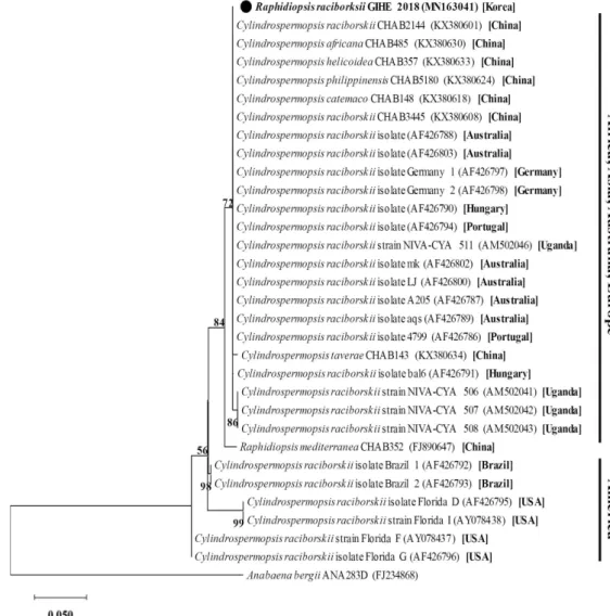
(5) 354. ∙ Jeong et al.. Fig. 2. Molecular phylogenetic analysis by maximum likelihood method based on cpcBA-IGS with Anabaena bergii as the outgroup. The evolutionary history was inferred by using the Maximum Likelihood method based on the Tamura 3-parameter model The percentage of trees in which the associated taxa clustered together is shown next to the branches. Initial tree(s) for the heuristic search were obtained automatically by applying Neighbor-Join and BioNJ algorithms to a matrix of pairwise distances estimated using the Maximum Composite Likelihood (MCL) approach, and then selecting the topology with superior log likelihood value. A discrete Gamma distribution was used to model evolutionary rate differences among sites (5 categories [+ G, parameter = 0.1911]).. 용한 연구는 현재까지 보고된 바가 없다. 이번 연구에서는 국. raciborskii 종의 cpcBC-IGS 유전자는 미국 ․ 남미에서 관찰된. 내에서 최초로 분리 ․ 배양된 R. raciborskii를 대상으로 cpcBA-. 개체들의 유전자 염기서열 보다, 중국 ․ 유럽 ․ 호주 ․ 아프리카. IGS와 nifH 유전자 염기서열의 계통분석법을 적용하여, 유전. 에서 분리된 개체들의 유전자 염기서열과 보다 높은 유연관계. 자의 염기서열의 차이를 토대로 지리적 분포 특성을 비교 분. 를 형성하였다.. 석하는, 미생물 지리학(microbial geography) 분석 연구를 수 행하였다.. 반면, 질소고정 기능을 보유한 모든 미생물에 존재하는 유전자인 nifH 유전자의 염기서열의 경우, 주로 uncultured. 연구 과정에서 순수배양 된 R. raciborskii GIHE 2018의. bacterium clone의 유전자 염기서열과 99% 이상 유사하였다. cpcBA-IGS 유전자 염기서열은 기존 문헌상에 보고된 R.. (Fig. 3). 안타깝게도, 본 연구 과정에서는 아시아에서 분리된. raciborskii 종의 유전자 염기서열들과 ~99% 상동성을 보이는. R. raciborskii 개체의 염기서열을 얻을 수 없어 직접적인 유연. 것으로 관찰되었으며, 이들은 분자계통학적 분석결과 크게 2. 관계를 파악할 수 없었다. Figure 3에서와 같이 계통수는 세 개. 개의 그룹으로 관찰되었다(Fig. 2). 특히, 이번에 분리된 R.. 의 그룹으로 분리되었으며, 본 연구를 통해 분리된 R. racibor-. 미생물학회지 제55권 제4호.
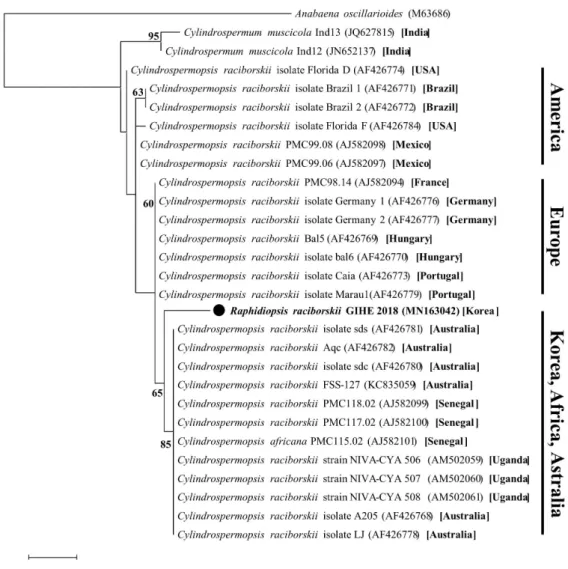
(6) Isolation of Unrecorded Harmful Algae R. raciborskii in Korea ∙. 355. Fig. 3. Molecular phylogenetic analysis by maximum likelihood method based on nifH gene with Anabaena oscillarioides as the outgroup. The evolutionary history was inferred by using the Maximum Likelihood method based on the Kimura 2-parameter model. The percentage of trees in which the associated taxa clustered together is shown next to the branches. Initial tree(s) for the heuristic search were obtained automatically by applying Neighbor-Join and BioNJ algorithms to a matrix of pairwise distances estimated using the Maximum Composite Likelihood (MCL) approach.. skii GIHE 2018의 nifH 유전자는 아프리카와 호주에서 분리된. 연구가 필요할 것으로 사료된다.. 종과 상대적으로 근접한 유연관계를 형성하였다. 또한, 미국 ․. R. raciborskii GIHE 2018이 보유한 16S rRNA 유전자 염기. 남미에서 분리된 개체의 nifH 유전자와는 확연하게 구분된. 서열은 기존 데이터베이스 내 보고된 R. raciborskii 종들의. 그룹을 형성하며, cpcBA-IGS 유전자와는 달리, 유럽 지역에. 16S rRNA 유전자와 ~99% 이상 상동성을 보였다(Fig. 4). 특. 서 분리된 개체의 유전자들보다 호주와 아프리카에서 분리된. 이하게도, cpcBA-IGS 및 nifH 유전자와는 달리, 16S rRNA 유. 개체들과 단일 그룹을 형성하는 것으로 관찰되었다. 이 결과. 전자 염기서열의 경우 지역적인 분포 특성이 관찰되지는 않았. 는, R. raciborskii가 아프리카 → 호주 → 아시아 → 중부 유럽. 다. 16S rRNA 유전자의 계통수 분석 결과, 모든 Raphidiopsis. 으로의 경로와, 남미 → 북미 → 지중해로의 2가지 경로를 통해. (Cylindrospermopsis) 속에 속하는 개체들이 분리된 지역과. 서 전파되었을 것이라는 선행 연구결과(Panou et al., 2018)와. 관련 없이 단일 클러스터를 형성하는 것으로 관찰되었다(Fig.. 일치한다. 향후, 추가적인 연구를 통해 R. raciborskii GIHE. 4). 이를 통해, Raphidiopsis 속의 각 종들은 16S rRNA 유전자. 2018 및 아시아에서 분리 동정된 종들의 유전체(Whole genome. 의 염기서열은 분리 동정된 지역과는 무관하게 서로 유사할. sequence) 분석 과정을 통한 nifH 유전자의 변이를 관찰하는. 것으로 사료되며, 분포 지역별 상이한 종이 아닌 단일 종의 개. Korean Journal of Microbiology, Vol. 55, No. 4.
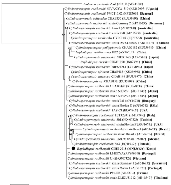
(7) 356. ∙ Jeong et al.. Fig. 4. Molecular phylogenetic analysis by maximum likelihood method based on 16S rRNA gene with Anabaena circinalis as the outgroup. The evolutionary history was inferred by using the Maximum Likelihood method based on the Kimura 2-parameter model. The percentage of trees in which the associated taxa clustered together is shown next to the branches. Initial tree(s) for the heuristic search were obtained automatically by applying Neighbor-Join and BioNJ algorithms to a matrix of pairwise distances estimated using the Maximum Composite Likelihood (MCL) approach, and then selecting the topology with superior log likelihood value. A discrete Gamma distribution was used to model evolutionary rate differences among sites (5 categories [+ G, parameter = 0.0720]).. 체가 지역 이동을 통해 현재의 분포양상을 보이고 있을 것임. 서열 유사성) 혹은 DNA-DNA hybridization 분석법(70% 염. 을 암시하는 결과로 사료된다. 또한, cpcBA-IGS 및 nifH 유전. 기서열 유사성)을 적용한 분석에서 모두 같은 종으로 분류될. 자의 경우, 균주의 지역 이동 과정에서 지역 특이적 유전자 염. 수 있다는 선행연구의 결과들과 상응하는 것으로 판단된다. 기서열들이 유전자 수평이동을 통해 genome 내부로 유입되. (Otsuka et al., 2001). 중국의 수계에서 분리된 Raphidiopsis. 었을 가능성이 있음을 암시한다.. (Cylindrospermopsis) 속에 속하는 6종(R. raciborskii, R. ta-. 또한, Raphidiopsis (Cylindrospermopsis)는 형태적 특징을. verae, R. africana, R. helicoidea, R. philippinensis)의 16S rRNA. 기반으로 전통적으로 5개의 종으로 분류되지만, 박테리아의. 유전자는 99.2~100%의 높은 유사도를 보이는 것으로 관찰되. 종 판단 기준인 16S rRNA 유전자 염기서열 분석법(97% 염기. 었다. 하지만 분자계통학적 분석 결과에서는 이들이 서로 유. 미생물학회지 제55권 제4호.
(8) Isolation of Unrecorded Harmful Algae R. raciborskii in Korea ∙. 357. 사한 종으로 분류되었다. 또한, 선행연구를 통해, 16S rRNA,. raciborskii가 관찰되었기 때문에, 이들이 중국의 경우와 유사. cpcBA-IGS, rpoC1, ITS-L의 4가지 유전자를 이용한 분석을. 하게 차후 높은 밀도로 전국적인 분포를 나타낼 가능성이 있. 통해서도 앞서 언급된 6종의 분자계통학적 특성을 명확하게. 다. 또한, 선행 연구 결과, 중국, 일본, 호주 지역에서 분리된 R.. 구별하지 못한 바 있다(Li et al., 2017). 따라서, 이들 유전자 염. raciborskii 종들이 조류 독소를 생산한다는 연구 결과가 보고. 기서열 분석법을 통해, R. raciborskii의 지리적 분포 특성에 대. 된 바 있기 때문에, 분자유전학적으로 매우 유사한 국내 종 역. 한 추론은 가능하지만, 종간의 분자계통학적 특성을 구분하기. 시 독소를 생산할 가능성이 클 것으로 추정된다. 따라서, 국내. 에는 한계가 있을 것으로 판단된다.. 에서도 R. raciborskii의 전국적인 분포 조사가 수행되어야 할. 결론적으로, 본 연구를 통해, 국내에서 처음 발견된 남조류. 것으로 사료되며, 국내에서 발견된 R. raciborskii를 유해남조. 는 말단 세포와 휴면포자의 형태에 따라 R. raciborskii로 동정. 류로 추가 지정하여 체계적인 관리가 필요할 것으로 판단된다.. 되었으며, cpcBA-IGS 및 nifH 유전자 염기서열을 이용한 분 자유전학적 분석결과, 아시아 ․ 호주 ․ 아프리카 지역에서 분 리된 개체들과의 유연관계가 상대적으로 높다는 결과를 관찰 할 수 있었다.. 적 요 온난화에 의한 수 생태계는 유해 조류의 발생이 증가되고 새로운 외래종이 유입되는 등 변화를 겪고 있다. R. raciborskii. 결 론. 는 열대지방의 부영양 수계에서 기원하였지만 기후변화 등 환 경요인에 따라 현재 온대지역까지 그 분포 영역을 확장하여. 본 연구를 통해, 인천지역의 저수지 시료로부터 휴면포자. 중국과 일본을 포함한 많은 지역에서 발견되고 있다. 이 남조. 와 사상체 말단 세포의 형태 분석을 통해, 국내 수계로부터 처. 류는 독소를 생산할 가능성이 있는 유해 조류의 일종으로, 현. 음으로 남조류인 R. raciborskii를 분리 ․ 배양 및 동정하였다.. 재까지 국내에서는 발견이 보고된 바가 없었다. 본 연구에서. 본 연구를 통해 분리된 R. raciborskii는 사상체 말단에 존재하. 는 국내 최초로 인천지역의 저수지에서 분리된 R. raciborskii. 는 이형세포를 제외하고는 다른 남조류와 형태적으로 매우 유. 를 대상으로 종 분리 배양 및 동정을 위해 현미경을 이용한 형. 사하였다. cpcBA-IGS, nifH 유전자 염기서열을 이용한 분자. 태관찰 및 cpcBA-IGS, nifH, 그리고 16S rRNA 유전자를 이용. 계통학적 연구를 통해, 본 연구에서 관찰된 R. raciborskii가 중. 하여 지리적 연관성을 추정할 수 있는 분자유전학적 분석을. 국에서 분리된 R. raciborskii와 염기서열이 매우 유사하며, 미. 수행하였다. 분리된 균주는 직선형의 사상체로, 말단에 질소. 국 ․ 남미에서 분리된 종 보다는 아시아 ․ 호주 ․ 아프리카에서. 고정을 담당하는 이형세포의 위치가 특징이며, 말단세포와 휴. 분리된 종과 상대적으로 높은 유연관계를 형성할 수 있다는. 면포자의 형태에 따라 R. raciborskii로 동정되었다. 이와 함께,. 것이 관찰되었다. 반면, 16S rRNA 유전자 염기서열은 Raphi-. cpcBA-IGS, 그리고 nifH 유전자를 이용한 분자유전학적 분석. diopsis (Cylindrospermopsis) 속 내에서 서로 유사하여, 지역. 결과 아시아, 유럽, 호주, 아프리카에서 분리된 개체와 밀접한. 별 특성이 관찰되지는 않았다. 이를 통해, 국내에서 발견된 R.. 유연관계를 형성하는 것으로 관찰되었다. 반면, 16S rRNA 유. raciborskii는 아프리카와 호주를 거쳐 전파되었을 가능성이. 전자의 계통학적 분석 결과에서는 지역별 분포 특성이 관찰되. 있으며, 이들이 보유한 cpcBA-IGS 및 nifH 유전자는 지역 특. 지 않았다. 2017년 이후 인천, 의왕, 화성에서 직선형 및 S자 형. 이적 유전자의 유전자 수평이동을 통해 genome 내부로 유입. 태의 R. raciborskii가 관찰되면서, 전국적으로 광범위한 분포를. 되었을 것으로 사료되었다.. 보일 가능성이 매우 높을 것으로 추정되며, 아시아와 호주에서. 현재까지 국내에서 발견된 R. raciborskii 종은 수계 내에 소 수의 개체만이 존재하기 때문에 미기록 종으로 남아있었을 것. 독소를 생산하는 종이 발견되고 있기 때문에 전국적인 분포조 사 및 생태학적 특성에 대한 연구가 필요할 것으로 사료된다.. 으로 추정된다. Raphidiopsis 속은 2012년 낙동강 수계 내 남 조류 유전자 조사 과정에서 유전자 관찰을 통해 분포 가능성 이 제기된 바는 있지만, 그 수가 매우 소수로 관찰되었으며, 실 제 현미경 관찰을 통해 분포가 확인되지는 않았다. 2017년부 터 수행된 남조류 분포 조사 과정에서 인천, 화성, 의왕 소재 저 수지에서도 수온이 높은 8월 다양한 형태적 특성을 갖는 R.. 감사의 말 본 연구는 국립환경과학원의 시도보건환경연구원 국고보 조사업의 일환으로 진행되었으며 이에 깊이 감사 드립니다.. Korean Journal of Microbiology, Vol. 55, No. 4.
(9) 358. ∙ Jeong et al.. References Aguilera A, Gómez EB, Kaštovský J, Echenique, RO, and Salerno GL. 2018. The polyphasic analysis of two native Raphidiopsis isolates supports the unification of the genera Raphidiopsis and Cylindrospermopsis (Nostocales, Cyanobacteria). Phycologia 57, 130–146. Altschul SF, Madden TL, Schaffer AA, Zhang J, Zhang Z, Miller W, and Lipman DJ. 1997. Gapped BLAST and PSI-BLAST: a new generation of protein database search programs. Nucleic Acids Res. 25, 3389–3402. Antunes JT, Leão PN, and Vasconcelos VM. 2015. Cylindrospermopsis raciborskii: review of the distribution, phylogeography, and ecophysiology of a global invasive species. Front. Microbiol. 6, 473. Chonudomkul D, Yongmanitchai W, Theeragool G, Kawachi M, Kasai F, Kaya K, and Watanabe MM. 2004. Morphology, genetic diversity, temperature tolerance and toxicity of Cylindrospermopsis raciborskii (Nostocales, Cyanobacteria) strains from Thailand and Japan. FEMS Microbiol. Ecol. 48, 345–355. Dyble J, Paerl HW, and Neilan BA. 2002. Genetic characterization of Cylindrospermopsis raciborskii (cyanobacteria) isolates from diverse geographic origins based on nifH and cpcBA-IGS nucleotide sequence analysis. Appl. Environ. Microbiol. 68, 2567–2571. Gugger M, Molica R, Le Berre B, Dufour P, Bernard C, and Humbert JF. 2005. Genetic diversity of Cylindrospermopsis strains (cyanobacteria) isolated from four continents. Appl. Environ. Microbiol. 71, 1097–1100. Griffiths DJ and Saker ML. 2003. The Palm Island mystery disease 20 years on: a review of research on the cyanotoxin cylindrospermopsin. Environ. Toxicol. 18, 78–93. Haande S, Rohrlack T, Ballot A, Røberg K, Skulberg R, Beck M, and Wiedner C. 2008. Genetic characterisation of Cylindrospermopsis raciborskii (Nostocales, Cyanobacteria) isolates from Africa and Europe. Harmful Algae 7, 692–701. John DM, Whitton BA, and Brook AJ. 2011. The freshwater algal flora of the british isles: An identification guide to freshwater and nd terrestrial algae 2 edn, Cambridge University Press, Cambridge. Kokociński M, Gągała I, Jasser I, Karosienė J, Kasperovičienė J, Kobos J, Koreivienė J, Soininen J, Szczurowska A, Woszczyk M, et al. 2017. Distribution of invasive Cylindrospermopsis raciborskii in the East-Central Europe is driven by climatic and local environmental variables. FEMS Microbiol. Ecol. 93, fix035. Komárek J and Kling H. 1991. Variation in six planktonic cyanophyte genera in Lake Victoria (East Africa). Arch. Hydrobiol. 61, 21–45. Komárek J and Komárková-Legnerová J. 2003. Phenotype diversity of the cyanoprokaryotic genus Cylindrospermopsis (Nostocales). Czech Phycol. 3, 1–30. Kumar S, Stecher G, Li M, Knyaz C, and Tamura, K. 2018. MEGA X: molecular evolutionary genetics analysis across computing. 미생물학회지 제55권 제4호. platforms. Mol. Biol. Evol. 35, 1547–1549. Lagos N, Onodera H, Zagatto PA, Andrinolo D, Azevedo SM, and Oshima Y. 1999. The first evidence of paralytic shellfish toxins in the fresh water cyanobacterium Cylindrospermopsis raciborskii, isolated from Brazil. Toxicon 37, 1359–1373. Lei L, Peng L, Huang X, and Han, BP. 2014. Occurrence and dominance of Cylindrospermopsis raciborskii and dissolved cylindrospermopsin in urban reservoirs used for drinking water supply, South China. Environ. Monit. Assess. 186, 3079–3090. Li R, Watanabe M, and Watanabe MM. 1997. Akinete formation in planktonic Anabaena spp. (cyanobacteria) by treatment with low temperature. J. Phycol. 33, 576–584. Li XC, Huo SL, Cai FF, Yang YM, Xi BD, and Li RH. 2017. The taxonomy and phylogeny of the genus Cylindrospermopsis (Cyanobacterium) evaluated by adding five new records from China. Phytotaxa 316, 224–238. Moreira C, Fathalli A, Vasconcelos V, and Antunes A. 2011. Genetic diversity and structure of the invasive toxic cyanobacterium Cylindrospermopsis raciborskii. Curr. Microbiol. 62, 1590– 1595. Nakdong River Environment Research Institute, National Institute of Environmental Research. 2013. Analysis of cyanobacteria on the Nakdong River by Molecular based analysis. Republic of Korea. Neilan BA, Jacobs D, and Goodman, AE. 1995. Genetic diversity and phylogeny of toxic cyanobacteria determined by DNA polymorphisms within the phycocyanin locus. Appl. Environ. Microbiol. 61, 3875–3883. Neilan BA, Jacobs D, Del Dot T, Blackall LL, Hawkins PR, Cox PT, and Goodman, AE. 1997. rRNA sequences and evolutionary relationships among toxic and nontoxic cyanobacteria of the genus Microcystis. Int. J. Syst. Bacteriol. 47, 693–697. Otsuka S, Suda S, Shibata S, Oyaizu H, Matsumoto S, and Watanabe MM. 2001. A proposal for the unification of five species of the cyanobacterial genus Microcystis Kützing ex Lemmermann 1907 under the rules of the Bacteriological Code. Int. J. Syst. Evol. Microbiol. 51, 873–879. Padisák J. 1997. Cylindrospermopsis raciborskii (Woloszynska) Seenayya et Subba Raju, an expanding, highly adaptive cyanobacterium: worldwide distribution and review of its ecology. Arch. Hydrobiol. 107, 563–593. Panou M, Zervou SK, Kaloudis T, Hiskia A, and Gkelis S. 2018. A Greek Cylindrospermopsis raciborskii strain: Missing link in tropic invader’s phylogeography tale. Harmful Algae 80, 96– 106. Saker ML, Neilan BA, and Griffiths DJ. 1999. Two morphological forms of Cylindrospermopsis raciborskii (cyanobacteria) isolated from Solomon Dam, Palm Island, Queensland. J. Phycol. 35, 599–606. Saker ML and Neilan BA. 2001. Varied diazotrophies, morphologies, and toxicities of genetically similar isolates of Cylindrospermopsis raciborskii (nostocales, cyanophyceae) from Northern.
(10) Isolation of Unrecorded Harmful Algae R. raciborskii in Korea ∙. Australia. Appl. Environ. Microbiol. 67, 1839–1845. Schembri MA, Neilan BA, and Saint CP. 2001. Identification of genes implicated in toxin production in the cyanobacterium Cylindrospermopsis raciborskii. Environ. Toxicol. 16. 413–421. Soares MCS, Lürling M, and Huszar VLM. 2013. Growth and temperature-related phenotypic plasticity in the cyanobacterium Cylindrospermopsis raciborskii. Phycol. Res. 61, 61–67. Tillett D and Neilan BA. 2000. Xanthogenate nucleic acid isolation from cultured and environmental cyanobacteria. J. Phycol. 36, 251–258. Thompson JD, Higgins DG, and Gibson TJ. 1994. CLUSTAL W: improving the sensitivity of progressive multiple sequence alignment through sequence weighting, position-specific gap penalties and weight matrix choice. Nucleic Acids Res. 22, 4673– 4680. Thompson AS, Rhodes JC, and Pettman I. 1988. Natural environmental research council culture collection of algae and protozoa-Catalogue of strains, p. 22. Ambleside: Freshwater Biology Association, UK. Water Environment Research Department, Gyeonggi-do Institute of. 359. Health and Environment. 2019. Research report of major reservoir phytoplanktons in Gyeonggi-do 2018. Republic of Korea. Wilk-Woźniak E, Solarz W, Najberek K, and Pociecha A. 2016. Alien cyanobacteria: an unsolved part of the “expansion and evolution” jigsaw puzzle? Hydrobiologia 764, 65–79. Woloszynska, J. 1912. Das Phytoplankton einiger javanischer Seen, mit Berucksichtigung des Sawa-Planktons. Bull. Acad. Sci. Cracovie, mat.-nat. ser. B 6, 649–709. Wood SA and Stirling DJ. 2003. First identification of the cylindrospermopsin‐producing cyanobacterium Cylindrospermopsis raciborskii in New Zealand. New Zeal. J. Mar. Fresh. 37, 821–828. Yilmaz M, Phlips EJ, Szabo NJ, and Badylak S. 2008. A comparative study of Florida strains of Cylindrospermopsis and Aphanizomenon for cylindrospermopsin production. Toxicon 51, 130– 139. Zarenezhad S, Sano T, Watanabe MM, and Kawachi M. 2012. Evidence of the existence of a toxic form of Cylindrospermopsis raciborskii (nostocales, cyanobacteria) in Japan. Phycol. Res. 60, 98–104.. Korean Journal of Microbiology, Vol. 55, No. 4.
(11)
수치


+2
관련 문서
First, the general characteristics of the two groups were analyzed by frequency analysis and correlation analysis, which is technical statistics, and the
Phylogenetic analysis of Orientia tsutsugamushi strains based on the sequence homologies of 56-kDa type-specific antigen genes. FEMS
We determined the nucleotide sequences of the mitochondrial DNA (mtDNA) control region using cloning and sequencing, and obtained the complete sequence from the cattle bones
In addition, using the properties of water shown in diverse shapes, Ki Hyung-do's recognition on the reality and future consciousness from regression of
Full genome cloning and nucleotide sequence analysis of hepatitis C virus from sera of chronic hepatitis patients in Korea. Detection of hepatitis C virus RNA
분리된 균주 ( Pseudoalteromonasagarovorans )를 이용하여 알긴산으로부터 효 율적 당화를 위한 최적 배양 조건을 찾기 위해 전 배양 시간 및 접종량,교반
Therefore, in this study, based on the media facade expression characteristics and expression techniques, evaluation factors through satisfaction analysis on
So, the result of the comparative analysis in the social studies object, curriculums and textbooks of South and North Korea indicate that the differences
