ISSN 1225-6552
http://dx.doi.org/10.7853/kjvs.2012.35.3.169
< Original Article >
Veterinary Service
Available online at http://kosves.re.kr
*Corresponding author: Dong-Kun Yang, Tel. +82-31-467-1783, Fax. +82-31-467-1797, E-mail. [email protected]
Evaluation of Japanese encephalitis virus vaccine strains currently used in pigs by molecular characterization
Jeong Ah Lee, Dong-Kun Yang*, Ha-Hyun Kim, Sun-Young Kim, Jin-Ju Nah, Soo-Dong Cho, Jae-Young Song
Animal, Plant and Fishery Quarantine Inspection and Agency, MIFAFF, Anyang 430-757, Korea (Received 7 August 2012; revised 27 August 2012; accepted 28 August 2012)
Abstract
Japanese encephalitis virus (JEV) is one of the main causes of viral encephalitis in human and animals.
For over 30 years, a live attenuated JEV vaccine strain has been used in the veterinary field, and it is required to conduct quality evaluation studies on the commercial vaccines. For the quality control of live attenuated JEV vaccine, we investigated the nucleotide sequence similarity of prME gene derived from five JEV vaccines commercially available in pigs in Korea. The Vero cells infected with JEV vac- cines showed specific cytopathic effect, which was characterized by rounding and detached cells. In the phylogenetic analysis, all of the vaccine strains showed a close relationship with the original vaccine seed strain (Anyang 300) and clustered into the genotype 3. In comparison of the nucleotide and de- duced amino acid sequences of prME genes with the original strain, all JEV vaccine strains showed high amino acid similarity ranging from 98.9% to 99.5%, but had several point mutations, probably due to high mutation rates of viral RNA polymerase by several virus passages. Even though the current JEV vaccine strains have been maintained and produced for a long period of time, the genetic character- ization of them have been rarely changed. However, since the mid 1990’s, molecular epidemiology of JEV has been changed sharply from genotype 3 to genotype 1 in Korea, further studies on new vaccine strains to genotype 1 is required for more effective prevention in the field.
Key words : Japanese encephalitis virus, Vaccine, prME
INTRODUCTION
Japanese encephalitis (JE) is one of the most important viral encephalitis caused by Japanese encephalitis virus (JEV) in human and animals. JEV is particularly danger- ous to domestic animals such as swine, horses, dogs, chickens, ducks and reptiles (Ghosh and Basu, 2009;
Gulati et al, 2012). After an incubation period of about 6 to 12 days, severe infection of JEV can cause febrile head- ache, aseptic meningitis, or encephalitis (Yang et al, 2005).
JEV is a member of the Flaviviridae family and Flavivirus genus (Yun et al, 2010). RNA genome of JEV contains a single open reading frame (ORF) encod-
ing a polyprotein approximately 11kb in length. The polyprotein is cleaved into three structural proteins - capsid (C), precursor to membrane (prM), envelope (E) and seven nonstructural proteins (NS1, NS2A, NS2B, NS3, NS4A, NS4B, NS5) (Sumiyoshi et al, 1987; Yang et al, 2004; Yang et al, 2005). The E protein (54 kDa) is the major envelope glycoprotein of virion as well as a determinant of viral neurovirulence and neuroinvasi- veness (Nitayaphan et al, 1990; Yang et al, 2004; Yang et al, 2005). It has a number of neutralization epitopes that mediate attachment to a host cell, along with a re- ceptor-binding domain that induces immune response.
The prM protein interacts with E protein to form prME heterodimers, which are important for formation of im- mature virion and play an important role in maintaining
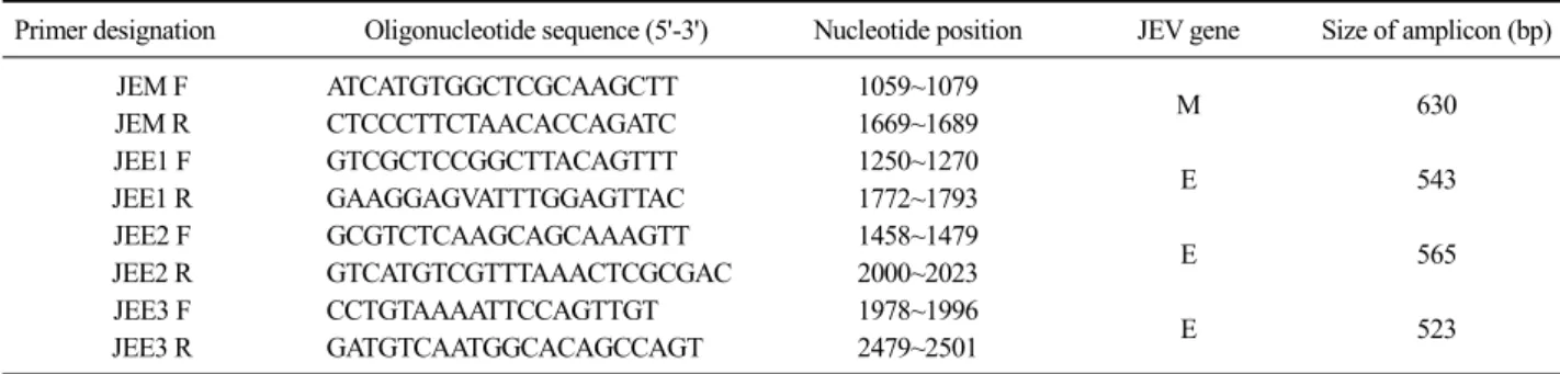
Table 1. List of oligonucleotide primers used in RT-PCR for JEV
Primer designation Oligonucleotide sequence (5'-3') Nucleotide position JEV gene Size of amplicon (bp)
JEM F ATCATGTGGCTCGCAAGCTT 1059~1079
M 630
JEM R CTCCCTTCTAACACCAGATC 1669~1689
JEE1 F GTCGCTCCGGCTTACAGTTT 1250~1270
E 543
JEE1 R GAAGGAGVATTTGGAGTTAC 1772~1793
JEE2 F GCGTCTCAAGCAGCAAAGTT 1458~1479
E 565
JEE2 R GTCATGTCGTTTAAACTCGCGAC 2000~2023
JEE3 F CCTGTAAAATTCCAGTTGT 1978~1996
E 523
JEE3 R GATGTCAATGGCACAGCCAGT 2479~2501
E protein (Nitayaphan et al, 1990). JEV is maintained in a zoonotic cycle involving pigs as major amplifica- tion/reservoir hosts, water birds as carriers, mosquitoes as vectors and human as dead end hosts (Unni et al, 2011). According to epidemiological investigations, every year at least 67,000 clinical JEV cases occurred world- wide and approximately 15,000 of which have resulted in death. The JEV has spread throughout Asia and as far as northern Australia (Deng et al, 2011; Solomon, 2006;
Wang et al, 2010). Since pigs serve as reservoirs and amplifiers of the virus, development of JEV vaccines for pigs could help prevent epidemics among human.
In order to protect sow from reproductive disorders, a live-attenuated JEV vaccine (Anyang 300 strain) was developed by continuous passage in chicken fibroblast cells in 1980 in Korea (Yang et al, 2004). Although at- tenuated JEV vaccine has been produced and applied to pigs over 30 years in Korea, commercial veterinary vac- cine strains have not properly been investigated until now. Therefore, it is necessary to evaluate JEV vaccine strains based on the molecular characterization.
In this study, we identified JEV vaccine strains using cytopathic effects (CPE) in cells, indirect fluorescence assay (IFA) test, reverse transcription - polymerase chain reaction (RT-PCR) technique and investigated nucleotide sequence analysis of prME gene of JEV among five commercial vaccines produced by veterinary pharma- ceutical companies in Korea.
MATERIALS AND METHODS
Vaccines
Five Korean commercial JEV vaccines produced by
different Korean animal vaccine companies were used in this study as follows: GreencrossⓇ JE (Greencross Co., Korea), DaesungⓇ JE (Daesung Co., Korea), SuiShotⓇ JE (ChoongAng Co., Korea), KomiphamⓇ JE (Komi- pharm Co., Korea), HimmvacⓇ JE (KBNP Co., Korea).
All JE vaccines have been licensed in Korea for the prevention of JE in pigs.
Identification of JEV in vaccine
Each commercial JEV vaccine was inoculated into Vero cells grown in alpha minimum essential medium (α-MEM) with 10% fetal bovine serum. The cells were incubated in a CO2 incubator at 37oC for 7 days until CPE was observed and viral titer was checked under microscopy by the Reed-Muench method. Subsequently the cells were fixed with 80% cold acetone and in- cubated with JEV-specific monoclonal antibody at 37oC for 1 h. Then they were stained with FITC-conjugated anti-mouse IgG (KPL, USA). The cells were then wash- ed in phosphate buffer saline (pH 7.2) and examined under fluorescence microscopy.
RNA extraction and RT-PCR
Viral RNA was extracted from 5 commercial JEV vaccines using an RNA extraction kit (Qiagen, USA) according to the manufacturer’s indications. The RNA samples were then amplified by RT-PCR using 4 kinds of specific primer pairs of prME genes of JEV (Table 1).
The reaction mixture contained 10 μl of denatured RNA, 1 μl of each primer (50 pmol), 10 μl of 5× buf- fer (12.5 mM MgCl2), 2 μl of enzyme mix of One-step RT-PCR kit (Qiagen, USA), 2 μl of 10 mM dNTP, and 24 μl of distilled water, for a 50 μl final volume. The
Fig. 1. Confirmation of cytopathic effects (CPE) and JEV-positive cells in the Vero cells infected with JEV vaccines. (A) CPE was shown in Vero cells infected with a JEV vaccine. (B) No CPE was shown in the mock-infected Vero cells. (C) Many JEV-positive cells were detected in the JEV vac- cine-infected Vero cells using indirect immunofluores- cence assay (IFA). (D) No JEV-positive cells were detected in the mock-infected Vero cells. IFA was performed with monoclonal antibodies against JEV.
Fig. 2. Amplification and confirmation of JEV prM, E1, E2 and E3 genes. (A) The prM, E1, E2 and E3 genes of JEV were am- plified using RT-PCR. (B) The prM, E1, E2 and E3 genes of plasmid DNA were confirmed by EcoR I enzyme digestion.
M: 100 bp ladder, Lane 1: prM gene, Lane 2: E1 gene, Lane 3: E2 gene, Lane 4: E3.
RT-PCR condition was as follows: cDNA synthesis at 42oC for 30 min; 35 cycles with denaturation at 95oC for 45 sec; annealing at 50oC for 45 sec and extension at 72oC for 45 sec; final extension at 72oC for 5 min.
The PCR products were detected on 1.5% agarose gel containing ethidium bromide. The purified PCR products were ligated with pGEM-TⓇ Easy Vector (Promega, USA) and used to transform into competent cells (DH5α). The plasmid DNA was isolated from Escherichia coli and identified by EcoRI enzyme digestion.
Sequencing and phylogenetic analysis After cloning the prM and E genes of JEV, the sequ- ences of the purified plasmids were analyzed using an MJ Research PTC-225 Peltier Thermal Cycler and ABI PRISMⓇ BigDyeTM Terminator Cycle Sequencing kits with AmpliTaq DNA polymerase (FS enzyme; Applied Biosystems, USA) according to the manufacturer's pro- tocols. The prME genes of JEV vaccine strains were com- pared with those of the other known JEV submitted to NCBI and seed strain (Anyang 300). The sequences em- ployed were obtained from the NCBI GenBank database [accession numbers: AY303795 (CC27-L1), AY303798
(CC27-S8), AB551990 (JaTAn1/75), M18370 (JaOArS982), EF543861 (SH0601), AF416457 (SA14-12-1-7), M55506 (SA14), AF254452 (CH1392), AY303792 (T1P1-L4), AF069076 (JaGAr01), AB551991 (JaTAn1/90), FJ185036 (B58), EF571853 (Nakayama), L48961 (Beijing-1), L78128 (Ling), EU880214 (XJ169), EU693899 (XJP613), AF045551 (K94P05), AY316157 (KV1899), GQ902062 (4790-85), AF217620 (FU)]. Nucleotide sequence sim- ilarity calculations were conducted using the DNASISⓇ (Hitachi Software, Japan) software. Individual sequences were initially aligned using BioEdit and Clustal X 1.81.
Phylogenetic reconstructions were generated using the neighbor-joining method by the computer program DNAstar. Phylogenetic trees were reconstructed on align- ed nucleotide sequences using ClustalW (version 2.0.12;
European Bioinformatics Institute, UK).
RESULTS
The Vero cells inoculated with five Korean commer- cial JEV vaccines showed specific CPE, which was characterized by the rounding and detachment of cells (Fig. 1A). The viral titers of the vaccines ranged from 105.3 to 106.0 TCID50/ml. Also specific cytoplasmic fluo- rescence was identified in the JEV vaccine strain-in- fected Vero cells using IFA with JEV specific mono- clonal antibody (Fig. 1C). No CPE and JEV-positive cells were detected in the mock-infected Vero cells (Fig.
1B). The prM and E genes from five vaccine strains
Fig. 4. Comparison of deduced amino acid sequences among five commercial JEV vaccine strains and Anyang 300 strain.
The dots and slashes indicate identical amino acids and omissions of amino acid sequences, respectively.
Fig. 3. Phylogenetic tree based on prME genes of JEV vaccine strains with another known sequences obtained from GenBank database to show genetic relationships.
were successfully amplified and cloned using RT-PCR with each gene-specific primer (Fig. 2).
Phylogenetic analysis was conducted using prME genes of JEV vaccine strains with other nucleotide se- quences obtained from GenBank database. Most of the Korean field strains have been divided into genotypes 1 or 3. Five commercial vaccine strains including the Anyang 300 strain were grouped in genotype 3, together with JEV strains isolated in China, Japan and Taiwan.
Other Korean strains such as KV1899 and K94P05 were clustered into genotype 1 together with strains isolated in China, the United Kingdom, and Australia (Fig. 3). In comparison of the deduced amino acid sequences of prME genes with the original strain, all JEV vaccine strains showed high amino acid similarity ranging from 98.9%
to 99.5%, but had several point mutations (Fig. 4).
DISCUSSION
JE is one of the most important viral encephalitis in Asia, especially in rural and suburban areas where rice culture and pig farming coexist (Campbell et al, 2011).
JEV is transmitted by a zoonotic cycle involving pigs as major amplification/reservoir hosts, water birds as car- riers, mosquitoes as vectors and human as dead end hosts (Unni et al, 2011). As pigs were considered am-
plification/reservoir hosts of JEV in zoonotic cycle, it was important to immunize pigs for the purpose of blocking the cycle. Therefore, a live-attenuated JEV vaccine (Anyang 300 strain) for pigs was developed by continuous passage in several cells including chicken fi- broblast cells in Korea in 1980 (Yang et al, 2004). The JEV vaccines manufactured by biologic companies in Korea have been applied to pigs for the prevention of JE epidemic in human and pigs. Developments of JEV vaccine based on the molecular biology have been re- ported by several scientists (Beasley et al, 2008; Hong et al, 1998; Yun et al, 2010; Zhang et al, 2011). We conducted biological methods on JEV vaccine and ob- tained that JEV in the vaccines was identified by specif- ic CPE and IFA in the Vero cells, indicating that five different Korean animal vaccine companies have pro- duced JEV vaccine without changing original seed.
Phylogenetic analyses based on nucleotide sequences of prME, NS1 and full length genes revealed that JEVs worldwide were classified into 5 genotypes (genotype 1,
2, 3, 4 and 5) (Solomon et al, 2003; Nitatpattana et al, 2008; Uchil and Satchidanandam, 2001). In this study, phylogenetic analysis based on the nucleotide sequences of the prME genes indicated that five commercial vac- cine strains showed a close relationship with the Anyang 300 strain grouping the genotype 3. All JEV vaccine strains showed high amino acid similarity rang- ing from 98.9% to 99.5%, but had several point muta- tions, probably due to high mutation rates of viral RNA polymerase by several virus passages. RNA viruses have a high mutation rate during replication due to both the lack of proofreading and post-replication error correction by RNA polymerase (Steinhauer and Holland, 1987). It also assumed that the current JEV vaccine strains have been produced for a long period of time and passaged in several kinds of primary cells in order to propagate high titer without strict management. On the basis of our results, the strict standards for the live vaccines in- cluding JEV vaccines are needed to be imposed for quality control. On the other hand, since molecular epi- demiology of JEV has been changed sharply from geno- type 3 to genotype 1 in Korea from the mid 1990’s, further studies on new vaccine strains to genotype 1 is required for more effective prevention in the field.
In conclusion, five Korean commercial JEV vaccines contained original JEV seed strain but showed some point mutations in prME gene. The biological and mo- lecular methods could be useful for identifying vaccine strains in attenuated JEV vaccines. Especially, the prME-based molecular method might be useful for de- tection of genetic similarity and variation among JEV vaccine strains.
ACKNOWLEDGMENTS
This work was supported financially by a grant (B-AD14-2010-11-03) from the Animal, Plant and Fisheries Quarantine and Inspection Agency, Ministry for Food, Agriculture, Forestry and Fisheries, Korea.
The authors thank all of the Korean vaccine companies that provided commercial vaccines for this research.
REFERENCES
Beasley DW, Lewthwaite P, Solomon T. 2008. Current use and development of vaccines for Japanese encephalitis.
Expert Opin Biol Ther 8: 95-106.
Campbell GL, Hills SL, Fischer M, Jacobson JA, Hoke CH, Hombach JM, Marfin AA, Solomon T, Tsai TF, Tsu VD, Ginsburg AS. 2011. Estimated global incidence of Japanese encephalitis: a systematic review. Bull World Health Organ 89: 766-774.
Deng X, Shi Z, Li S, Wang X, Qiu Y, Shao D, Wei J, Tong G, Ma Z. 2011. Characterization of nonstructural protein 3 of a neurovirulent Japanese encephalitis virus strain iso- lated from a pig. Virol J 8: 209.
Ghosh D, Basu A. 2009. Japanese encephalitis-a pathological and clinical perspective. PLoS Negl Trop Dis 3: e437.
Gulati BR, Singha H, Singh BK, Virmani N, Kumar S, Singh RK. 2012. Isolation and genetic characterization of Japanese encephalitis virus from equines in India. J Vet Sci 13: 111-118.
Hong SP, Chung YJ, Moon SB, Shin YC, Lee SH, Kim SO.
1998. Growth characteristics of an attenuated Japanese encephalitis virus in a monkey kidney cell(Vero).
Korean J Biotechnol Bioeng 13: 231-237.
Nitatpattana N, Dubot-Pérès A, Gouilh MA, Souris M, Barbazan P, Yoksan S, de Lamballerie X, Gonzalez JP. 2008.
Change in Japanese encephalitis virus distribution, Thailand. Emerg Infect Dis 14: 1762-1765.
Nitayaphan S, Grant JA, Chang GJ, Trent DW. 1990. Nucleotide sequence of the virulent SA-14 strain of Japanese ence- phalitis virus and its attenuated vaccine derivative, SA-14-14-2. Virology 177: 541-552.
Solomon T. 2006. Control of Japanese encephalitis-within our grasp? N Engl J Med 355: 869-871.
Solomon T, Ni H, Beasley DW, Ekkelenkamp M, Cardosa MJ, Barrett AD. 2003. Origin and evolution of Japanese en- cephalitis virus in southeast Asia. J Virol 77: 3091-3098.
Steinhauer DA, Holland JJ. 1987. Rapid evolution of RNA viruses. Annu Rev Microbiol 41: 409-433.
Sumiyoshi H, Mori C, Fuke I, Morita K, Kuhara S, Kondou J, Kikuchi Y, Naqamatu H, Iqarashi A. 1987. Complete nucleotide sequence of the Japanese encephalitis virus genome RNA. Virology 161: 497-510.
Uchil PD, Satchidanandam V. 2001. Phylogenetic analysis of Japanese encephalitis virus: envelope gene based analy- sis reveals a fifth genotype, geographic clustering, and multiple introductions of the virus into the Indian subcontinent. Am J Trop Med Hyg 65: 242-251.
Unni SK, Růžeek D, Chhatbar C, Mishra R, Johri MK, Singh SK.
2011. Japanese encephalitis virus: from genome to infectome. Microbes Infect 13: 312-321.
Yang DK, Kim BH, Kweon CH, Kwon JH, Lim SI, Han HR.
2004. Molecular characterization of full-length genome of Japanese encephalitis virus(KV1899) isolated from
pigs in Korea. J Vet Sci 5: 197-205.
Yang DK, Kweon CH, Kim BH, Lim SI, Kwon JH, Kim SH, Song JY, Han HR. 2005. Immunogenicity of baculovirus expressed recombinant proteins of Japanese encephalitis virus in mice. J Vet Sci 6: 125-133.
Yun SM, Cho JE, Ju YR, Kim SY, Ryou J, Han MG, Choi WY, Jeong YE. 2010. Molecular epidemiology of Japanese encephalitis virus circulating in South Korea, 1983-2005.
Virol J 7: 127.
Wang L, Fu S, Zhang H, Ye X, Yu D, Deng Z, Yuan J, Zhai
Y, Li M, Lv Z, Chen W, Jiang H, Gao X, Cao Y, Wang H, Tang Q, Liang G. 2010. Identification and isolation of genotype-1 Japanese encephalitis virus from encepha- litis patients. Virol J 7: 345.
Zhang Y, Chen P, Cao R, Gu J. 2011. Mutation of putative N-linked glycosylation sites in Japanese encephalitis vi- rus premembrane and envelope proteins enhances hu- moral immunity in BALB/C mice after DNA vacci- nation. Virol J 8: 138.