• 검색 결과가 없습니다.

Molecular characterization of Korean rabies virus isolatesDong-Kun Yang

N/A
N/A
Protected

Academic year: 2022

Share "Molecular characterization of Korean rabies virus isolatesDong-Kun Yang"

Copied!
7
0
0

로드 중.... (전체 텍스트 보기)

전체 글

(1)

Veterinary Science

DOI: 10.4142/jvs.2011.12.1.57

Received: 23 Mar. 2010, Accepted: 05 Jun. 2010

Original Article

*Corresponding author

Tel: +82-31-467-1783; Fax: +82-31-467-1797 E-mail: [email protected]

Molecular characterization of Korean rabies virus isolates

Dong-Kun Yang

1,

*, Young-Nam Park

2

, Gyeong-Soo Hong

2

, Hee-Kyung Kang

1

, Yoon-I Oh

1

, Soo-Dong Cho

1

, Jae-Young Song

1

1

National Veterinary Research and Quarantine Service, Anyang 430-824, Korea

2

Gangwon-do Veterinary Service Laboratory, Chuncheon 200-822, Korea

The nucleoprotein (N) and glycoprotein (G) of 11 Korean rabies virus (RABV) isolates collected from animals diagnosed with rabies between 2008 and 2009 were subjected to molecular and phylogenetic analyses. Six isolates originated from domestic animals (cattle and dogs) and five were obtained from wild free-ranging raccoon dogs. The similarities in the nucleotide sequences of the N gene among all Korean isolates ranged from 98.1 to 99.8%, while those of the G gene ranged from 97.9 to 99.3%. Based on the nucleotide analysis of the

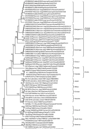
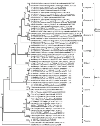
N and G genes, the Korean RABV isolates were confirmed as

genotype I of Lyssavirus and classified into four distinct subgroups with high similarity. Phylogenetic analysis showed that the Korean isolates were most closely related to the non-Korean NeiMeng1025B and 857r strains, which were isolated from rabid raccoon dogs in Eastern China and Russia, respectively. These findings suggest that the Korean RABV isolates originated from a rabid raccoon dog in Northeastern Asia. Genetic analysis of the Korean RABV isolates revealed no substitutions at several antigenic sites, indicating that the isolates circulating in Korea may be pathogenic in several hosts.

Keywords: characterization, genotype I, molecular epidemiology, rabies virus

Introduction

Rabies is an almost invariably fatal viral disease in animals and humans. According to the World Health Organization, rabies infections result in approximately 55,000 human deaths worldwide annually [23]. More than 29,000 human deaths from rabies were reported in Far East Asia including Thailand and China in 2006. However, there were no cases of human rabies reported in some countries such as Japan, Singapore, Korea, Malaysia and Taiwan in 2006 [5]. Rabies

is transmitted by vector species such as bats, red foxes, skunks, raccoon dogs, dogs, wolves, and mongooses, depending on the country [9,12]. Dogs were known to be the main vectors for the transmission of rabies virus (RABV) to humans or other animals in Korea prior to 1980. Raccoons such as Nyctereutes procyonoide and Procyon lotor have been involved in RABV circulation in eastern Europe and the eastern United States since the late 1990s [2,13,23].

RABV belongs to the genus Lyssavirus in the family Rhabdoviridae and consists of a non-segmented negative single-stranded RNA genome that encodes five structural proteins: nucleoprotein (N), phosphoprotein, matrix protein, glycoprotein (G), and large protein [12]. Recent advances in technology have contributed to better understanding of the molecular epidemiology and geographic relationships of RABV isolates [20,21,26]. The nucleoprotein, which contains antigenic sites such as NI and NIII, is associated with encapsidation of the genomic RNA and the formation of an active cytoplasmic ribonucleoprotein complex that is essential for viral replication [25]. The N gene or protein has been targeted for diagnosing wild rabies using RT-PCR or indirect fluorescent antibody tests because the gene is highly conserved and the protein is produced in large quantities in infected brain tissue. Moreover, the nucleotide sequence of the N gene has been used extensively as a molecular marker to explain the patterns of the geographic distribution of RABV at the regional and global levels [20]. The glycoprotein plays an important role in attachment of the virus to the host cell surface, pathogenicity, immunogenicity, and neurovirulence [6,14,19]. Additionally, the RABV G gene has been used as another target for studying genetic diversity and antigenic typing because it contains several major antigenic sites designated as I∼VI, ‘a’, and G1 on the ectodomain (1∼439 amino acids) and is related to pathogenicity.

In Korea, the first case of rabies was reported in 1907, and

a number of rabies cases were identified nationwide until

1945 [8]. After implementing an effective RABV control

program using live and inactivated vaccine, the number of

rabies cases decreased dramatically to an average of 32 cases

per year by 1984, and there were no reports of rabies between

(2)

Fig. 1. Map of Far East Asia and Korea showing the geographical location from which the rabies viruses were obtained. The isolates circled in Far East Asia showed high nucleotide similarity.

1985 and 1992 [7,11]. However, rabies was identified in a dog in Gyeonggi-do Province in 1993. Since then, a few rabies cases have been reported annually. Although a renewed national RABV control program involving the distribution of bait vaccine was implemented in early 2000, rabies still occurs in some regions of Korea. Korean RABV isolates collected between 1998 and 2004 have been identified as genotype I and found to be closely related to the Arctic lineage virus, but distantly related to other Asian strains including Chinese and Sri Lanka strains [8,16].

Therefore, we analyzed the N and G gene diversity of the recent strain of RABV circulating in Korea as well as the correlation among rabies genes and phylogenetic relationships. The genetic characterization of RABV is considered important for developing diagnostic and preventive measures, including a more effective vaccine. In this study, we cloned and sequenced the N and G genes of 11 RABV isolates obtained between 2008 and 2009 in Korea and analyzed the phylogenetic relationships of the isolates with other available sequences retrieved from GenBank.

Materials and Methods Samples

Eleven RABV isolates were obtained from brain samples that had tested positive for rabies using indirect immunofluorescence with monoclonal antibody against the N gene in Gangwon-do Province of Korea between 2008 and 2009. Five isolates (KRVR0801, KRVR0803, KRVR0804, KRVR0901, and KRVR0906) originated from raccoon dogs (Nyctereutes procyonoides koreensis), while another five isolates (KRVB0902, KRVB0903, KRVB0904,

KRVB0905, and KRVB0907) were from cattle, and one (KRVC0802) was from a dog. These Korean RABV isolates are described in Fig. 1 and Table 1.

Extraction of viral RNA and RT-PCR

Viral RNA was extracted from the 11 isolates using an RNA extraction kit (Qiagen, Germany) according to the manufacturer’s instructions. The extracted RNA was eluted in 50 μ L of RNase- and DNase-free water. RT-PCR was conducted using primers specific for the N and G regions of RABV (RVN1F, RVN1R, RVN2F, RVN2R, RVG1F, RVG1R, RVG2F, and RVG2R; Table 2). RT-PCR was conducted in a reaction mixture containing 10 μ L of denatured RNA, 1 μ L of each primer (50 pmol), 10 μ L of 5 × buffer (12.5 mM MgCl

2

), 2 μ L of dNTP mix, 2 μ L of enzyme mix (reverse transcriptase and Taq polymerase), and 24 μ L of distilled water (Qiagen, Germany). The cycling profile consisted of cDNA synthesis at 42

o

C for 30 min, followed by 35 cycles of 95

o

C for 45 sec, 50

o

C for 45 sec, and 72

o

C for 1 min, with a final extension at 72

o

C for 5 min.

The PCR products were visualized using electrophoresis on 1.8% agarose gels containing ethidium bromide.

Cloning and sequencing

All of the PCR products that were purified using the gel extraction kit were ligated with pGEM-T easy vector (Promega, USA) according to the manufacturer’s protocol.

Plasmid DNA was isolated from amplified Escherichia coli

(DH5 α ), and recombinant plasmids were identified by

EcoRI enzyme digestion (Bioneer, Korea). The sequences

of the purified plasmids were analyzed using an MJ

Research PTC-225 Peltier Thermal Cycler and ABI PRISM

(3)

Table 1. The Korean rabies virus isolates analyzed in this study

Isolates Locality Species Year of isolation Accession number

N* gene G

gene

KRVR0801 Sokcho Raccoon dog 2008 GU937035 GU937024 

KRVC0802 Inje Dog 2008 GU937036 GU937025 

KRVR0803 Sokcho Raccoon dog 2008 GU937037 GU937026 

KRVR0804 Sokcho Raccoon dog 2008 GU937038 GU937027 

KRVR0901 Goseong Raccoon dog 2009 GU937039 GU937028 

KRVB0902 Goseong Cattle 2009 GU937040 GU937029 

KRVB0903 Hongcheon Cattle 2009 GU937041  GU937030 

KRVB0904 Goseong Cattle 2009 GU937042  GU937031 

KRVB0905 Inje Cattle 2009 GU937043  GU937032 

KRVR0906 Goseong Raccoon dog 2009 GU937044  GU937033 

KRVB0907 Inje Cattle 2009 GU937045  GU937034 

SKRBV9801YC Yeoncheon Cattle 1998 DG076119 DQ076105

SKRRD9901PJ Paju Raccoon dog 1999 DQ076123 DQ076103

SKRRD9902PJ Paju Raccoon dog 1999 DQ076121 DQ076102

SKRRD9903YG Yanggu Raccoon dog 1999 DQ076131 DQ076099

SKRDG9901GY Goyang Dog 1999 DQ076122 DQ076100

SKRDG9902GY Goyang Dog 1999 DQ076120 DQ076101

SKRRD0204CW Cheorwon Raccoon dog 2002 DQ076125 DQ076094

SKRRD0205HC Hwacheoun Raccoon dog 2002 DQ076127 DQ076096

SKRDG0203CW Cheorwon Dog 2002 DQ076124 DQ076104

SKRDG0204HC Hwacheon Dog 2002 DQ076128 DQ076093

SKRRD0406CC Chuncheon Raccoon dog 2004 DQ076126 DQ076098

SKRBV0403CW Cheorwon Cattle 2004 DQ076129 DQ076095

SKRBV0404HC Hwacheon Cattle 2004 DQ076130 DQ076097

KRH2-04 Hongcheon Raccoon dog 2004 AY730595 -

KRH3-04 Hongcheon Raccoon dog 2004 AY730596 -

KRC5-04 Hongcheon Dog 2004 AY730597 -

*N: nucleoprotein, G: glycoprotein.

Table 2. List of the oligonucleotide primers used for RT-PCR of rabies virus Primer Nucleotide sequences (5´ - 3´) Nucleotide

Position* Sense Rabies virus gene

Size of amplicon (bp)

RVN1F ATGGATGCCGACAAGATTGTATTC 71-94 +

N

1,047

RFN1R GAATTCCTCTCCCAGATAGCC 1097-1118 –

RVN2F CTA GGGGGCTATCTGGGAGA 1091-1110 +

N

472

RVN2R CGGCCAGACCGGCTCTAACAC 1539-1562 –

RVG1F ATGGTTCCTCAGGCTCTCCTGTTTGT 3317-3342 +

G

1,052

RVG1R GACTGACTTGTAGTGAGCATCGGC 4346-4369 –

RVG2F GTCCATCATGACAACCAAGTC 4237-4257 +

G

711

RVG2R GGCCAGTCCTCACAGTCTGGTCTC 4877-4900 –

*The positions of primers are based on the ERA strain (GenBank accession no. EF206707).

(4)

Fig. 2. Phylogenetic analysis based on the complete N gene nucleotide sequences of the isolates and other sequences obtained from the GenBank database. Numbers at each key node indicate the degree of bootstrap support and only those with >70%

support are shown.

BigDye Terminator Cycle Sequencing kits with AmpliTaq DNA polymerase (FS enzyme; Applied Biosystems, USA) according to the manufacturers’ protocols. Single-pass sequencing was conducted for each template using universal primers (e.g., SP6 and T7). The fluorescent-labeled fragments were purified from the unincorporated terminators using an ethanol precipitation protocol. The samples were resuspended in distilled water and subjected to electrophoresis in an ABI 3730xl sequencer (Applied Biosystems, USA). Both DNA strands were sequenced to verify the sequences.

Phylogenetic analysis

The nucleotide sequences, accession numbers, and names of the strains used for the phylogenetic analysis in this study were obtained from the GenBank database. Nucleotide sequence similarity calculations were conducted using the DNASIS (Hitachi Software, Japan) software. Individual sequences were initially aligned using BioEdit and Clustal X 1.81. Phylogenetic reconstructions were generated using the neighbor-joining method by the computer program PHYLIP 3.572c. Phylogenetic trees were reconstructed on aligned nucleotide sequences using ClustalW (version 2.0.12; European Bioinformatics Institute, UK). The robustness of the phylogenetic analysis was determined by bootstrap analysis with 1,000 replications. Graphic output was produced by TreeView (version 1.6.6; Institute of Biomedical and Life Science, UK).

Results

Phylogenetic analysis of the complete N gene sequences A total of 1,350 bp nucleotide sequences of the complete N gene encoding the nucleoprotein from 11 isolates were determined, and their amino acids sequences were deduced. The N gene sequences of 51 other RABVs obtained from GenBank were compared with those of the 11 Korean isolates to analyze molecular epidemic relationships. The nucleotide sequence analysis showed that all of the Korean isolates were classified into genotype I of Lyssavirus, and the nucleotide similarity among the 27 Korean isolates ranged from 98.1 to 99.8% (Fig. 2). The highest sequence similarity of the 11 Korean isolates with non-Korean RABV corresponded to the Chinese strains NeiMeng1025B and NeiMeng927 (96.1 and 96.7% at the nucleotide level and 98.8 and 99.5% at the amino acid level, respectively). The nucleotide similarity of the Korean isolate (KRVB0902) with Russian strain (857r) and Sri Lanka strain (SRL1145) were 95.7 and 86.8%, respectively [13]. The lowest nucleotide similarity of the Korean isolates with 35 non-Korean RABV strains was with the 99ABV3306 strain ranging from 82.7 to 82.9%. A phylogenetic analysis based on the N gene classified the Korean isolates into four subgroups (Gangwon I, II, III and Gyeonggi) with high similarity (Fig. 2).

Phylogenetic analysis of the complete G gene sequences

The 1,575 bp nucleotide sequences of the G gene encoding the glycoprotein from the 11 RABV isolates were identified and their amino acids were also deduced.

The nucleotide sequences of the 11 Korean isolates were compared with those of 42 RABVs retrieved from GenBank to analyze the evolutionary relationships of RABVs. The phylogenetic tree based on the nucleotide sequence analysis of the G gene revealed that all of the Korean isolates were also divided into four groups (Gangwon I, II, III and Gyeonggi) and were most closely related to the Chinese strains designated as Chinese I in Fig. 3, similar to the results for the N gene. The similarity of the G gene nucleotide sequences of RABV ranged from 97.9 to 99.3% among all Korean isolates and 95.4 to 96.2%

between Korean isolates and Chinese strains such as

NeiMeng927A. Nucleotide similarity between the Korean

isolates and Arctic strain ranged from 89.6 to 90.6% and

(5)

Fig. 3. Phylogenetic analysis based on the complete G gene nucleotide sequences of the isolates and other sequences obtained from the GenBank database. Numbers at each key node indicate degree of bootstrap support and only those with >70% support are indicated.

comparison of Korean isolates with the NY516 and USA7-BT strains showed 78.4 and 78.9% similarity, respectively. Alignment of the deduced amino acid sequences revealed 98.3 to 99.8% similarity among the 24 Korean isolates.

Comparison of the proteins coding the N and G genes

The deduced amino acid sequences of the nucleoprotein and glycoprotein were compared to analyze the genetic variability among Korean RABVs. Antigenic sites NI (374

∼383) and NIII (313∼337) and the phosphorylation site (389) mapping to serine were conserved in the 11 Korean RABVs. Comparison of the nucleoprotein among the 11 Korean RABVs revealed eight amino acid substitutions (data not shown). The deduced amino acid sequence demonstrated that RABV circulating in Korea consisted of the SP (−19∼1), Ecto (20∼439), TM (440∼461), and Endo (462∼524) domains. Among the 524 residues in the deduced amino acid sequences of the G gene were 38 substitutions among 24 Korean RABV isolates (data not shown). The arginine at position 333 of the residues within antigenic site III, which is essential for virulence, was not changed in the 11 Korean isolates (data not shown). All 11

Korean RABV isolates had two N-glycosylation sites at amino acid positions 37 and 319 of the ectodomain of the glycoprotein.

Discussion

Since rabies recurred in the northern part of Korea in 1993, many RABV cases have been reported in Gyeonggi- do and Gangwon-do Provinces [8,11]. According to national data regarding rabies, no cases were reported in Gyeonggi-do Province in 2009, whereas 18 rabies cases were reported in Gangwon-do Province. These findings suggest that our efforts to prevent rabies should be focused on limited regions.

Among the 11 Korean RABV isolates used in this study, five were obtained from domestic cattle and one was obtained from a dog infected by contact with rabid raccoon dogs, while five isolates were obtained from naturally infected raccoon dogs. Although various vector species of RABV are known, the raccoon dog has been the main carrier between domestic and wild animals in Korea.

Raccoon dogs, which are the only canids known to hibernate in winter, were introduced to Far East Asia from Russia for the production of fur and pelts in 1928 [1].

However, the raccoon dog industry decreased due to the rise in popularity of silver foxes and goats. Raccoon dogs eventually escaped from fur farms and became wild animals in Korea. The Korean raccoon dog population increased until 2007 due to lack of predators. However, in 2008, the population of raccoon dogs decreased in Gyeonggi-do Province due to various parasitic diseases involving Demodex spp., tremadoes, cestodes and rapid urbanization of the region.

As advanced technology expanded, the nucleotide sequences of the RABVs circulating in various regions of the world have been submitted to several genetic information banking systems [8,16,18]. Using recent genetic information, we determined the nucleotide sequences and conducted a phylogenetic analysis of the N and G genes of Korean isolates obtained between 2008 and 2009 to understand the evolution of RABV in Korea. The genetic relationships among various RABV strains have been described elsewhere [10,15,26]. Analysis of the N and G genes provided similar results, suggesting that either gene can be used for phylogenetic analysis. Comparing the nucleotide similarity with non-Korean isolates, the Korean RABV isolates formed a close phylogenetic relationship with the NeiMeng1025B strain, which was isolated from a naturally RABV-infected raccoon dog in Jilin Province of China and with 857r strain, which was isolated from Chabarovsk of Russia and classified into group B [13].

However, it was only distantly related to other Asian

RABV strains (8764THA, 9702INDI and SRL1145). The

phylogenetic analysis revealed that the Korean isolates

(6)

belonged to four subgroups (Gangwon I, II, III and Gyeonggi) that shared high homology, indicating that the genetic features of rabies are related more to geographic relationships and isolation year than to the species infected [2]. A previous study showed that Korean isolates obtained from 1998 to 2004 had the closest relationship with Arctic-like virus, such as Canadian strains [8,16]. The results of this study indicate that RABV in Korea was transmitted from the northeastern region of China.

The deduced amino acid sequences of the N and G genes of the 11 RABV isolates were aligned to investigate the antigenic variation in RABV, as reported earlier [17]. The genetic analysis of the N and G genes of the 11 Korean isolates showed that all of the antigenic sites were conserved, and the putative phosphorylation site at amino acid position 389 in the N gene was also conserved. It is well known that the arginine or the lysine at residue 333 on the ectodomain of glycoprotein are essential for neurovirulence within antigenic site III, and that virus variants that substitute glutamine, isoleucine, and glycine at that position show less phathogenic or avirulent symptoms [3,19,22]. Genetic analysis of the compared Korean RABVs showed no substitutions at these positions.

Therefore, the Korean isolates recently circulating in Korea are pathogenic in several hosts such as dogs and cattle and have the ability to infect neural cells, such as neuroblastoma NG-108 cells [4]. Wunner et al. [24]

reported that glycosylation is essential for complete folding and there are three or four putative glycosylation sites on the glycoprotein depending on the virus strain. The 11 Korean isolates had two putative N-glycosylation sites (Asparagine-X-Serine or Asparagine-X-Threoine) at amino acids 37 and 319 within the ectodomain, indicating the existence of antigenic variants of RABV with altered glycosylation in rabies viruses.

In conclusion, our results indicate that Korean RABV isolates are closely related with northeastern Asian RABV strains based on a phylogenetic analysis. Based on these findings, raccoon dogs play an important role as a vector species for rabies and raccoon dog movement could be responsible for the distribution of RABV. Current strategies to prevent rabies in Korea should be reconsidered and new tactics such as point infection control and trap-vaccinate- release programs should be reviewed to eradicate rabies.

Application of bait vaccine for wild animals should also be scaled up around the place of outbreaks and genetic surveillance of RABV circulating in Korea is needed to monitor the epidemiological status of rabies in Korea.

Acknowledgments

We thank Dr. KW Lee for critically reviewing our manuscript. This work was supported financially by a grant from the National Veterinary Research and Quarantine

Service, Ministry for Food, Agriculture, Forestry and Fisheries, Korea.

References

1. Botvinkin AD, Savitskiĭ VP, Sidorov GN, Iudin VG.

Importance of the racoon dog in the epidemiology and epizootiology of rabies in the Far East. Zh Mikrobiol Epidemiol Immunobiol 1981, 12, 79-82.

2. Bourhy H, Kissi B, Audry L, Smreczak M, Sadkowska- Todys M, Kulonen K, Tordo N, Zmudzinski JF, Holmes EC. Ecology and evolution of rabies virus in Europe. J Gen Virol 1999, 80, 2545-2557.

3. Coulon P, Ternaux JP, Flamand A, Tuffereau C. An avirulent mutant of rabies virus is unable to infect motoneurons in vivo and in vitro. J Virol 1998, 72, 273-278.

4. Dietzschold B, Wunner WH, Wiktor TJ, Lopes AD, Lafon M, Smith CL, Koprowski H. Characterization of an antigenic determinant of the glycoprotein that correlates with pathogenicity of rabies virus. Proc Natl Acad Sci USA 1983, 80, 70-74.

5. Fu ZF. The rabies situation in Far East Asia. Dev Biol (Basel) 2008, 131, 55-61.

6. Guyatt KJ, Twin J, Davis P, Holmes EC, Smith GA, Smith IL, Mackenzie JS, Young PL. A molecular epidemiological study of Australian bat lyssavirus. J Gen Virol 2003, 84, 485-496.

7. Hwang EK. Outbreak and control of rabies in animals in Korea. Korean J Vet Public Health 1995, 19, 281-293.

8. Hyun BH, Lee KK, Kim IJ, Lee KW, Park HJ, Lee OS, An SH, Lee JB. Molecular epidemiology of rabies virus isolates from South Korea. Virus Res 2005, 114, 113-125.

9. Johnson N, Black C, Smith J, Un H, McElhinney LM, Aylan O, Fooks AR. Rabies emergence among foxes in Turkey. J Wildl Dis 2003, 39, 262-270.

10. Khawplod P, Shoji Y, Ubol S, Mitmoonpitak C, Wilde H, Nishizono A, Kurane I, Morimoto K. Genetic analysis of dog rabies viruses circulating in Bangkok. Infect Genet Evol 2006, 6, 235-240.

11. Kim CH, Lee CG, Yoon HC, Nam HM, Park CK, Lee JC, Kang MI, Wee SH. Rabies, an emerging disease in Korea. J Vet Med B Infect Dis Vet Public Health 2006, 53, 111-115.

12. Knipe DM, Howley PM, Griffin DE, Lamb RA, Martin MA, Roizman B, Straus SE. Field Virology. pp.

1221-1277, Lippincott Williams & Wilkins, Philadelphia, 2001.

13. Kuzmin IV, Botvinkin AD, McElhinney LM, Smith JS, Orciari LA, Hughes GJ, Fooks AR, Rupprecht CE.

Molecular epidemiology of terrestrial rabies in the former Soviet Union. J Wildl Dis 2004, 40, 617-631.

14. Morimoto K, Hooper DC, Spitsin S, Koprowski H, Dietzschold B. Pathogenicity of different rabies virus variants inversely correlates with apoptosis and rabies virus glycoprotein expression in infected primary neuron cultures.

J Virol 1999, 73, 510-518.

15. Nagarajan T, Mohanasubramanian B, Seshagiri EV,

Nagendrakumar SB, Saseendranath MR, Satyanarayana

ML, Thiagarajan D, Rangarajan PN, Srinivasan VA.

(7)

Molecular epidemiology of rabies virus isolates in India. J Clin Microbiol 2006, 44, 3218-3224.

16. Park YJ, Shin MK, Kwon HM. Genetic characterization of rabies virus isolates in Korea. Virus Genes 2005, 30, 341- 347.

17. Sacramento D, Badrane H, Bourhy H, Tordo N. Molecular epidemiology of rabies virus in France: comparison with vaccine strains. J Gen Virol 1992, 73, 1149-1158.

18. Sato G, Itou T, Shoji Y, Miura Y, Mikami T, Ito M, Kurane I, Samara SI, Carvalho AA, Nociti DP, Ito FH, Sakai T. Genetic and phylogenetic analysis of glycoprotein of rabies virus isolated from several species in Brazil. J Vet Med Sci 2004, 66, 747-753.

19. Seif I, Coulon P, Rollin PE, Flamand A. Rabies virulence:

effect on pathogenicity and sequence characterization of rabies virus mutations affecting antigenic site III of the glycoprotein. J Virol 1985, 53, 926-934.

20. Smith JS, Orciari LA, Yager PA, Seidel HD, Warner CK.

Epidemiologic and historical relationships among 87 rabies virus isolates as determined by limited sequence analysis. J Infect Dis 1992, 166, 296-307.

21. Susetya H, Sugiyama M, Inagaki A, Ito N, Mudiarto G,

Minamoto N. Molecular epidemiology of rabies in Indonesia. Virus Res 2008, 135, 144-149.

22. Tuffereau C, Leblois H, Bénéjean J, Coulon P, Lafay F, Flamand A. Arginine or lysine in position 333 of ERA and CVS glycoprotein is necessary for rabies virulence in adult mice. Virology 1989, 172, 206-212.

23. World Health Organization (WHO). WHO expert committee on rabies. World Health Organ Tech Rep Ser 1992, 824, 1-84.

24. Wunner WH, Dietzschold B, Smith CL, Lafon M, Golub E. Antigenic variants of CVS rabies virus with altered glycosylation sites. Virology 1985, 140, 1-12.

25. Yang J, Hooper DC, Wunner WH, Koprowski H, Dietzschold B, Fu ZF. The specificity of rabies virus RNA encapsidation by nucleoprotein. Virology 1998, 242, 107- 117.

26. Zhang YZ, Xiong CL, Lin XD, Zhou DJ, Jiang RJ, Xiao

QY, Xie XY, Yu XX, Tan YJ, Li MH, Ai QS, Zhang LJ,

Zou Y, Huang C, Fu ZF. Genetic diversity of Chinese

rabies viruses: evidence for the presence of two distinct

clades in China. Infect Genet Evol 2009, 9, 87-96.

수치

Fig. 1. Map of Far East Asia and Korea showing the geographical location from which the rabies viruses were obtained
Table 2. List of the oligonucleotide primers used for RT-PCR of rabies virus Primer Nucleotide sequences (5´ - 3´) Nucleotide
Fig. 2. Phylogenetic analysis based on the complete N gene  nucleotide sequences of the isolates and other sequences obtained from the GenBank database
Fig. 3. Phylogenetic analysis based on the complete G gene  nucleotide sequences of the isolates and other sequences obtained from the GenBank database

참조

관련 문서

Genetic analysis indicated that the nucleotide sequences of swine HEV-3 and rotavirus A detected in this study were closely related to those of human isolates.. However, swine

A phylogenetic tree, based on the helicase gene and HVR nucleotide sequences, revealed the newly detected viruses and other avian HEVs were classified similarly..

This study revealed that the P genes of Korean RABVs are genetically similar to those of RABV strains of lyssavirus genotype I including V739 (dogs, Korea), NNV-RAB-H

Phylogenetic and structure prediction analyses of DEV pUL49.5 Phylogenetic analysis based on gN protein sequences of different herpesvirus strains clearly demonstrated that DEV

Phylogenetic analysis of different parapoxviruses based on nucleotide sequences of the B2L gene (A) and partial virus interferon resistance gene (B). Nucleotide sequences of

Phylogenetic relationships among four species of Mullidae (Perciformes) inferred from DNA sequences of mitochondrial cytochrome b and 16S rRNA genes.. Systematic

Phylogenetic tree based on nucleotide sequences of the 16s rRNA (1,400-bp) genes of Borrelia miyamotoi isolates from humans and ticks in northeastern China, May 2013–June 2015,

Molecular phylogeny of infectious hematopoietic necrosis virus (IHNV). A) molecular phylogeny among 49 isolates based on nucleotide sequences of the glycoprotein