저작자표시-비영리-변경금지 2.0 대한민국 이용자는 아래의 조건을 따르는 경우에 한하여 자유롭게 l 이 저작물을 복제, 배포, 전송, 전시, 공연 및 방송할 수 있습니다. 다음과 같은 조건을 따라야 합니다: l 귀하는, 이 저작물의 재이용이나 배포의 경우, 이 저작물에 적용된 이용허락조건 을 명확하게 나타내어야 합니다. l 저작권자로부터 별도의 허가를 받으면 이러한 조건들은 적용되지 않습니다. 저작권법에 따른 이용자의 권리는 위의 내용에 의하여 영향을 받지 않습니다. 이것은 이용허락규약(Legal Code)을 이해하기 쉽게 요약한 것입니다. Disclaimer 저작자표시. 귀하는 원저작자를 표시하여야 합니다. 비영리. 귀하는 이 저작물을 영리 목적으로 이용할 수 없습니다. 변경금지. 귀하는 이 저작물을 개작, 변형 또는 가공할 수 없습니다.
A THESIS
FOR THE DEGREE OF DOCTOR OF PHYLOSOPHY
TRANSCRIPTOME-WIDE DISCOVERY OF INNATE
IMMUNE-RELATED GENES AND MOLECULAR
CHARACTERIZATION OF THE
INFLAMMATORY/APOPTOTIC PATHWAY IN MANILA
CLAM (Ruditapes philippinarum)
Youngdeuk Lee
Department of Marine Life Science
GRADUATE SCHOOL
JEJU NATIONAL UNIVERSITY
2012-07
TRANSCRIPTOME-WIDE DISCOVERY OF INNATE
IMMUNE-RELATED GENES AND MOLECULAR
CHARACTERIZATION OF THE
INFLAMMATORY/APOPTOTIC PATHWAY IN MANILA
CLAM (Ruditapes philippinarum)
Youngdeuk Lee
(Supervised by Professor Jehee Lee)
A thesis submitted in partial fulfillment of the requirement for the degree of
DOCTOR OF PHILOSOPHY
2012-07
The thesis has been examined and approved by
Thesis Director, Gi-Young Kim, Professor of Marine Life Sciences
Moon-Soo Heo, Professor of Marine Life Sciences
Joon Bum Jeong, Professor of Marine Life Sciences
Mahanama De Zoysa, Professor of Veterinary medicine
Jehee Lee, Professor of Marine Life Science
2012-07-07
Department of Marine Life Science Date
GRADUATE SCHOOL
JEJU NATIONAL UNIVERSITY
- I -
CONTENTS
요약문 III
SUMMERY VI
LIST OF FIGURES V
LIST OF TABLES VII
CHAPTER I: Characterization of manila clam transcriptome using 454-pyrosequencing approaches
1
1.1 INTRODUCTION 2
1.2 MATERIALS AND METHODS 5 1.3 RESULTS AND DISCUSSION 5
CHAPTER II: Characterization of novel molluscan MyD88 family protein from manila clam
11
2.1 INTRODUCTION 12
2.2 MATERIALS AND METHODS 14
2.3 RESULTS 18
2.4 DISCUSSION 20
CHAPTER III: Immune response-related gene expression of a novel molluscan IκB protein member from manila clam
31
3.1 INTRODUCTION 32
3.2 MATERIALS AND METHODS 34
3.3 RESULTS 38
3.4 DISCUSSION 40
CHAPTER IV: Molecular characterization and expression analysis of molluscan TNFα homologue from manila clam
51
4.1 INTRODUCTION 52
- II -
4.3 RESULTS 57
4.4 DISCUSSION 60
CHAPTER V: Characterization of cDNAs of key genes involved in apoptosis in manila clam
70
5.1 INTRODUCTION 71
5.2 MATERIALS AND METHODS 72
5.3 RESULTS 75
5.4 DISCUSSION 79
Reference 92
- III -
요약문
바지락은 전세계적으로 매우 중요한 양식 이매패류 종으로 한국을 비롯한 동아시아 지역과, 유럽 및 북미에서 넓게 서식하고 있다. 최근 바지락의 대량 폐사에 대한 연구가 보고되고 있다. 이러한 바지락의 대량 패사는 다양한 환경 요인, 환경오염, 그리고 감염 등이 그 원인으로 보고되었으며, 그중에서 특히 brown ring disease (BRD)와 perkinsus 감염증이 중요 원인으로 알려져 있다. BRD 와 perkinsus 감염증과 같은 병원체에 의한 대량패사는 바지락의 생산량에 커다란 영향을 미치고 있어 경제적 손실 또한 커지고 있기 때문에 이러한 문제의 해결을 위하여 바지락의 면역체계에 대한 연구에 대한 관심이 증가하고 있다. 병원체에 의한 바지락내의 면역반응을 이해하기 위해서는 분자 수준에서의 접근이 필요한데, 이는 해당 종에 대한 유전정보가 필수적이다. 하지만 NCBI GenBanK database 에 등록되어 있는 바지락의 유전정보는 다른 양식 이매패류종인 굴이나 가리비 등에 비하여 매우 적은 7%에 불과한 실정이다. 바지락을 포함한 연체동물은 후천면역이 결핍되어 있다고 알려져 있다. 그렇기 때문에 바지락과 같은 연체동물에서의 선천면역은 매우 중요한 역할을 하지만 해양연체동물의 선천면역반응에 대한 메커니즘 연구는 매우 미비한 실정이다. 반면 포유류에서는 광범위하게 연구가 이루어지고 있으며, 이러한 자료는 연체동물의 면역 시스템을 연구하기 위한 방향을 제시하고 있다. 바지락과 같은 무척추동물에서는 선천면역이 외부 병원체감염으로부터 방어하기 위한 주요 시스템으로 작용한다. 이런 이유로, 분자수준에서 다양한 병원체의 감염에 대한 면역반응에 대한 이해는 매우 중요하다. 이 연구에서는 바지락의 면역체계에 대한 이해를 위하여 부족한 유전정보를 확보하고자 454 Genome Sequencer Flex titanium 을 이용하여 바지락의 transcriptome 을 분석하였고, 이를 통하여 117,347 개의 유전자에 대한 정보를 확보하였고, 이중에서 NCBI 에 등록된 단백질들과의 비교를 통하여 24,327 개의 unigene 을 확보하였다. 확보한 24,327 개의 unigene 에 대한 data 로부터 중요한 면역 관련 유전자로 알려진 MyD88, IκB, TNFα 와 세포사멸에 중요한 조절자로 작용하는 것으로 알려진 두 개의 Bcl-2 family 단백질의 전체서열을 확보하고 분자적 특성 및 면역자극을 통한 발현변화를 관찰하였다.
바지락의 RpMyD88 은 1416 bp 의 ORF 로 구성되어 있으며, 471 개의 아미노산을 암호화하고 있으며, RpMyD88 의 아미노산 서열에서 MyD88 단백질의 전형적인 특징인 death doman 과 TIR 도메인을 확인 할 수 있었다. RpMyD88 의 아미노산 서열은 가리비 MyD88 과 27%로 가장 높은 identity 를 나타내었다. RpMyD88 은 바지락의 모든 조직에서 발현이 관찰되었으며, 특히 주요 면역 유전자인 hemocyte 와 gill 그리고 mantle 에서 높은
- IV -
발현량을 나타내었다. 반면 siphon, foot, adductor muscle 에서는 매우 적은 양의 발현을 나타내었다. RpMyD88 은 Vibrio tapetis 와 LPS 를 주사한 바지락의 hemocyte 와 gill 모두에서 유의적인 발현 증가를 나타내었으며, 이러한 서열특성 및 유전자 발현에 관련된 결과들은 바지락에서도 포유류와 같은 MyD88 dependent signaling pathway 가 존재하며 RpMyD88 역시 박테리아에 대한 선천 면역반응에서 중요한 역할을 수행하고 있을 것으로 예상할 수 있었다.
NF-kB signaling pathway 는 선천면역 시스템에서 가장 중요한 구성요소 중의 하나로, NF-kB 의 활성은 inhibitor of NF-κB (IκB)와의 물리적 상호작용에 의하여 조절되게 된다. IκB 가 NF-κB 에 결합하게 되면 NF-κB 의 nuclear localization signal 을 막음으로써 NF-κB 를 불활화 상태로 Cytoplasm 에 가둬두게 된다. 3 장에서는 NF-κB 의 활성을 조절하는 IκB 의 분자적 특성과 발현 정보를 나타내었다. 바지락 IκB (Rp-IκB)의 cDNA 는 1032 bp 의 ORF 로 구성되어있고, 343 개의 아미노산을 암호화 하였다. Rp-IκB 단백질은 IκB 단백질의 전형적인 특징인 IκB degradation motif, PEST sequence 그리고 6 개의 ankyrin repeats 로 구성되어 있었다. 또한 계통분류학적 분석을 통하여 무척추동물의 IκB 단백질들과 그룹을 이루는 것을 확인 하였다. Rp-IκB 는 모든 조직에서 지속적으로 발현을 나타내었으며, 주요 면역 세포인 hemocyte 와 gill 에서 V. tapetis 와 LPS 자극에 의하여 유의적인 발현 증가를 나타내었다. 이러한 결과는 포유류에서 NF-κB 의 key 조절자인 IκB 와 같이 Rp-IκB 가 박테리아의 침임에 대하여 중요한 역할을 수행할 것으로 예상할 수 있었고, 이는 Rp-IκB 는 포유류의 IκB orthologs 들과 비슷한 기능을 가지고 있음을 뒷받침하고 있다.
포유류의 TNFα 의 homology 를 바지락으로부터 확인하였고, RpTNFα 로 명명하였다. RpTNFα 는 다른 선행 연구에서 보고된 TNFα 의 전형 적인 특징인 Transmembrane 과 TNF-signature 를 가지고 있었다. qRT-PCR 을 통하여 조직 특이적 발현을 분석한 결과 모든 조직에서 발현이 관찰되었으며, V. tapetis, LPS 그리고 poly I:C 를 주사하였을 때 이에 반응하여 hemocyte 와 gill 에서 유의적인 발현 증가를 나타내었다.
Bcl-2 는 Mitochondrial membrane permiabilzation (MMP)의 억제를 통하여 세포 사멸을 억제하는 Anti-apoptotic Bcl-2 sub-family 다. Bax 는 pro-apoptotic Bcl-2 subfamily 다. GS-flx transcriptome 서열에 기초하여, Bcl-2 와 Bax like 단백질의 orthologue 로 예상되는 cDNA 전체 서열을 바지락에서 확인하였고, 이 서열들은 Bcl-2 homology domain 과 transmembrane (only RpBcl-2)을 포함하고 있었다. Real time PCR 을 통하여 분석한 조직특이적 발현에서는 모든 조직에서 발현이 관찰되었으며, gill 에서 가장 높은 발현을
- V -
나타내었다. V. tapetis 를 주사한 후 gill 과 hemocyte 에서 유의적인 발현 변화를 나타내었다. 이러한 결과는 RpBcl2 와 RpBax 가 바지락의 면역반에서 포유류의 apoptotic regulator 들과 비슷한 기능 수행과 함께 면역에 매우 깊이 관련 있는 것을 나타내고 있다.
- VI -
Summary
Manila clam, Ruditapes philippinarum, is an important aquaculture species worldwide. Recent mass mortalities of manila clam landings, believed to be due to microbial infection, have significantly impacted production and sparked interest in immune-related research of this species. Genetic information is important to better understand the molecular basis for the host immune response against pathogen. However, genomic data of clam form just 7% in bivalve genomic database. The manila clam is an economically important species, but to date only a few fully-annotated genomic data are available in genomic databases.
The manila clam is known to lack an adaptive immune system, but its mechanisms of innate immunity have yet to be fully elucidated. In contrast, the innate immune system in mammals has been extensively studied and this data may be used to provide significant insights into the immune system used by molluscs. The innate immune system is the main host defense in manila clam like invertebrates. Hence, it is important to understand the immune responses and their mechanisms against different microbial challenge at molecular level. In this study, in order to fill the gap in transcriptome sequence data abailable for the manila clam, transcriptome analysis was performed by Next-Generation Sequenicng (NGS) technology, furthermore molecular analysis of immune genes in manila clam, Ruditapes
phillipinarum was investigated using transcription profiling.
Myeloid differentiation factor 88 (MyD88) is a universal adaptor protein which is required for signal transduction of TLR/IL-1R family. In this study, a novel molluscan MyD88 family member protein (named as RpMyD88) was identified from
- VII -
manila clam, Ruditapes philippinarum. It was identified using BLAST algorithm from GS-FLXTM sequencing data. The cDNA of RpMyD88 consists of 1416 bp open reading frame (ORF) encoding 471 amino acid residues. The RpMyD88 contains death domain and Toll/interleukin-1 receptor (TIR) domain which are typical features of MyD88 family proteins. The predicted amino acid sequence of RpMyD88 shares 27% identity with scallop MyD88. The expression level of RpMyD88 mRNA was investigated in healthy and challenged clams by quantitative real-time RT-PCR. The RpMyD88 gene expression is ubiquitous in all selected tissues. The RpMyD88 mRNA was strongly expressed in hemocyte, gill and mantle. In contrast, it was weakly expressed in siphon, foot and adductor muscle. RpMyD88 was up-regulated in gill and hemocyte after immune challenge with both Vibrio tapetis and LPS challenge. All results considered, sequence characterization, comparison and gene expression data suggesting that MyD88 dependent signaling pathway is presence in manila clam and RpMyD88 plays an important role in innate immune response against bacteria.
The nuclear factor-kappaB (NF-κB) signaling pathway is one of the most importantcomponentsof the innate immune system and its activity is regulated by physical interaction with the inhibitor of NF-κB (IκB). Upon binding, IκB protein masks the nuclear localization signal (NLS) of NF-κB, thereby sequestering inactive NF-kB inthe cytoplasm. Thus, elucidating the expression profile of such NF-kB inhibitors in the agriculturally-important manila clam will advance our understanding of the species’ immune response to pathogenic threats for preventing and controlling molluscan diseases. Our investigations led to the identification a novel IκB (Rp-IκB)
- VIII -
from the manila clam, Ruditapes philippinarum. The Rp-IκB cDNA is comprised of a 1032 bp ORF, which encodes 343 amino acids residues with a predicted molecular mass of 38 kDa. Rp-IκB protein exhibited typical features of IκB protein family members, including the IκB degradation motif, PEST sequence and six ankyrin repeats. Phylogenetic analysis showed that manila clam and other known molluscan IκB proteins grouped together in the invertebrate cluster. Tissue specific expression analysis revealed that Rp-IκB was ubiquitiously expressed in all tested tissues. Significant up-regulation of Rp-IκB expression was observed in gill and hemocytes following bacterial immune challenge with Vibrio tapetis and purified lipopolysaccharide endotoxin. These results indicated that as a key regulator of NF-κB in mammals, Rp-INF-κB might play an important role in manila clam defense against bacterial infection. Thus, Rp-IκB appears to function similarly to mammalian IκB family orthologs.
The RpTNF amino acid sequences showed their characteristic TNF family signature and N-terminal transmembrane domains. Phylogenic analysis results showed that AbTNF-α was closely related with their invertebrate counterparts. qRT-PCR results showed that RpTNF-α transcripts were constitutively expressed in Clam hemocytes, gills, mantle, adductor muscle, Siphon and foot in a tissue-specific manner. Transcription level of RpTNF-α was significantly (p<0.05) up-regulated in gills and hemocytes by Vibrio tapetis and LPS challenge.
Bcl-2 is conserved anti-apoptotic protein that belongs to the anti-apoptotic Bcl-2 sub-family, which inhibits cell death by preventing mitochondrial membrane permeabilization (MMP). Bax is belongs to the pro-apoptotic Bcl-2 sub-family
- IX -
protein. Given the anti-apoptotic functions of these proteins in vertebrate and the involvement of apoptotic regulation in immune responses, we studied the sequences of this gene and its transcript expression in manila clam during innate immune responses to V. tapetis stimuli. Based on GS-FLX transcriptome sequence data, we identified full-length cDNA sequences of putative orthologues of manila clam Bcl-2 and Bax-like protein. The analysis of manila clam cDNA sequences, and comparisons of the clam deduced amino acid sequences to putative orthologues in other species, revealed the presence of highly conserved Bcl-2 homology (BH) and trnasmembrane (TM) domain (only RpBcl-2) in manila clam sequences. Quantitative RT-PCR was used to study tissue specific expression in six tissues of un-challenged clam. We observed that constitutively expression of these genes in all examined tissues and both genes highly expressed in gill. In challenge of V. tapetis , RpBcl-2 and RpBax mRNA expression were significantly up-regulated in gill and hemocytes.
- X -
List of Figures Figure 1: Global bivalve production
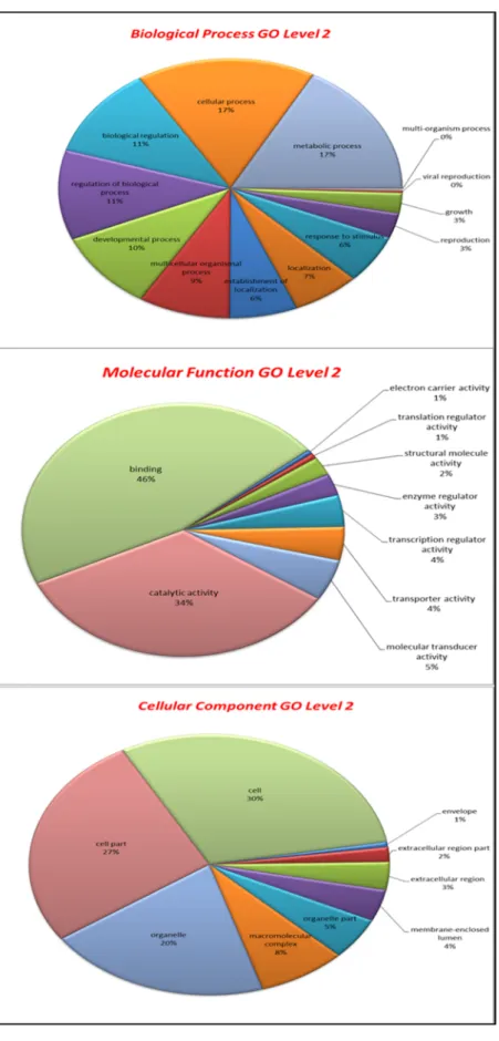
Figure 2: Gene ontology analysis
Figure 3: Nucleotide and deduced amino acid sequence of RpMyD88
Figure 4: Multiple alignment of RpMyD88 with other known MyD88 amino acid sequences
Figure 5: Phylogenetic analysis of RpMyD88
Figure 6: Tissue-specific expression analysis of RpMyD88 mRNA
Figure 7: Expression profiles of RpMyD88 mRNA in manila clam hemocyte after V. tapetis and LPS challenge
Figure 8: Expression profiles of RpMyD88 mRNA in manila clam gill tissue after V. tapetis and LPS challenge
Figure 9: The complete nucleotide and deduced amino acid sequences of the manila clam IκB
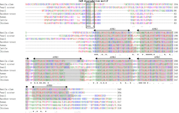
Figure 10: Multiple alignment of Rp-IκB with other selected species IκB amino acid sequences
Figure 11: The phylogenetic relationship of Rp-IκB with other selected species IκB family memebers
Figure 12: Tissue-specific expression of Rp-IκB mRNA
Figure 13: RpIκB mRNA expression in clam gill and hemocytes after LPS and V. tapetis challenge
Figure 14: The complete nucleotide and deduced amino acid sequences of the RpTNFα
Figure 15: Multiple alignment of RpTNFα with other selected species TNF family amino acid sequences
- XI -
Figure 17: Tissue-specific expression analysis of RpTNFα
Figure 18: RpTNFα mRNA expression in clam hemocyte (A) and gill (B) after V.
tapetis, LPS and poly I:C challenge
Figure 19: The complete nucleotide and deduced amino acid sequences of the RpBcl-2
Figure 20: The complete nucleotide and deduced amino acid sequences of the RpBax
Figure 21: Multiple alignment of RpBcl-2 with other selected species Bcl-2 family amino acid sequences
Figure 22: Multiple alignment of RpBax with other selected species Bax family amino acid sequences
Figure 23: Phylogenetic analysis of RpBcl-2 and RpBax
Figure 24: Tissue-specific expression analysis of RpBcl-2 and RpBax mRNA
Figure 25: RpBcl-2 mRNA expression in clam hemocyte and gill after V. tapetis challenge
Figure 26: RpBax mRNA expression in clam hemocyte and gill after V. tapetis challenge
- XII -
LIST OF TABLES
Table 1: Reads analysis summary GS-FLX sequencing
Table 2: The innate immune-related genes from GS-FLX sequencing data
Table 3: Pair wise ClustalW analysis and comparison of the RpMyD88 deduced amino acid sequence
Table 4: Primers used in RpIκB expression analysis
Table 5: Primers used in RpTNFα expression analysis
Table 6: Pair wise ClustalW analysis and comparison of the RpTNFα deduced amino acid sequence
- 1 -
CHAPTER 1
Characterization of manila clam transcriptome using
454-pyrosequencing approaches
- 2 - 1.1. Introduction
Manila clam Ruditapes philippinarum is an infaunal bivalve mollusk constitutes natural population in intertidal zones of Yellow Sea bordering countries such as Korea, China and Japan and has worldwide distribution including the countries of Canada, America, Italy, France, the UK and Ireland [Goullertquer, 1997; Coughlan et al., 2009]. Manila clam is commercially important shellfish in Korea as well as in worldwide aquaculture. One of the most serious constraints to the clam aquaculture industry in Korea and worldwide is the occurrence of high mortality and poor growth due to several known infectious disease (brown ring disease, perkinsus infection), oxidative stress, physical factors and other unknown reasons resulting massive economic loss [Paillard et al., 2004; Park et al., 2001; Park et al., 2006].
In GenBank, more than 206,000 and 14,600 expressed Sequence Tags (ESTs) are available for the Pacific oyster Crassostrea gigas and the eastern oyster
Crassostrea virginica, respectively, and more than 42,500 and 19,500 ESTs for the
California mussel Mytillus californiacus and the Mediterranean mussel Mytilus
galloprovincialis, respectively. Recently, functional analysis of large-scale EST
sequencing efforts from C. gigas and M. galloprovincialis led to the publication of many ESTs that were compiled in publish-available databases such as the Gigas database and the MytliBase [Fleury et al., 2009; Venier et al., 2009]. In addition to these large-scale sequencing efforts, a project has been initiated to sequence the entire genome of the Pacific oyster using next-generation sequencing technique [Zhang et al., 2011a].
- 3 -
species, there is still a lack of data available. The manila clam R. philippinarum, is also an economically important shellfish, but to date only a few fully-annotated genomic data are available in genomic databases.
In the last few years, global genomic approaches have been applied successfully to the immune cells of bivalve species to better understand the molecular basis for the host immune response to challenges by Vibrio species [Gueguen et al., 2003; Roberts et al., 2009; Araya et al., 2010; De Lorgeril et al., 2011]. The presence of previously-undescribed genes in these organisms has provided new insights into the molecular basis of host-pathogen interactions.
Invertebrate mollusk including clam defense mechanisms rely on an innate immune system, which employs circulating cells (hemocytes) and a large variety of molecular effectors. Bivalve immune defense against pathogens mainly occurs by major innate immune systems or components and pathways which include; pattern recognition receptors (PRRs), hemolymph coagulation, antimicrobial peptides (AMP), and respiratory burst activity. The mechanisms underlying these host defense components depend on the presence of functional proteins in appropriate quantities, within a crucial time window. These proteins are encoded by genes whose transcription is tightly coordinated by complex programs of gene expression. However, immune and stress response mechanism of manila clam is not understood well even though several immune genes have been cloned and functionally characterized.
- 4 -
clam using Roche 454 GS-FLX platform. Annotation and gene ontology analysis were then performed on assembled contigs, providing the valuable resource for future genetic and genomic research on manila clam and closely related species.
1.2. Materials and methods
1.2.1. Biological materials
Manila clams were collected from the Eastern coastal region of Jeju Island (Republic of Korea) and there after maintained in 80 L tank with aerated seawater until tissues collection.
Hemolymph (1–2 mL/clam) was collected from the pericardial cavity of healthy, unchallenged clams using a 22G syringe and the samples were immediately centrifuged (3000g at 4 °C for 10 min) to harvest the hemocytes. In order to determine the tissue specific expression, adductor muscle, mantle, siphon, gill and foot tissues were collected from three healthy, unchallenged animals and snap frozen in liquid nitrogen before storage at −80 °C.
1.2.2. RNA isolation, cDNA synthesis and normalization
The total RNA was isolated from tissues using the TRi Reagent (Sigma– Aldrich, USA). The mRNA was purified by using an mRNA isolation kit (FastTrack® 2.0; Invitrogen, USA) according to the manufacturer’s instructions. Concentration was determined at 260 nm in a UV-spectrophotometer (BioRad, USA).
- 5 -
Purified RNA samples, were diluted to 1 μg/μL and pooled to perform multi-tissue first strand cDNA synthesis using Creator SMART cDNA library construction kit (Clontech, USA). Then, amplification was carried out with the Advantage 2 PCR kit (Clontech, USA) and then resultant products were normalized using the Trimmer-Direct cDNA normalization kit (Evrogen, Russia) according to the manufacturer’s instructions.
1.2.3. Next generation cDNA sequencing and assembling of 454 sequencing data
Sequencing of normalized manila clam cDNA was carried out on a GS-FLX titanium instrument (DNA linker, Korea). Sequencing yielded 1,240,820 raw reads. Low quality 454-sequencing reads were removed by the Roche signal processing software. The remaining 797,858 (64.30%) reads were then assembled into contigs. The output reads were processed and assembled by using the Arachne whole-genome shotgun assembler program. The basic local alignment search tool (BLAST) [Altschul et al., 1990] was used to find sequence similarity to entries in public databases, including the non-redundant (NR) protein database from NCBI (http://www.ncbi.nlm.nih.gov).
1.3. Results and discussion
Samples sequenced on three full picotitre plates using the Roche 454 GS FLX platform. Analysis summary of NGS sequencing was shown in table 1. After filtering the two three data sets (L1, L2 and L3), 428,668 reads of 123,616,280
- 6 -
nucleotides were obtained in Lane 1 (L1), 422,234 reads of 130,486,649 nucleotides were obtained in Lane 2 (L2) and 389,914 reads of 111,309,109 nucleotides were obtained in Lane 3 (L3). A total of 1,240,820 raw sequencing reads with average length of 340 bp were generated. After removal of partially assembled sequences (300,429 reads), singletons (314,408), repeat sequences (9,554 reads), too short sequences (62,993 reads) and outlier sequences (55,489 reads), a total of 496,947 reads ranging from 100 bp to 600 bp were assembled average length of 351.
After assemble, 117,347 contigs were obtained, after blastx 24,327 contigs hit with NCBI database sequence. The number of bases was 19,113,449 nucleotides was matched with protein in NCBI database, which showed 785 nucleotides of average contig size and the largest contig size was 7,063 nucleotide length.
Functional genomics technologies have been widely adopted in the biological research of both model and non-model species. An efficient functional annotation of DNA or protein sequences is a major requirement for the successful application of these approaches as functional information on gene products is often the key to the interpretation of experimental results. Therefore, there is an increasing need for bioinformatics resources which are able to cope with large amount of sequence data, produce valuable annotation results and are easily accessible to laboratories where functional genomics projects are being undertaken [Gotz et al., 2008].
Functional annotation of transcripts of the combination assembly was carried out by BLASTX search against the NCBI non-redundant protein database.
- 7 -
Blast result accessions were used to retrieve associated gene names and Gene ontology (GO) terms. Gene Ontology is widely used to standardize representation of genes across species and provides a controlled vocabulary of terms for describing gene product [Salem et al., 2010]. Molecular function constituted the largest class of GO assignment of the transcripts (17,290), followed biological process (14,959) and cellular components (13,611). Level 2 GO assignments within the three categories are summarized in Fig 2. From the molecular function categories, 46% of the genes are associated with binding and 34 % are related to catalytic activity. Within the biological process category, 17% are related to cellular process followed by 17 % metabolic process. Of the cellular components, 30% of the genes are related to the cell and 27% were related to cell parts.
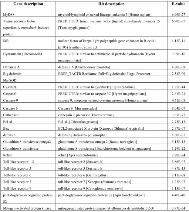
The identified major immune related genes by the contigs are presented in table 3. Table 3 lists the predicted gene name of manila clam, followed by the putative identification sequence, and E-value. We found pattern-recognition receptors (PGRPs, TLRs), inflammatory response genes (TNF, MyD88 and IκB), apoptosis-related gene (Bcl-2, Bax like gene and caspases), antimicrobial peptides (defensins and theromacin) and anti-oxidant genes (GSTs, Mn-SOD).
- 8 -
- 9 -
- 10 -
Table 2. The innate immune-related genes from GS-FLX sequencing data
Gene description Hit description E-value
MyD88 myeloid/lymphoid or mixed-lineage leukemia 5 [Homo sapiens] 6.96E-27 Tumor necrosis factor
superfamily memeber5-induced protein
PREDICTED: tumor necrosis factor (ligand) superfamily, member 15 [Taeniopygia guttata]
6.90E-07
IkB nuclear factor of kappa light polypeptide gene enhancer in B-cells 1 (p105) [synthetic construct]
1.12E-11
Hydramacin (Theromacin) PREDICTED: similar to antimicrobial peptide hydramacin [Hydra magnipapillata]
7.09E-16
Defensin A defensin A [Ornithodoros moubata] 4.80E-08
Big defensin BDEF_TACTR RecName: Full=Big defensin; Flags: Precursor 2.51E-09 Mn-SOD
CystatinB PREDICTED: similar to cystatin B [Equus caballus] 1.25E-14 Caspase3 PREDICTED: similar to caspase 3C [Hydra magnipapillata] 2.61E-23 Caspase-9 caspase 9, apoptosis-related cysteine protease [Homo sapiens] 9.51E-08
Caspase 6 Caspase 6 [Mus musculus] 8.04E-07
CathepsinC cathepsin C precursor [Ixodes ricinus] 3.67E-77
Bcl-xL Bcl-xL [Cricetulus griseus] 2.75E-13
Bax BCL2-associated X protein [Xenopus (Silurana) tropicalis] 2.97E-07
defensin defensin [Dreissena polymorpha] 1.60E-07
Glutathion S-transferase omega2 glutathione S-transferase omega 2 [Rattus norvegicus] 5.13E-13 Glutathion S-transferase glutathione S-transferase [Branchiostoma belcheri tsingtauense] 1.29E-22
Relish relish [Apis andreniformis] 2.20E-24
Toll-like receptor 2 toll-like receptor 2 [Sus scrofa] 3.86E-07 Toll-like receptor 3 toll-like receptor 3 [Sus scrofa] 4.87E-13 Toll-like receptor 6 toll-like receptor 6 [Gallus gallus] 2.11E-08 Toll-like receptor 7 toll-like receptor 7 [Xenopus (Silurana) tropicalis] 1.22E-07 Toll-like receptor 9 toll-like receptor 9 [Cynoglossus semilaevis] 1.15E-07 peptidoglycan-recognition protein
S2
peptidoglycan-recognition protein S2 [Apis koschevnikovi] 4.40E-40
- 11 -
CHAPTER II
Characterization of a novel molluscan MyD88 family
protein from manila clam, Ruditapes philippinarum.
- 12 - 2.1. Introduction
Innate immunity is evolutionarily ancient, and predates adaptive immunity. As such, it is an organism’s first line of defense against invading pathogens [Akira et al., 2006]. Innate immune responses are triggered when pathogen-associated molecular patterns (PAMPs) come into contact with host-expressed pattern recognition receptors (PRRs) [Medzhitov and Janeway, 2000; Qiu et al., 2007], which can detect and distinguish PAMPs derived from various microbial pathogens [Kawai and Akira, 2007]. These microbial components include bacterial membrane-expressed lipopolysaccharide (LPS), viral RNA, CpG DNA, flagellin, and lipoprotein.
The myeloid differentiation factor 88 (MyD88) is an evolutionarily conserved host expressed adaptor protein that is essential for proper TLR/Interluekin -1 receptor (IL-1R) immune-response signaling [Janssen and Beyaert 2002]. MyD88 consists of a Toll/IL-1 receptor (TIR) domain, located in the N-terminal region, and a death domain, at its C-terminus. TLRs are known to interact with the TIR domain and be activated through the death domain. MyD88 itself can also interact with the death domains of interleukin-1 receptor associated kinase (IRAK) family members, including IRAK1, IRAK2, IRAK4 and IRAK-M [Kawai and Akira, 2007, Akira and Takeda, 2004; West et al., 2006] to trigger downstream signaling cascades that lead to the activation of the transcription factor nuclear factor (NF)-κB.
MyD88 was first identified in 1990 as a protein that was induced during the terminal differentiation of M1D+ myeloid precursors in response to IL-6 [Lord et al.,
- 13 -
1990]. Subsequently, orthologues of this protein have been found in several species, including human [Bonnert et al., 1997], mouse [Strausberg et al., 2002], frog [Prothmann et al., 2000], zebra fish [Wheaton et al., 2007], rock bream [Whang et al., 2011], and fruit fly [Tauszig-Delamasure et al., 2001]. However, to our knowledge, molluscan MyD88 has been only reported in scallop [Qiu et al., 2007].
Manila clam, Ruditapes philippinarum, is an infaunal bivalve mollusc that is an important constituent of the intertidal zones of the Yellow Sea bordering countries such as Korea and China and Japan [Goullet, 1997; Coughlan et al., 2009]. Its worldwide distribution as a human food commodity has made it an economically-important species of the Korean aquaculture industry. Recent mass mortalities of manila clam landings, believed to be due to microbial infection, have significantly impacted production and sparked interest in immune-related research of this species [Park and Choi, 2004; Lee et al., 2001]. The manila clam is known to lack an adaptive immune system, but its mechanisms of innate immunity have yet to be fully elucidated. In contrast, the innate immune system in mammals has been extensively studied and this data may be used to provide significant insights into the immune system used by molluscs. In mammals, the MyD88-dependent TLR signaling pathway relies completely on the presence and proper activity of MyD88 adaptor protein. Therefore, we presumed that MyD88 play a similar role in the immune system of manila clam.
This study was designed to first determine the presence of a MyD88 orthologue in manila clam and subsequently characterize the function of such in response to immune challenge. We describe here our successful cloning of the
- 14 -
complete ORF sequence of manila clam MyD88 and the results of comparative analysis with other known MyD88 genes to establish the phylogenetic and evolutionally relationship. In addition, we defined the tissue-specific expression and temporal expression profile in response to immune stimulation (Vibrio tapetis and LPS).
2.2. Materials and methods
2.2.1. Next-generation sequencing and identification of manila clam MyD88
We have previously established a manila clam cDNA sequence database based upon pyrosequencing data obtained from the 454 Genome Sequencer FLX platform (GS-FLXTM; Roche, USA). Briefly, total RNA was isolated from whole body of healthy manila clams using the Trizol reagent (Sigma, USA). Poly(A) mRNA was then isolated using the FastTrack® 2.0 kit (Invitrogen, USA). First-strand cDNA was generated from 1.5 g of poly(A+) RNA using a CreatorTM SMARTTM cDNA library construction kit (Clontech, USA), which was then amplified with the 50X Advantage 2 polymerase mix (Clontech). The resultant cDNA library was normalized using the Trimmer-Direct cDNA normalization kit (Evrogen, Russia). Sequencing of normalized manila clam cDNA was carried out on a GS-FLX Titanium instrument (DNA linker, Korea), and the output reads were processed and assembled by using the Arachne whole-genome shotgun assembler program [Batzoglou et al, 2002; Jaffe et al, 2003; Gnerre et al., 2009]. MyD88 gene from
- 15 -
manila clam (designated as RpMyD88) was identified by using the Basic Local Alignment Search Tool (BLAST) algorithm [Altchul et al, 1990].
2.2.2. Sequence analysis of RpMyD88
The RpMyD88 gene ORF sequence was identified using DNAssist software (Version 2.2; http://www.dnassist.com). To determine the conserved domains of the RpMyD88 predicted protein, motif scan Pfam hidden Markov models (Local models) were used (http://hits.isb-sib.ch/cgi-bin/PFSCAN). Multiple alignment of protein sequences from different species was performed by the ClustalW program (http://www.ebi.ac.uk/Tools/clustalw2/index.html), and the identity and similarity percentage was calculated using EMBOSS pairwise alignment algorithms (http://www.ebi.ac.uk/Tools/emboss/align/). The phylogenetic tree was constructed by the Neighbor-Joining method using molecular evolutionary genetic analysis software (MEGA, v5.03; http://www.megasoftware.net).
2.2.3. Experimental animals and immune challenge
Manila clams, with average shell length of 3.5-4.0 cm, were collected from the Eastern coastal region of Jeju Island (Republic of Korea). In the laboratory, the clams were maintained at 21±1°C in a flat bottom tank with recirculating seawater. All animals were allowed to acclimatize for one week before any experimentation.
To determine the normal tissue-specific expression of the RpMyD88 gene the following tissues and hemocytes were collected from three un-challenged manila clams: adductor muscle, mantle, siphon, gill and foot. The clam hemolymph was
- 16 -
collected from the posterior adductor muscle sinus using 1 ml syringes with 26-gauge needles through the shell hinge.The hemolymph was immediately centrifuged (3500
rpm for 10 min at 4 °C) and the hemocytes were obtained after removal of supernatant.
To evaluate the immune-responsive expression of RpMyD88 gene, whole bacteria (Vibrio tapetis, a common manila clam gram-negative pathogen) and purified endotoxin (LPS; E. coli 0127:B8; Sigma, USA) were selected for controlled exposure. The V. tapetis [KCTC no. 12728] was obtained from the Korean Collection for Type Culture (KCTC). V. tapetis and LPS were diluted in phosphate buffer saline (PBS) to make stock. Clams were intramuscularly injected with 100 μL (3.2 X 10⁷ cfu individual-1) of V. tapetis and LPS (50 μg individual-1) stock into live clam adductor muscle. A negative control group was established as un-injected, while positive control group was injected with an equal volume (100 μL) of PBS. Gill and hemocyte samples were taken from three animals at 3, 6, 12, 24 and 48 h post-challenge. PBS control samples also were isolated at each time point. All samples were immediately snap-frozen in liquid nitrogen and stored at -70°C until use.
2.2.4. RNA extraction and cDNA synthesis
Total RNA was extracted from isolated tissues (50 mg each) using the Trizol reagent (Sigma). First-strand cDNA synthesis was carried out using 1 μg of total RNA as template with the PrimeScriptTM first strand synthesis kit (TaKaRa, Japan) by following the manufacturer’s instruction. The cDNA product was diluted 10-fold and stored at -20°C until further use. Subsequent, quantitative real-time reverse transcriptase polymerase chain reaction (qRT-PCR) was performed to determine
- 17 -
tissue-specific expression and expression changes in response to immune stimulation.
2.2.5. qPCR analysis
Two PCR primers were designed based on the clam MyD88 coding sequence (forward primer, 5’-TGA GCT GGA AGT TAA ACG AGG GCT-3’; reverse primer, 5’-TCA CCA CTC TAC GGC ATC TTG CTT-3’). Two internal control primers were synthesized to amplify β-actin (forward primer, 5’-CTC CCT TGA GAA GAG CTA CGA-3’; reverse primer, 5’-GAT ACC AGC AGA TTC CAT ACC C-3’) [Kim et al., 2008, Zhang et al., 2011]. qPCR was performed in the Thermal Cycler Dice real-time system (TP800; TaKaRa, Japan) under the following conditions: one denaturation cycle of 95°C for 3 min, followed by 45 amplification cycles of 95°C for 20 s, 58°C for 20 s, 72 °C for 30 s. The baseline was set automatically by the accompanying system software (version 2.0). The relative expression of each gene was determined by the Livak (2-∆∆CT) method [Livak and Schmittgen, 2001]. For tissue-specific expression analysis, relative expression of RpMyD88 mRNA was normalized to the expression detected in healthy adductor muscle samples. To determine expression fold-change after immune challenge, the relative expression was further compared to tissue-matched un-injected controls. All data are presented as relative mRNA expressed as means ± standard deviation (SD). Statistical difference between the control and treatment groups were determined by t-test using the SPSS 11.5 program, and differences were considered significant at P < 0.05.
- 18 - 2.3. Results
2.3.1. RpMyD88 sequence characterization
The nucleotide and predicted amino acid sequences of RpMyD88 (GenBank accession No: JF773572) are shown in Fig. 3. The RpMyD88 cDNA contains a 1,416 base pair (bp) open reading frame (ORF), which encodes a putative protein of 471 amino acid (aa) residues. The calculated molecular mass of RpMyD88 is 54 kDa and the predicted isoelectric point is 6.5.
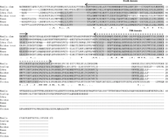
Motif scan analysis was used to identify characteristic domains of MyD88 family members. RpMyD88 was found to contain the highly conserved death domain (56DSD- RPI154) and TIR domain (180AFI-ALA263). Multiple alignment analysis of RpMyD88 to other known MyD88 family members from different species revealed a distinct nature of RpMyD88 (Fig. 4). RpMyD88 possesses the longest sequence of all MyD88s examined but also has significant sequence gaps at several positions.
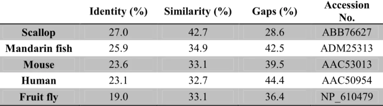
The calculated amino acid sequence identity and similarity demonstrated that RpMyD88 had the highest identity (27.0%) and similarity (42.7%) with Chlamys
farreri MyD88. Identities with mandarin fish (25.9%), mouse (23.6%), and human
(23.1%) were also significant (Table. 3.)
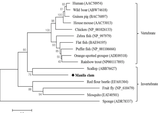
To determine the evolutionary position of RpMyD88 among the other MyD88 family members, a phylogenetic tree was constructed using Sponge MyD88 as the out-group (Fig. 5). Several other known MyD88 family members were selected from mammals, fish, and insects, along with the scallop. The results showed that MyD88 family proteins comprised two clades, those being populated by vertebrate and invertebrate MyD88s. The vertebrate clade consisted of two
sub-- 19 sub--
clades, which held the mammalian and teleost MyD88 proteins, respectively. The invertebrate clade also consisted of two sub-clades, which held mollusc and insect MyD88 proteins, respectively. The mullusc MyD88s grouped together, indicating that RpMyD88 was phylogenetically closest to scallop MyD88.
2.3.2. Tissue-specific expression of RpMyD88
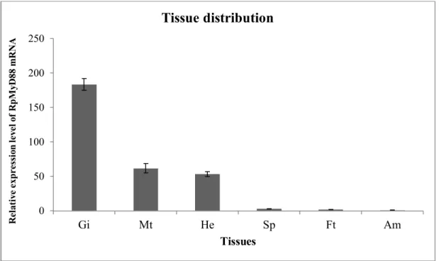
To investigate the basal transcription level of RpMyD88 in several tissues of healthy manila clams, the mRNA level was determined by means of qRT-PCR. RpMyD88 was found to be constitutively expressed in all tissues examined (hemocytes, gill, mantle, siphon, foot and adductor muscle), although the expression level was variable among each (Fig 6). The most robust level of RpMyD88 expression was detected in gill tissue. Hemocytes and mantle also showed high levels of expression, and the lowest levels were in siphon, foot and adductor muscle.
2.3.3. Expression profile of RpMyD88 after V. tapetis and LPS challenge
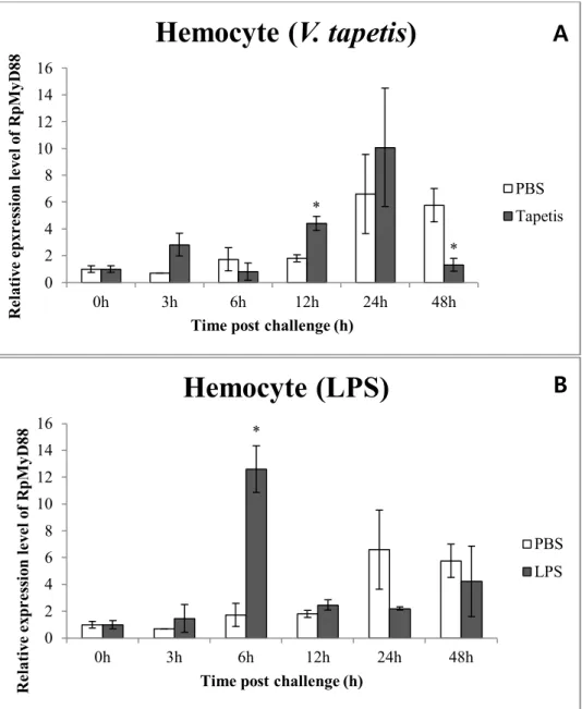
Changes in the expression profile of RpMyD88 were determined in hemocyte and gill tissues following V. tapetis and LPS challenge of live, intact animals. Following V. tapetis and LPS challenge, transcriptional up-regulation of RpMyD88 was detected by qRT-PCR for both hemocytes and gill. The expression levels of RpMyD88 in hemocyte after V. tapetis and LPS challenge, they have similar but slightly different expression patterns. RpMyD88 mRNA level was significantly up-regulated after both V. tapetis and LPS stimulation (Fig. 7). The significant difference of expression was observed at 12h (p<0.05) and 6 h (p < 0.05) after V.
- 20 -
tapetis and LPS challenge, respectively. At the other time points after challenge, no
significant difference in RpMyD88 level was found between the challenge and positive control group (p > 0.05), except that at 48 h after LPS challenge.
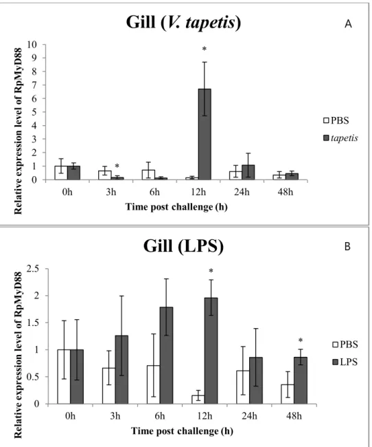
In gill tissue, mRNA of RpMyD88 was significantly up-regulated at 12 h after V. tapetis and LPS challenge, although it was down-regulated at 3 h after V.
tapetis challenge (Fig. 8). Peak expression was 6.7 (p<0.05) and 2.0 (p<0.05) -fold
increased compared to the control at 12 h after challenge. At the other time points after both V. tapetis and LPS challenge, we found no significant difference of RpMyD88 mRNA expression level.
2.4. Discussion
Molluscs have not evolved the adaptive immune system. Therefore, gaining a detailed understanding of the innate immune system is vital to developing methods to control species-related diseases. The TLR signaling pathway is one of the most important components of the mammalian and insect innate immune system; however, the molluscan Toll-like receptor signaling pathways remain under-researched and relatively unknown. When activated, all TLRs (except TLR3) recruit the adaptor protein MyD88 in order to propagate the signal. MyD88 activates IRAKs, which subsequently regulate downstream gene expression and modulate the immune response. In molluscs, only one MyD88 orthologue has been identified to date, that being from the scallop Chlamys farreri [Qiu et al., 2007].
In this study, we identified and characterized MyD88 cDNA (RpMyD88) from manila clam. Expression of this gene appears to play an important role in
TLR-- 21 TLR--
mediated signal transduction. The novel RpMyD88 gene is comprised of a 1,416 bp sequence that encodes 471 aa residues. When compared to other known MyD88 aa sequences, RpMyD88 demonstrated relatively low identity and similarity. Detailed homology analysis also indicated the presence of many gaps, indicating significant genomic deviation throughout evolution. The RpMyD88 peptide is longer than other vertebrate MyD88s. Human (AAC50954), scallop (ABB76627) and chicken (NP_001026133) MyD88 proteins are comprised of 296, 367 and 376 amino acids, respectively. Nevertheless, RpMyD88 harbors two highly conserved MyD88 characteristic regions: the death domain and TIR domain.
MyD88 is known as a universal adaptor protein having an N-terminal death domain and a C-terminal TIR domain. These two conserved domains facilitate MyD88 function as an adaptor between members of the TLR/Il-1R superfamily and downstream signaling proteins, such as IRAK [Wesche et al., 1997; Medzhitove et al., 1998; Bums et al., 1998]. The TIR domain is found almost exclusively in eukaryotic host organisms and governs dimerization between TLRs and the association between TLRs and TIR domain–containing adaptors [Barton and Medzhitov, 2003]. The death domain-containing proteins mediate protein-protein interaction, and are critically involved in apoptosis and inflammatory response-related processes [Bums et al., 1998]. Death domains are typically located in the C-terminal region [Feinstein et al., 1995], however the death domain in MyD88 and IRAKs of TLR signaling pathway are located in the N-terminal regions [Medzhitove et al., 1998]. In this study, the RpMyD88 death domain was found to be located at positions 56-154 of the N-terminal sequence. Presence of both the death domain and TIR domain in RpMyD88
- 22 -
suggest that this newly-identified protein may function in the TLR signaling pathway of the clam immune defense system.
The tissue-specific expression analysis revealed that RpMyD88 gene expression was ubiquitous in healthy clams, although its level of expression was variable among different tissues. Several reports by others have indicated that MyD88 from various species is also ubiquitously expressed. Chicken MyD88 was demonstrated to be expressed in all tested tissues, including spleen, bursa of fabricius, liver, kidney, lung, and brain [Qiu et al., 2008]. In the lower order vertebrate rock bream, MyD88 transcripts were detected in blood, gill, liver, spleen, head kidney, skin muscle, brain and intestine [Whang et al., 2011]. Another Chlamys farreri, was found to express MyD88 in normal hemocytes, muscle, mantle, heart, gonad and gill [Qiu et al., 2007].
In order to better understand the functional role of RpMyD88, especially in regards to bacterial infection and endotoxin exposure, we investigated the expression profile of RpMyD88 mRNA after immune stimulation. V. tapetis is a known pathogen of manila clam and causative agent of brown ring disease (BRD). BRD has been implicated in previous mass mortalities in cultured clam beds [Fulye-Sainte-Marie et al., 2008]. The gram-negative endotoxin LPS has also been reported as a powerful stimulator of innate immunity in various eukaryotic organisms [Qiu et al., 2007; Iwanaga, 2002]. Rock bream MyD88 has been evidenced to become up-regulated in blood, spleen and head kidney in response to experimental challenge with LPS and Edwardsiella tarda [Whang et al., 2011]. In Japanese flounder peripheral blood leukocytes, MyD88 was also found to be strongly up-regulated in
- 23 -
response to experimental exposure to LPS, PGN, and poly I:C [Takano et al., 2006]. In scallop, MyD88 was up-regulated in primary cultured hemocytes after LPS and PGN treatment [Qiu et al., 2007]. In the present study, we also found that RpMyD88 was strongly up-regulated in both hemocytes and gill after V. tapetis and LPS challenge. These findings collectively indicate that MyD88 plays an important role in vertebrate and invertebrate species in transducing the immune-response signal during pathogen invasion.
In conclusion, we identified the cDNA of MyD88 from manila clam,
Ruditapes philippinarum. The RpMyD88 contains death and TIR domains, which are
typical features of MyD88 family proteins. RpMyD88 was ubiquitously expressed in healthy manila clam tissues, and was significantly up-regulated in immune-related tissues (hemocytes and gill) in response to whole bacteria (V. tapetis) and gram-negative endotoxin (LPS) challenge. This suggested that the manila clam MyD88 may be involved in immune defense against V. tapetis infection. Sequence characterization and phylogenetic analysis further suggested that RpMyD88 may have a similar function to known vertebrate MyD88s. However, MyD88 has only been identified in one other mollusc species to date and its detailed molecular role in marine invertebrates is not yet completely understood. In order to attain a better understanding of the immunological function of RpMyD88, studies should be carried out to identify its functional interactions with other molecules of the molluscan innate immune system.
- 24 -
Table 3. Pairwise ClustalW analysis and comparison of the RpMyD88 deduced amino acid sequence
Identity (%) Similarity (%) Gaps (%) Accession No.
Scallop 27.0 42.7 28.6 ABB76627
Mandarin fish 25.9 34.9 42.5 ADM25313
Mouse 23.6 33.1 39.5 AAC53013
Human 23.1 32.7 44.4 AAC50954
- 25 -
Figure 3. Nucleotide and deduced amino acid sequence of RpMyD88. The ORF is shown in uppercase letters, and the 5’- and 3’-untranslated regions are in lowercase. Translated amino acid sequence is shown under the nucleotide sequence. The start codon (ATG) is underlined.
gaaggcggaagttatgtagcattgagttaatctt -76 taatcatttgaaatagttatagtggtcagaaacagttgtattttatgaacatttgatcaagctatatagaaaaac -1 ATGGCTACGAGAATGGATGATGATGATACACTTCAACCAGATCTTGTCCTGCCAGAAATATATACTTTTACGCCT 75 M A T R M D D D D T L Q P D L V L P E I Y T F T P 25 TTAAGGGCTCTAAGAGTAGCTTCACGACGGAAGTTGTCGGGGTTTCTGGATTTGGAAGGAGCTCTTGTTGTAGTT 150 L R A L R V A S R R K L S G F L D L E G A L V V V 50 AACCATGAGGGTGGAGACTCCGACGTTGTCAATGACTATAGTGGTTTGGCAGAACTTGCTGGATTTGATTACAAC 225 Death domain N H E G G D S D V V N D Y S G L A E L A G F D Y N 75 AGTATCATGAACATGAAAAGACAGAAAAGTCCAACTTTTGAATTACTTGAGAAGTGGACAGGACGGAATGGAACC 300 S I M N M K R Q K S P T F E L L E K W T G R N G T 100 GTGGGAAATCTATGGACGTATCTATTTTTGATGGAAAGATTTGATGTTCTTGTTGACTGCCGGCGACCAATAATA 375 V G N L W T Y L F L M E R F D V L V D C R R P I I 125 AGAGACTGTGAGAAATATGAAGAAAATCTGCAGCTAGCAGAAGATGCCGAAAGATACAGAATGGAACAAGATCCT 450 R D C E K Y E E N L Q L A E D A E R Y R M E Q D P 150 ACAGTTACAACATCTGAGATGAGAGTTGATGAGACGAAATTTGCAGCAAAAGGAGATGTTATGACCGGGAAGAAA 525 T V T T S E M R V D E T K F A A K G D V M T G K K 175 ACATTTTATGACGCCTTCATCTGTTGGTCATCTGAGGACGAGAAAGACCGCCAATTTCTGTATGAAATGATAAAT 600 TIR domain T F Y D A F I C W S S E D E K D R Q F L Y E M I N 200 GAGCTGGAAGTTAAACGAGGGCTAAAATTGTTTGTACCTGGAAGAGACGACCTGCCTGGTTCAGCTGAACACACA 675 E L E V K R G L K L F V P G R D D L P G S A E H T 225 ATCACAGCTTATCTCATAGAAGCAAGATGCCGTAGAGTGGTGATACTGATGTCCAGAACGTTTTTGCAGAGTTCT 750 I T A Y L I E A R C R R V V I L M S R T F L Q S S 250 GCGTGTGACTTTCAGGTGAAATTCGCTCATGCTCTAGCACCAGGTGCAAGAAGCAAAAAACTTATACCAGTTATC 825 A C D F Q V K F A H A L A P G A R S K K L I P V I 275 CGTGAAAAAAATACACCAATACCCAGAATCCTTCGCTTTCTGGCATTATGTGACTTCGGCAAGTCCGACATGGTT 900 R E K N T P I P R I L R F L A L C D F G K S D M V 300 GAATGGGTTTGGGACAGACTTTCTACAGCCATAATTGCGCCAATAGGACCAAACACATATTTTGAACCGGAAGAT 975 E W V W D R L S T A I I A P I G P N T Y F E P E D 325 GAGCACAATCCATTTGAACAGAGTGATGAAAGTTTAAAAGCTATAAGATTTCCAACACCCAGGCGTCTAGATGAG 1050 E H N P F E Q S D E S L K A I R F P T P R R L D E 350 CATGATGAGAGATCGGTTAATTCCGGAGCCGAACGCATACCGACAAATGTAAGCAATAGGTCCCAATTGAGAGCG 1125 H D E R S V N S G A E R I P T N V S N R S Q L R A 375 CCAACAGAACACGGTATGTCTTCAGAGGTACGGGGCCAACCAACATCCAATTATCAATCTCTTTCTTCATCTCCC 1200 P T E H G M S S E V R G Q P T S N Y Q S L S S S P 400 ACAACTTTTACTGATCACACCAAGAAGGGGGCAACTAATAAATCAAAGACCGCTACATCACAAAAGCCAAGCAAA 1275 T T F T D H T K K G A T N K S K T A T S Q K P S K 425 AAGGATAAGAAACGTGAGTCGGGCGGCATGTTCAATAAATTTAAAGAAAATTTGTGTGATCTCATTTGTACATTA 1350 K D K K R E S G G M F N K F K E N L C D L I C T L 450 AAATCAGTATTGAAAAAACCTTTTAAAGAAGTAATTTTGCTTATTTATTTTTCCATTTCTCATTGACGCTATGTA 1425 K S V L K K P F K E V I L L I Y F S I S H * 471 Gtcaagctcaatcaatctgattaatgatttaaacactttttgtgaatcattgcagagcattttgtatgagcgttc 1500 tagatgacgatacaaatgacgtcagatacacaccaagaacaaaacaaacaacattattttatgtgttcgttgttc 1575 tagccagactcattaact 1593
- 26 -
The stop codon at the end of the ORF is indicated by asterisk. Numbers to the right of each row refer to nucleotide or amino acid position, accordingly. The death domain is shaded and the TIR domain is boxed.
Figure 4. Multiple alignment of RpMyD88 with other known MyD88 amino acid sequences. The death and TIR domainsare indicated with continuous and discontinuous arrows, respectively. Scallop (ABB76627), mouse (AAC53013), human (AAC50954), chicken (NP_001026133), zebrafish (NP_997979), and fruit fly (NP_610479) are shown.
- 27 -
Figure 5. Phylogenetic analysis of RpMyD88. The number at each node indicates the percentage of bootstrapping after 1000 replications. The GenBank accession numbers of selected sequences are indicated within brackets.
- 28 -
Figure 6. Tissue-specific expression analysis of RpMyD88 mRNA. The relative mRNA expression of each tissue was normalized with that in adductor muscle. Data are presented as the mean relative expression ± standard error (SE) for three individual real-time
reactions using pooled tisssues from three individual clams. Gi, gill; Mt, mantle; He, hemocyte; Sp, Siphon; Ft, foot; Am, adductor muscle.
0 50 100 150 200 250 Gi Mt He Sp Ft Am R e lat iv e e xp r e ss io n l e v e l o f R p M y D 88 m R N A Tissues Tissue distribution
- 29 -
Figure 7. Expression profiles of RpMyD88 mRNA in manila clam hemocytes after V. tapetis (A) and LPS (B) challenge. The relative level of expression for each time point was compared to that of un-injected controls. Data are presented as the mean relative expression± SE. Significant difference (p < 0.05) across control were indicated with
an asterisk. * * 0 2 4 6 8 10 12 14 16 0h 3h 6h 12h 24h 48h R e lati v e e p x r e ss io n l e v e l o f R p M yD 88
Time post challenge (h)
Hemocyte (V. tapetis)
PBS TapetisA
* 0 2 4 6 8 10 12 14 16 0h 3h 6h 12h 24h 48h R e lati v e e x p r e ss io n l e v e l o f R p M yD 88Time post challenge (h)
Hemocyte (LPS)
PBS LPS
B
- 30 -
Figure 8. Expression profiles of RpMyD88 mRNA in manila clam gill tissue after V. tapetis (A) and LPS (B) challenge. The relative level of expression for each time point was compared to that of un-injected controls. Data are presented as the mean relative expression± SE. Significant difference (p < 0.05) across control were indicated with
an asterisk. * * 0 1 2 3 4 5 6 7 8 9 10 0h 3h 6h 12h 24h 48h R e lati v e e x p r e ss io n l e v e l o f R p M yD 88
Time post challenge (h)
Gill (V. tapetis)
PBS tapetis A * * 0 0.5 1 1.5 2 2.5 0h 3h 6h 12h 24h 48h R e lati v e e x p r e ss io n l e v e l o f R p M yD 88Time post challenge (h)
Gill (LPS)
PBS LPS B
- 31 -
Chapter III
Immune response-related gene expression of a novel
molluscan IκB protein member from manila clam,
- 32 - 3.1. Introduction
The nuclear factor-kappa B (NF-κB) proteins are dimeric transcription activators that contribute to regulation of many biological processes, such as immune response, inflammation, apoptosis, cell growth, differentiation, proliferation, tumorigenesis and embryonic morphogenesis [Pahl, 1999; Whang et al., 2009; Barkett and Gilmore, 1999]. In resting cells, NF-κB exists in its inactive form, sequestered in the cytoplasm by physical interaction with a member of the inhibitor of NF-κB (IκB) protein family. Upon IκB degradation by the IκB kinase (IKK) [Brown et al., 1993; Cordle et al., 1993; Traenckner et al., 1995], the unbound NF-κB rapidly translocates into the nucleus, where it binds to gene promoters in a sequence-specific manner and activates gene transcription [Kawagoe et al., 2009; Verma et al., 1995; Wang et al., 2001; Huxford et al., 1998].
IκB family members are characterized by the presence of three domain features: ankyrin repeats that mediate binding to NF-κB; an N-terminal regulatory region that contains two conserved serine residues and the IκB degradation motif; and, a region of sequence rich in proline (P), glutamine (E), serine (S), and threonine (T) residues (known as PEST) [Traenckner et al., 1995; Huxford et al., 1998; Rechsteiner et al., 1996]. In mammals, seven IκB members have been identified, including IκBα, IκBβ, IκBε, IκBγ, IκBδ, IκBζ, and Bcl-3 [Kawai and Akira 2007; Karin and Ben-Neriah, 2000]. IκB genes have also been found in several non-mammalian species, including the molluscs Crassostrea gigas [Montagnani et al., 2008, Zhang et al., 2011], Pinctada fucata [Zhang et al., 2009] and Euprymna
- 33 -
remain to be fully characterizaed.
The manila clam Ruditapes philippinarum represents one of the most economically-important shellfish species of the aquaculture industry worldwide, and especially in Korea [Park et al., 2006]. Over the last decade, Korean clam landings have been decreasing dramatically, presumably due to the associated upsurge in prevalence of the marine protist Perkinsus olseni [Park et al., 1999; Park et al., 2001]. However, incidence of Gram-negative bacteria Vibrio tapetis infection has risen remarkably along the west coast of Korea over this same time period [Park et al., 2006]. V. tapetis is the causative agent of brown ring disease (BRD), named as such for the obvious brown conchiolin deposits that accumulate on the infected clam’s inner shell surface [Borrego et al., 1996]. The underlying mechanism of this phenotype has been defined as V. tapetis-mediated disruption of normal periostracal lamina production, which leads to anomalous deposition of peristracum. In addition to this morphological symptom, V. tapetis infected clams experience significant weight loss, altered biochemical composition [Plana et al., 1996], depressed defense-associated activities [Allam et al., 2000; Paillard et al., 2004], and ultimately death [Allam et al., 2002].
In order to control such pathogen-associated diseases in the aquacultural mollosc population, and to enhance the yields of manila clam landings, it is necessary to research the innate immune defense mechanisms that protect clams. Since the NF-κB/IκB pathway is a very important mediator of innate immunity in molluscs, we investigated the factors involved in activating or suppressing this particular immune response. Using a genome-based sequencing approach, we
- 34 -
isolated a novel full-length IκB cDNA from manila clam, designated as Rp-IκB. The Rp-IκB mRNA expression profiles in response to bacterial challenge were determined in the immune-related tissues of gill and hemocytes.
3.2. Materials and methods
3.2.1. Next-generation sequencing
Total RNA was extracted from various tissues of manila clams using the Trizol reagent (Sigma-Aldrich, USA) and following the manufacturer’s protocol. Poly(A) mRNA was isolated using the FastTrack® 2.0 kit (Invitrogen, USA), according to the manufacturer’s instructions. Purified mRNA from various tissues was diluted to 1 g/μL and then pooled in equal amounts for use as template to synthesize multi-tissue cDNA. The full-length, enriched first-strand cDNA was generated using the CreatorTM SMARTTM cDNA library construction kit (Clontech, USA) and following the manufacturer’s instructions. Resultant cDNA was amplified in a reaction mix containing 50X Advantage 2 polymerase (Clontech) and normalized using the Trimmer-Direct cDNA normalization kit (Evrogen, Russia). The normalized cDNA was used for parallel pyrosequencing in a GS-FLX titanium automated sequencing instrument (454 Life Sciences, USA). The creation of the cDNA GS-FLX shotgun library and subsequent pyrosequencing on the next-generation sequencing platform were performed by DNA Linker (Korea). Sequencing reads were processed and assembled by the Arachne assembler algorithm [Batzoglou et al., 2002; Jaffe et al., 2003; Gnerre et al., 2009]. Unique cDNAs that
- 35 -
showed homology to known IκB family members were identified by the Basic Local Alignment Tool (BLAST) algorithm [Altchul et al., 1990].
3.2.2. Sequence characterization of Rp-IκB
The nucleotide sequence was translated to protein sequence by DNAssist 2.2 software (http://www.dnassist.soft32.com). The conserved serine domain and PEST sequence were identified by the SMART sequence analysis program
(http://smart.embl-heidelberg.de) and ePESTfind
(http://emboss.bioinformatics.nl/cgi-bin/emboss/epestfind), respectively. Multiple alignment of protein sequences was carried out by the ClustalW program (http://www.ebi.ac.uk/Tools/clustalw2/index.html), and the identity and similarity percentage of Rp-IκB were calculated using EMBOSS pairwise alignment algorithms (http://www.ebi.ac.uk/Tools/emboss/align/). The phylogenetic tree was constructed by means of the Neighbor-Joining method using molecular evolutionary genetic analysis (MEGA) software (http://www.megasoftware.net).
3.2.3. Manila clam care and tissue collection
Manila clams (shell lengths of 3.5-4 cm) were collected from the Eastern coastal region of Jeju Island (Republic of Korea). Clams were maintained in the laboratory in 80 L flat-bottom tank filled with aerated and sand-filtered seawater having salinity of 34‰ and kept at 21±1°C. All animals were acclimatized to laboratory conditions for at least one week prior to initiation of any experimental
- 36 -
procedures.
To investigate the tissue-specific expression of Rp-IκB mRNA, hemocytes, adductor muscle, siphon, mantle, gill, and foot were dissected. The clam hemolymph was harvested from the posterior adductor muscle sinus by using 1 mL syringes with 26-gauge needles placed through the shell hinge. The hemolymph was immediately centrifuged (3000 x g for 10 min at 4°C) and the pelleted hemocytes were collected after removal of supernatant. All of the tissue samples and hemocytes were immediately snap-frozen in liquid nitrogen and stored at -70°C until RNA isolation.
3.2.3. Immune challenge
In order to determine the immune response of Rp-IκB, intact V. tapetis (no. 12728; Korean Collection for Type Culture) and purified lipopolysaccharide (LPS from Escherichia coli 0127:B8; Sigma-Aldrich) were injected directly into adductor muscle of manila clams. The V. tapetis was initially grown by culturing on solid marine agar (Difco, USA) at 25°C for 48 h, after which a single colony was selected and used to inoculate 15 mL of marine broth. The liquid culture was then grown at 25°C with shaking at 180 rpm; twenty-two hours later, the entire sample volume was centrifuged at 3500 rpm for 10 min. The resulting supernatant was removed and discarded, and the bacterial pellet was resuspended in 0.9% saline. Clams were intramuscularly injected with 100 μL (1.3 X 10⁸ cfu individual-1) of V. tapetis and LPS (100 μg individual-1) stock into live clam adductor muscle. A negative control group established as un-injected, while positive control group was injected with an