Organ Correlation in IgG4-Related Diseases
IgG4-related disease (IgG4-RD) is a potentially multiorgan disorder. In this study, clinical and serological features from 132 IgG4-RD patients were compared about organ
correlations. Underlying pathologies comprised autoimmune pancreatitis (AIP) in 85 cases, IgG4-related sclerosing cholangitis (IgG4-SC) in 12, IgG4-related sialadenitis (IgG4-SIA) in 56, IgG4-related dacryoadenitis (IgG4-DAC) in 38, IgG4-related lymphadenopathy (IgG4- LYM) in 20, IgG4-related retroperitoneal fibrosis (IgG4-RF) in 19, IgG4-related kidney disease (IgG4-KD) in 6, IgG4-related pseudotumor (IgG4-PT) in 3. Sixty-five patients (49%) had multiple IgG4-RD (two affected organs in 36 patients, three in 19, four in 8, five in 1, and six in 1). Serum IgG4 levels were significantly higher with multiple lesions than with a single lesion (P < 0.001). The proportion of association with other IgG4-RD was 42% in AIP, the lowest of all IgG4-RDs. Serum IgG4 level was lower in AIP than in other IgG4-RDs.
Frequently associated IgG4-RDs were SIA (25%) and DAC (12%) for AIP; AIP (75%) for IgG4-SC; DAC (57%), AIP (38%) and LYM (27%) for IgG4-SIA; AIP (26%) and LYM (26%) for IgG4-DAC; SIA (75%), DAC (50%) and AIP (45%) for IgG4-LYM; SIA (58%), AIP (42%) and LYM (32%) for IgG4-RF; AIP (100%) and SIA (67%) for IgG4-KID; and DAC (67%) and SIA (67%) for IgG4-PT. Most associated IgG4-RD lesions were diagnosed simultaneously, but IgG4-SIA and IgG4-DAC were sometimes identified before other lesions. About half of IgG4-RD patients had multiple IgG4-RD lesions, and some associations were seen between specific organs.
Keywords: IgG4; Pancreatitis; Cholangitis, Sclerosing Satomi Koizumi, Terumi Kamisawa,
Sawako Kuruma, Taku Tabata, Kazuro Chiba, Susumu Iwasaki, Go Kuwata, Takashi Fujiwara, Junko Fujiwara, Takeo Arakawa, Koichi Koizumi, and Kumiko Momma Department of Internal Medicine, Tokyo Metropolitan Komagome Hospital, Tokyo Japan Received: 31 October 2014
Accepted: 28 January 2015 Address for Correspondence:
Terumi Kamisawa, MD
Department of Internal Medicine, Tokyo Metropolitan Komagome Hospital, 3-18-22 Honkomagome, Bunkyo-ku, Tokyo, Japan
Tel: +81.3-3823-2101, Fax: +81.3-3824-1552 E-mail: [email protected]
http://dx.doi.org/10.3346/jkms.2015.30.6.743 • J Korean Med Sci 2015; 30: 743-748
INTRODUCTION
IgG4-related disease (IgG4-RD) is a newly recognized concept presenting a potentially multiorgan disorder. IgG4-RD is a fi- broinflammatory condition characterized by a tendency toward the formation of tumefactive lesions, elevated serum IgG4 lev- els, abundant infiltration of IgG4-positive plasma cells and lym- phocytes with fibrosis, and steroid responsiveness (1-3). The pancreas was the first organ in which IgG4-RD was identified, but the disease has now been described in virtually every organ system, including the biliary tract, salivary glands, lacrimal glands, kidneys, lungs, lymph nodes, and retroperitoneum. Clinical man- ifestations are identified in single organ in some cases, whereas others show effects on two or more organs simultaneously or metachronously (4, 5).
Although many reports have described the clinical and path- ological characteristics of individual IgG4-RDs, no investigations appear to have discussed correlations between organs involved in IgG4-RD. In this study, clinical and serological features of IgG4- RD were retrospectively examined with a focus on correlations between affected organs.
MATERIALS AND METHODS
Participants in this retrospective study were 132 patients diag- nosed with and managed for IgG4-RD at Tokyo Metropolitan Komagome Hospital from 1991 to 2013. The underlying pathol- ogies were autoimmune pancreatitis (AIP) in 85 cases, IgG4-re- lated sclerosing cholangitis (IgG4-SC) in 12, IgG4-related sial- adenitis (IgG4-SIA) in 56, IgG4-related dacryoadenitis (IgG4- DAC) in 38, IgG4-related lymphadenopathy (IgG4-LYM) in 20, IgG4-related retroperitoneal fibrosis (IgG4-RF) in 19, IgG4-re- lated kidney disease (IgG4-KD) in 6, IgG4-related pseudotumor (IgG4-PT) in 3 (pleura, n = 1; breast, n = 1; and dura, n = 1). Di- agnosis was established by a multidisciplinary approach includ- ing imaging, serological, and pathological tests. When the pa- tient was diagnosed as having one IgG4-RD, systemic examina- tion by ultrasonography and computed tomography (CT) and/
or magnetic resonance imaging were prospectively performed in each patient. Although whole-body 18F-fluorodeoxyglucose (FDG)-PET was performed in 10 AIP patients, findings from FDG-PET were not included in this study. Histopathological examinations of resected or biopsy specimens were performed for 64 organs.
Gastroenterology & Hepatology
Koizumi S, et al. • Organ Correlation in IgG4-related Diseases
All patients met definite, probable or possible criteria in the following diagnostic criteria. AIP type 1 was diagnosed using international consensus diagnostic criteria (6). IgG4-related SC was diagnosed using diagnostic criteria for IgG4-related SC 2012 (7), when the stenosis was located in the hilar and/or intrahe- patic bile duct and limited intrapancreatic bile duct stricture associated with AIP was excluded. IgG4-related RF was diag- nosed using our previously proposed criteria (8). IgG4-related KD was diagnosed using the diagnostic criteria for IgG4-related KD (9). All other IgG4-RDs were diagnosed using comprehen- sive diagnostic criteria for IgG4-RD (2). IgG4-LYM was diagnosed when lymph node swellings occurred at multiple sites, such as the neck, mediastinum, axilla and abdomen. Clinical and sero- logical features of each IgG4-RD were compared to clarify or- gan correlations.
For statistical analysis, the Mann-Whitney U test and unpaired t-test were used. When repeated comparisons were made, P val- ues were corrected using the Bonferroni method. Values of P <
0.05 were considered statistically significant.
Ethics statement
This study was approved by the institutional review board of To- kyo Metropolitan Komagome Hospital (IRB No. 7299). Inform- ed consent for invasive modalities and comprehensive informed consent for the study had been obtained.
RESULTS
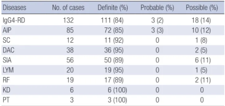
Of the 132 IgG4-RD patients, diagnoses in 111 patients were clas- sified as definite, 3 as probable, and 18 as possible (Table 1).
Sixty-seven patients had only one IgG4-RD, while the remain- ing 65 (49%) showed multiple IgG4-RD (two diseases in 36 pa- tients, three diseases in 19, four diseases in 8, five diseases in 1, and six diseases in 1). Serum IgG4 levels were significantly high- er in patients with multiple lesions (mean, 783.2 mg/dL) than in those with a single lesion (307.4 mg/dL, P < 0.001) (Table 2).
The proportion of association with other IgG4-RDs was 42%
for AIP, the lowest for all IgG4-RDs. Serum IgG4 level was lower in AIP (491 mg/dL) than in other IgG4-RDs. Steroid therapy was performed mainly for symptomatic IgG4-RD patients (62%) (Table 3).
Frequently associated IgG4-RDs were: SIA (25%) and DAC (12%) in AIP; AIP (75%) in IgG4-SC; DAC (57%), AIP (38%) and LYM (27%) in IgG4-SIA; SIA (84%), AIP (26%) and LYM (26%) in Table 1. Diagnosis of IgG4-related disease
Diseases No. of cases Definite (%) Probable (%) Possible (%)
IgG4-RD 132 111 (84) 3 (2) 18 (14)
AIP 85 72 (85) 3 (3) 10 (12)
SC 12 11 (92) 0 1 (8)
DAC 38 36 (95) 0 2 (5)
SIA 56 50 (89) 0 6 (11)
LYM 20 19 (95) 0 1 (5)
RF 19 17 (89) 0 2 (11)
KD 6 6 (100) 0 0
PT 3 3 (100) 0 0
AIP, autoimmune pancreatitis; SC, IgG4-related sclerosing cholangitis; DAC, IgG4-re- lated dacryoadenitis; SIA, IgG4-related sialadenitis; LYM, IgG4-related lymphadenop- athy; RF, IgG4-related retroperitoneal fibrosis; KD, IgG4-related kidney disease; PT, IgG4-related pseudotumor.
Table 2. Comparison of clinical and serological features between single and multiple lesions
Parameters Single
lesion Double
lesions Triple
lesions Four or more lesions No.
Male:Female 67
46:21 36
22:14 19
11:8 10
8:2 IgG4 (mg/dL)
IgG (mg/dL)
307.4 2,003.6
655.2*
2,190.9
930.9*
2,432.8
976.6*
2,644.2
*P < 0.001 compared to single lesion. Serum IgG4 levels were significantly higher in patients with multiple lesions (783.2 mg/dL) than in those with a single lesion (P < 0.01).
Table 3. Clinical feature of each organ for IgG4-related disease
Features IgG4-RD AIP SC DAC SIA LYM RF KID PT
No. association Single lesion Multiple lesion Double lesions Triple lesions Four lesions Five lesions Six lesions Rate (%)
132 67 65 36 19 8 1 1 0
85 49 36 17 13 4 1 1 42
12 3 9 6 2 1 0 0 75
38 3 35 16 10 8 1 0 92
56 7 49 24 15 8 1 1 88
20 2 18 2 9 5 1 1 90
19 2 17 6 6 3 1 1 89
6 0 6 1 2 2 0 1 100
3 1 2 0 0 1 0 1 67 Serum levels
IgG4 (mg/dL)
IgG (mg/dL) 548.3
2,094.2 491.0
2,082.0 568.0
2,402.0 724.8
2,107.8 821.3
2,432.8 979.0
2,687.4 769.1
2,526.8 524.5
1,838.3 600.0
1,967.0 Treatment
Steroid Observation Operation
82 36 14
60 (71%) 15 10
10 (83%) 1 1
21 (55%) 17
0
30 (54%) 22
4
7 (35%) 12
1
13 (68%) 6 0
5 (83%) 1 0
1 (33%) 0 2 AIP, autoimmune pancreatitis; SC, IgG4-related sclerosing cholangitis; DAC, IgG4-related dacryoadenitis; SIA, IgG4-related sialadenitis; LYM, IgG4-related lymphadenopathy; RF, IgG4-related retroperitoneal fibrosis; KD, IgG4-related kidney disease; PT, IgG4-related pseudotumor.
Fig. 1. Venn diagram showing correlation between autoimmune pancreatitis and as- sociated other IgG4-related diseases.
Autoimmune pancreatitis
IgG4-related sclerosing cholangitis
Figure 1. Correlation between autoimmune pancreatitis and associated other IgG4‐related diseases.
n= 6
n= 11 n= 9
n= 11 n= 8
n= 28 n= 10
n= 9 n= 3
n= 35 n= 21
IgG4-related lymphadenopathy
IgG4-related retroperitoneal
fibrosis IgG4-related
dacryoadenitis
IgG4-related kidney disease IgG4-related
sialadenitis
n= 85
Fig. 2. Venn diagram showing correlation between IgG4-related sialadenitis and as- sociated other IgG4-related diseases.
Figure 2. Correlation between IgG4‐related sialadenitis and associated other IgG4‐related diseases.
IgG4-related sclerosing cholangitis
Autoimmune pancreatitis IgG4-related
lymphadenopathy
IgG4-related retroperitoneal
fibrosis IgG4-related dacryoadenitis
IgG4-related kidney disease
IgG4-related sialadenitis
n= 6 n= 32
n= 11 n= 8
n= 64 n= 21 n= 56
n= 10 n= 15
n= 4 n= 5 n= 2
n= 2
Fig. 3. Venn diagram showing correlation between IgG4-related dacryoadenitis and associated other IgG4-related diseases.
other IgG4‐related diseases.
Autoimmune pancreatitis IgG4-related
sclerosing cholangitis
IgG4-related lymphadenopathy IgG4-related
retroperitoneal fibrosis
IgG4-related dacryoadenitis
IgG4-related kidney disease
IgG4-related sialadenitis
n= 32 n= 23 n= 11 n= 8
n= 11n= 1 n= 2 n= 4
n= 9 n= 11
n= 10 n= 75
n= 38
IgG4-DAC; SIA (75%), DAC (50%) and AIP (45%) in IgG4-LYM;
SIA (58%), AIP (42%) and LYM (32%) in IgG4-RF; AIP (100%) and SIA (67%) in IgG4-KID; and DAC (67%) and SIA (67%) in IgG4-PT (Fig. 1-3). Most associated IgG4-RD lesions were diag- nosed simultaneously, but IgG4-DAC and IgG4-SIA were some- times identified before other lesions (Table 4).
DISCUSSION
IgG4-RD can occur, either synchronously or metachronously, in a variety of organs throughout the body. Clinical symptoms
of IgG4-RD depend on the location of the lesion (1-5). However, the prevalence, distribution and correlation of each IgG4-RD are unknown.
This is the first study to clarify correlations between affected organs in IgG4-RD patients. A total of 49% of the 132 IgG4-RD patients had multiple IgG4-RDs. The proportion of association with another IgG4-RD was 42% in AIP, but ≥ 75% in other IgG4- RDs. Proportions of association with other IgG4-RD in AIP have been reported as 23% (10) and 63% (11), but the latter study in- cluded diabetes and rheumatoid arthritis as other organ involve- ments. According to the report by Hamano et al. (12), the pro- portion of association among AIP patients was as high as 91%, but they included hilar lymph node swelling (80%) examined by gallium scintigraphy as extrapancreatic lesions of AIP. In our study, the other IgG4-RD pathology most commonly associated with AIP was IgG4-SIA (25%), followed by IgG4-DAC (12%), IgG4- SC (11%), IgG4-LYM (10%), IgG4-RF (9%), and IgG4-KD (7%).
According to a nationwide survey of AIP in Japan, prevalence of other organ involvements in AIP patients was 53% for SC, 14%
for SIA, 13% for LYM, 8% for RF, and 7% for DAC, but SC restrict- ed to the lower bile duct was included among extrapancreatic lesions in that survey (13). The present study excluded SC re- stricted to the lower bile duct from IgG4-RD associated with AIP, as this pathology is sometimes affected by inflammation of the pancreatic head in AIP. The other prevalence is broadly compa- rable to our own results.
AIP was present in 75% of IgG4-SC, and isolated IgG4-SC was rare. IgG4-DAC and AIP were associated in 57% and 38% of pa- tients with IgG4-SIA, respectively, and IgG4-SIA and AIP were present in 84% and 26% of patients with IgG4-DAC, respective- ly. Symmetrical swelling of the salivary and/or lacrimal glands has been recognized as Mikulicz’s disease, and swelling of sali- vary glands and lacrimal glands is thus known to frequently oc-
Koizumi S, et al. • Organ Correlation in IgG4-related Diseases
cur together. In the study by Moriyama et al. (14) into Mikulicz’s disease, swelling of the salivary glands was detected in 83% of patients with swelling of the lacrimal glands, and swelling of the lacrimal glands was detected in 28% of patients with swelling of the salivary glands. Swelling of both salivary and lacrimal glands was also reportedly detected in 14% and 7% of AIP patients (13).
IgG4-SIA and IgG4-DAC were associated in 75% and 50% of pa- tients with IgG4-LYM. Salivary and lacrimal gland swelling were detected in 33% of patients with systemic IgG4-related lymph- adenopathy (15), and swelling of the lacrimal glands was detect- ed in all 3 patients with systemic IgG4-related lymphadenopa- thy (16). Although IgG4-SIA and AIP were associated in 58% and 42% of our patients with IgG4-related RF, respectively, IgG4-SIA, IgG4-DAC and AIP were reportedly associated only in 6% of 17 patients with IgG4-RF, respectively (17). AIP and IgG4-SIA were seen in 100% and 67% of our IgG4-KD patients. According to the report by Saeki et al. (18), IgG4-SIA, IgG4-DAC and AIP were present in 74%, 26%, and 35% of 43 patients with IgG4-KD. Al- though each IgG4-RD is frequently associated with other IgG4- RD, some patterns of association seem to exist between specific organs (Fig. 1-3). IgG4-SC is likely to be present with AIP, IgG4- SIA and IgG4-DAC are likely to coexist, and IgG4-LYM is likely to be associated with IgG4-DAC and IgG4-SIA.
Whole-body 18F-FDG PET is useful for detecting IgG4-RD lesions (19, 20). According to a study examining the utility of FDG-PET/CT for diagnosing IgG4-RD, 97% of 35 patients with IgG4-RD had more than one organ and 68% showed the involve- ment of three or more organs (20). In that study, IgG4-LYM, IgG4- SIA and AIP were involved in 86%, 66%, and 51% of the 35 pati- ents. Our study did not include FDG-PET findings, due to the small number of cases examined. The detection rate for IgG4- RD lesions appears rather different according to imaging mo- dalities used to screen for other IgG4-RD lesions.
Most associated IgG4-RD lesions were diagnosed simultane- ously with screening of associated other IgG4-RDs, but IgG4- DAC, and IgG4-SIA were sometimes identified before other le- sions. Whether the onset periods for each lesion differ between IgG4-RDs is unclear. However, compared with AIP or RF, swell- ing of the salivary or lacrimal glands is easily noticed even in the absence of symptoms. When IgG4-SIA or IgG4-DAC is diag- nosed, other IgG4-RDs such as AIP or RF might be present at a subclinical level.
Mean serum IgG4 level in AIP patients was 491 mg/dL, the lowest of all the IgG4-RDs. Mean serum IgG4 level in patients with Mikulicz’s disease has been reported as 1,110.0 mg/dL (21), and AIP patients with salivary or lacrimal gland involvement show higher serum IgG4 levels than those without (12, 22). How- ever, at this time, it is difficult to determine which organ involve- ment led to the elevation of serum IgG4 because there are cases with many complications. AIP patients with other IgG4-RD are also reported to have higher serum IgG4 levels than those with- Table 4. Synchronous and metachronous association of other IgG4-related diseases Associated IgG4-RD
AIP (n=85)SC (n=12)DAC (n=38)SIA (n=56)LYM (n=20)RF (n=19)KID (n=6)PT (n=3) n (%)BSAn (%)BSAn (%)BSAn (%)BSAn (%)BSAn (%)BSAn (%)BSAn (%)BSA AIP9 (75)09010 (26)1 5421 (38)21459 (45)0 728 (42)1616 (100)0601 (33)010 SC9 (11)0 901 (3)0 102 (4)0 111 (5)0 100 (0)0000 (0)0000 (0)000 DAC10 (12)4 511 (8)01032 (57)327210 (50)0 918 (43)3302 (33)0201 (33)100 SIA21 (25)51422 (17)01032 (84)227315 (75)212111 (58)3804 (67)0402 (67)020 LYM9 (10)2 701 (8)01010 (26)1 9015 (27)11226 (32)1501 (17)0102 (67)020 RF8 (9)1 610 (0)0008 (21)0 5311 (20)0 836 (3)0 512 (33)0201 (33)010 KID6 (7)0 600 (0)0002 (5)0 204 (7)0 401 (5)0 102 (11)0201 (33)010 PT1 (1)0 100 (0)0001 (3)0 012 (4)0 202 (10)0 201 (5)0101 (17)010 B, before; S, synchronous; A, after; AIP, autoimmune pancreatitis; SC, IgG4-related sclerosing cholangitis; DAC, IgG4-related dacryoadenitis; SIA, IgG4-related sialadenitis; LYM, IgG4-related lymphadenopathy; RF, IgG4-related retroperi- toneal fibrosis; KD, IgG4-related kidney disease; PT, IgG4-related pseudotumor.
out, reflecting higher disease activity (12).
There are some limitations of this study that must be consid- ered when interpreting the results. First limitation is that the study was retrospective and we could not include FDG-PET findings.
Second is that bias might be exist due to our special area of gas- troenterology. Third is that identical screening method was not applied to all the patients due to the long duration of enrolment period. However, this represents the first study to assess organ correlation in IgG4-RDs.
In conclusion, about half of IgG4-RD patients displayed mul- tiple IgG4-RD lesions, and some patterns of association appear to exist between specific affected organs.
DISCLOSURE
The authors declare that they have no conflicts of interest to dis- close.
AUTHOR CONTRIBUTION
All the authors contributed to conception and design, acquisi- tion of data, analysis and interpretation of data, drafting the ar- ticle, and revising it critically. All have approved the submission of this version.
ORCID
Satomi Koizumi http://orcid.org/0000-0002-3840-1542 Terumi Kamisawa http://orcid.org/0000-0002-2237-2767 Sawako Kuruma http://orcid.org/0000-0002-4113-2718 Taku Tabata http://orcid.org/0000-0003-1207-1087 Kazuro Chiba http://orcid.org/0000-0003-0324-859X Susumu Iwasaki http://orcid.org/0000-0001-8390-6032 Go Kuwata http://orcid.org/0000-0003-2368-230X Takashi Fujiwara http://orcid.org/0000-0002-6790-8713 Junko Fujiwara http://orcid.org/0000-0001-5820-1137 Takeo Arakawa http://orcid.org/0000-0002-3739-0386 Koichi Koizumi http://orcid.org/0000-0002-6441-5841 Kumiko Momma http://orcid.org/0000-0002-4697-1780 REFERENCES
1. Stone JH, Zen Y, Deshpande V. IgG4-related disease. N Engl J Med 2012;
366: 539-51.
2. Umehara H, Okazaki K, Masaki Y, Kawano M, Yamamoto M, Saeki T, Matsui S, Yoshino T, Nakamura S, Kawa S, et al. Comprehensive diag- nostic criteria for IgG4-related disease (IgG4-RD), 2011. Mod Rheumatol 2012; 22: 21-30.
3. Kamisawa T, Zen Y, Pillai S, Stone JH. IgG4-related disease. Lancet 2015;
385: 1460-71.
4. Kamisawa T, Funata N, Hayashi Y, Eishi Y, Koike M, Tsuruta K, Okamo-
to A, Egawa N, Nakajima H. A new clinicopathological entity of IgG4-re- lated autoimmune disease. J Gastroenterol 2003; 38: 982-4.
5. Kamisawa T, Takuma K, Egawa N, Tsuruta K, Sasaki T. Autoimmune pan- creatitis and IgG4-related sclerosing disease. Nat Rev Gastroenterol Hep- atol 2010; 7: 401-9.
6. Shimosegawa T, Chari ST, Frulloni L, Kamisawa T, Kawa S, Mino-Ke- nudson M, Kim MH, Klöppel G, Lerch MM, Löhr M, et al.; Internation- al Association of Pancreatology. International consensus diagnostic cri- teria for autoimmune pancreatitis: guidelines of the International Asso- ciation of Pancreatology. Pancreas 2011; 40: 352-8.
7. Ohara H, Okazaki K, Tsubouchi H, Inui K, Kawa S, Kamisawa T, Tazuma S, Uchida K, Hirano K, Yoshida H, et al.; Research Committee of IgG4- related Diseases; Research Committee of Intractable Diseases of Liver and Biliary Tract; Ministry of Health, Labor and Welfare, Japan; Japan Biliary Association. Clinical diagnostic criteria of IgG4-related sclerosing cholangitis 2012. J Hepatobiliary Pancreat Sci 2012; 19: 536-42.
8. Chiba K, Kamisawa T, Tabata T, Hara S, Kuruma S, Fujiwara T, Kuwata G, Egashira H, Koizumi K, Koizumi S, et al. Clinical features of 10 patients with IgG4-related retroperitoneal fibrosis. Intern Med 2013; 52: 1545-51.
9. Kawano M, Saeki T, Nakashima H, Nishi S, Yamaguchi Y, Hisano S, Ya- manaka N, Inoue D, Yamamoto M, Takahashi H, et al. Proposal for di- agnostic criteria for IgG4-related kidney disease. Clin Exp Nephrol 2011;
15: 615-26.
10. Ohara H, Nakazawa T, Sano H, Ando T, Okamoto T, Takada H, Hayashi K, Kitajima Y, Nakao H, Joh T. Systemic extrapancreatic lesions associat- ed with autoimmune pancreatitis. Pancreas 2005; 31: 232-7.
11. Okazaki K. Autoimmune pancreatitis: etiology, pathogenesis, clinical findings and treatment. The Japanese experience. JOP 2005; 6: 89-96.
12. Hamano H, Arakura N, Muraki T, Ozaki Y, Kiyosawa K, Kawa S. Preva- lence and distribution of extrapancreatic lesions complicating autoim- mune pancreatitis. J Gastroenterol 2006; 41: 1197-205.
13. Kanno A, Nishimori I, Masamune A, Kikuta K, Hirota M, Kuriyama S, Tsuji I, Shimosegawa T; Research Committee on Intractable Diseases of Pancreas. Nationwide epidemiological survey of autoimmune pan- creatitis in Japan. Pancreas 2012; 41: 835-9.
14. Moriyama M, Tanaka A, Maehara T, Ohyama Y, Shimizu M, Nakashima H, Hayashida JN, Shinozaki S, Kubo Y, Furukawa S, et al. Clinical char- acteristics of Mikulicz’s disease as an IgG4-related disease. Clin Oral In- vestig 2013; 17: 1995-2002.
15. Sato Y, Kojima M, Takata K, Morito T, Asaoku H, Takeuchi T, Mizobuchi K, Fujihara M, Kuraoka K, Nakai T, et al. Systemic IgG4-related lymph- adenopathy: a clinical and pathologic comparison to multicentric Cas- tleman’s disease. Mod Pathol 2009; 22: 589-99.
16. Cheuk W, Yuen HK, Chu SY, Chiu EK, Lam LK, Chan JK. Lymphadeno- pathy of IgG4-related sclerosing disease. Am J Surg Pathol 2008; 32: 671- 81.
17. Zen Y, Onodera M, Inoue D, Kitao A, Matsui O, Nohara T, Namiki M, Kasashima S, Kawashima A, Matsumoto Y, et al. Retroperitoneal fibro- sis: a clinicopathologic study with respect to immunoglobulin G4. Am J Surg Pathol 2009; 33: 1833-9.
18. Saeki T, Kawano M, Mizushima I, Yamamoto M, Wada Y, Nakashima H, Homma N, Tsubata Y, Takahashi H, Ito T, et al. The clinical course of pa- tients with IgG4-related kidney disease. Kidney Int 2013; 84: 826-33.
19. Ozaki Y, Oguchi K, Hamano H, Arakura N, Muraki T, Kiyosawa K, Mo- mose M, Kadoya M, Miyata K, Aizawa T, et al. Differentiation of autoim-
Koizumi S, et al. • Organ Correlation in IgG4-related Diseases
mune pancreatitis from suspected pancreatic cancer by fluorine-18 fluo- rodeoxyglucose positron emission tomography. J Gastroenterol 2008; 43:
144-51.
20. Zhang J, Chen H, Ma Y, Xiao Y, Niu N, Lin W, Wang X, Liang Z, Zhang F, Li F, et al. Characterizing IgG4-related disease with 18F-FDG PET/CT: a prospective cohort study. Eur J Nucl Med Mol Imaging 2014; 41: 1624-34.
21. Yamamoto M, Takahashi H, Ohara M, Suzuki C, Naishiro Y, Yamamoto
H, Shinomura Y, Imai K. A new conceptualization for Mikulicz’s disease as an IgG4-related plasmacytic disease. Mod Rheumatol 2006; 16: 335- 40.
22. Kubota K, Wada T, Kato S, Mozaki Y, Yoneda M, Fujita K, Takahashi H, Inamori M, Abe Y, Kobayashi N, et al. Highly active state of autoimmune pancreatitis with mikulicz disease. Pancreas 2010; 39: e6-10.