Journal of Life Science 2010 Vol. 20. No. 3. 453~456 ⓒJLS / ISSN 1225-9918
Identification of Novel Mutations in Adenosine Deaminase Gene in Korean Leukemia Patients
Kiho Park*
Department of Pediatrics, Wallace Memorial Baptist Hospital, Pusan, South Korea
Received October 13, 2009 /Accepted October 30, 2009Leukemia is the abnormal increase of hematopoietic progenitor cells in tissues, resulting in anemia, increased susceptibility to infection and impaired blood clotting. The adenosine deaminase (ADA) gene is an important druggable target for the treatment of leukemia patients. Genetic and molecular analyses were performed to determine the effects of ADA gene mutations in 20 leukemia patients in the Korean population. To analyze the relationship between genotype and phenotype, the ADA ge- nomic DNAs - including 1,092 bp of 12 exons and partial intron sequences flanking each exon - were sequenced and compared. In this study, the known mutations in other diseases, more than 50 muta- tions already reported in patients with severe combined immunodeficiency disease (SCID) and autism, were not found, but two novel mutations in leukemia patients were discovered. They include one nonsense mutation (A>C at nt position 478, F101F) and one missense mutation (G>A at nt position 778, E260K). One missense mutation (G>A at nt position 22, D8Y) was also detected in 20 normal con- trol patients (allelic frequency of 7.5%). Interestingly, subjects in the Korean population retained 2 bp insertion at the intron 6 (IVS6-52insGC), something that has never been shown in other populations.
The genetic study to find out the correlation between the mutant alleles and leukemia patients re- vealed no association statistically (p>0.05). The mutation found in leukemia needs further study to de- termine its possibility as a molecular marker for the diagnosis of leukemia.
Key words : Adenosine deaminase, mutation, leukemia
*Corresponding author
*Tel:+82-51-580-1307, Fax:+82-51-513-9166
*E-mail : [email protected]
Introduction
Adenosine deaminase (EC 3.5.4.4; ADA) is a ubiquitous enzyme that catalyzes the irreversible deamination of ad- enosine and deoxyadenosine in the purine catabolic path- way [5] and mainly participating the development and function of lymphocyte [4]. Partial sequence of ADA gene was isolated from a human T-cell lymphoblast cDNA li- brary [16] and a full-length ADA cDNA encoding a 363-amino acid protein with a molecular mass of 40 kD was also isolated [15]. In 1985, Valerio et al. determined that the ADA gene spans 32 kb and contains 12 exons [14].
By means of in situ hybridization to high resolution spreads of somatic and pachytene chromosomes, the ADA gene was confirmed to localized on 20q12-q13.11 [8].
Mutations at the ADA gene locus, in humans result in a spectrum of phenotypes.
Mutations in ADA gene causing the complete absence of enzyme activity in all cell types are associated with the fatal infantile onset syndrome of severe combined im-
munodeficiency (SCID). Mutations causing the absence of enzyme activity in erythrocytes but with retention of varia- ble amounts of enzyme activity in other cell types (partial ADA deficiency) are usually associated with grossly normal immune function. Mutations resulting in partial ADA defi- ciency but with extremely low residual enzyme activity usu- ally present as a late-onset milder and slowly progressive immunodeficiency [1,4,6,7,12]. Mutations in ADA gene have also been reported ADA activity was reduced in sera of au- tistic children compared with normal controls [2,10]. In gen- eral, the degree of enzyme deficiency, accumulation of the toxic metabolite deoxyATP and urinary excretion of the sub- strate deoxyadenosine grossly correlate with severe of disease. Consistent with this diversity, >50 variants have been identified in ADA gene, including deletions, missense, nonsense and splicing mutations, but most variants are het- eroallelic, and most alleles are rare [1].
The correlation between ADA genotypes and SCID phe- notype is well known but never examined this concept in leukemia population. The objectives of this study are to iden- tify the genetic variants of ADA gene in Korean population and to determine whether the ADA gene polymorphisms are associated with leukemia.
- Note -
454 생명과학회지 2010, Vol. 20. No. 3
Materials and Methods Study subjects
A total of 40 individuals were sampled throughout the leukemia patients and non-patients as controls. Of these, 20 were verified as leukemia patients as follows. Seven patients with acute lymphoblastic leukemia (ALL), 11 pa- tients with acute myeloid leukemia (AML), one patient with chronic myeloid leukemia (CML), and one patient with hairy cell leukemia (HCL). The other 20 were chos- en from the people has no symptoms of leukemia. The controls with a family history of leukemia were excluded.
Both parents of each study participant were reported to be of Korean descent. Without exception the participants reported both parents to be of Korean descent. This study was approved by the Scientific-Ethical Review Board of participating institution approved the experimental protocols.
PCR and Sequencing
For mutation analysis, genomic DNA was isolated from peripheral leukocytes from each patient by the QIAamp DNA blood Mimi kit (Qiagen, CA, USA) according to the manufacturer’s specification. DNA was amplified by PCR using sense and antisense primer set designed to amplify the exons 1, 2, 3, 4-5, 6, 7-9 and 10-11 from genomic DNA. Total 8 sets of primers were used from previous re- port (Jiang et al. 1997) [9] and sequenced using cycle se- quencing with a PCR products were amplified from 50 ng of genomic DNA in a 25 μl reaction system containing 2.0 mM MgCl
2, 1.0 mM dNTPs, 0.25 units of AmpliTag Gold DNA polymerase (Applied Biosystems, CA, USA), and 1X PCR buffer II; the amplification protocol was: denatura- tion 10 min at a 95
oC, then 40 amplification cycles of 95
oC for 30 sec, 65
oC for 30 sec, and 72
oC for 45 sec, followed by an extension of 10 min at 72
oC. The PCR products were purified using Microcon PCR filters (Millipore, FL, USA) and were directly sequenced in both directions us- ing BigDye Terminator kits (Applied Biosystems, CA, USA) and run on ABI 377 sequencing apparatus (ABI, MI, USA). Sequences were inspected using DNAstar (DNAstar Inc., CA, USA), unambiguously aligned using Clustal-X (UCD, Belfield, Ireland) [13] and visually inspected. PCR product includes full sequence of each exon and partial flanking intron sequences.
Results and Discussion
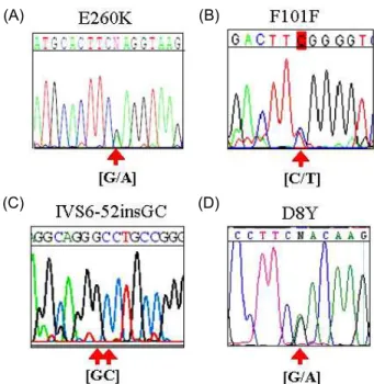
By direct sequencing analysis, all 40 Korean subjects con- sisting 20 patients with sporadic leukemia and 20 with non leukemia showed no statistical differences of the allele fre- quencies which were known in ADA gene. I initially found that three novel mutations from three sites on exon 5 (2) and 8 (2), and 2 bp insertion at intron 6 near exon 7 in Korean leukemia population (Fig. 1, Table 1). This was con- firmed by reverse direction PCR reactions. These mutations are G to A transition at the nt 778, that results in a glutamic acid to lysine substitution at codon 260 (G778A, E260K) in exon 8 (Fig. 1A) in one ALL patient and one AML patient.
The other mutation is a C to T transition at the nt 432 (C432T, F101F) in exon 5 of genomic DNA (Fig. 1B) in one AML patient. 2 bp insertion (IVS6-52insGC) was found all leuke- mia patients and also found all normal control samples (Fig.
1D). These mutations never showed in other populations.
I also detected one missense mutations in normal control, a G to A transition at the nt 22 (G22A, missense) predicting
(A)
(C)
(B)
(D)
Fig. 1. Determination of the ADA gene by sequence analysis.
A) E260K, C/G heterozygote at 778 position in two leu- kemia patients. B) F101F, T/C heterozygote at 432 posi- tion in two leukemia patients. C) IVS6-52insGC, 2 bp insertion was found in all Korean samples, but this was not reported in other ethnic groups studied before. D) D8Y, G/A mutant allele at 22 position of ADA exon 1 was found in three non-leukemia patient samples only but not in patients. The location of the mutation is in- dicated by arrow.
Journal of Life Science 2010, Vol. 20. No. 3 455
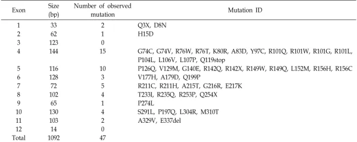
Table 1. The ADA mutations
Exon Size
(bp) Number of observed
mutation Mutation ID
12 34
56 78 109 11 Total12
3362 123144
116128 10272 13065 103 109214
21 150
103 54 14 2 470
Q3X, D8N H15D
G74C, G74V, R76W, R76T, K80R, A83D, Y97C, R101Q, R101W, R101G, R101L, P104L, L106V, L107P, Q119stop
P126Q, V129M, G140E, R142Q, R142X, R149W, R149Q, L152M, R156H, R156C V177H, A179D, Q199P
R211C, R211H, A215T, G216R, E217K T233I, R235Q, R253P, Q254X P274L
S291L, P197Q, L304R, M310T A329V, E337del
an aspartic acid to tyrosine substitution at codon 8 (D8Y).
This D8Y mutation was revealed in three non-leukemia pa- tients as heterozygote (allelic frequency of 5.0%). As men- tioned in other studies, the mutations were found as hetero- zyte and showed low frequencies [1]. The variants showed statistically non significant signal of the associations between leukemia patients group (case) and non-leukemia group (control) (p>0.05).
The sequences from 12 exons showed very conservative patterns among the samples without any statistical significance. The whole ADA genomic DNA sequences were blasted to the sequences reported [3,14]. Most of the se- quences were identical among the samples and there was no signal of the mutant alleles which were characterized as risk alleles in SCID patients [1,11] and in autism patients [2,10] (Table 1). These observations suggest that the known ADA mutations associated to the SCID are unlikely to affect genetic susceptibility to leukemia in Korean population.
In conclusion, the known about 50 mutations in the ADA gene are undetectable in Korean leukemia patients and non-leukemia patients groups. All previously reported ADA mutations in SCID, in consecutively ascertained child, this ADA mutation study is the first report related to leukemia.
In this study I found four novel mutations, and the possible involvement of these novel variations to the development of leukemia in Korean population remains to be investigated.
References
1. Arredondo-Vega, F. X., I. Santisteban, S. Daniels, S. Toutain,
and M. S. Hershfield. 1998. Adenosine deaminase defi- ciency: genotype-phenotype correlations based on ex- pressed activity of 29 mutant alleles.
Am. J. Hum. Genet.
63, 1049-1059.2. Bottini, N., D. De Luca, P. Saccucci, A. Fiumara, M. Elia, M. C. Porfirio, P. Lucarelli, and P. Curatolo. 2001. Autism:
evidence of association with adenosine deaminase genetic polymorphism.
Neurogenetics
3, 111-113.3. Bottini, N., F. Gloria-Bottini, P. Borgiani, E. Antonacci, P.
Lucarelli, and E. Bottini. 2004. Type 2 diabetes and the ge- netics of signal transduction: a study of interaction between adenosine deaminase and acid phosphatase locus 1 polymorphisms.
Metabolism
53, 995-1001.4. Cassani, B., M. Mirolo, F. Cattaneo, U. Benninghoff, M.
Hershfield, F. Carlucci, A. Tabucchi, C. Bordignon, M. G.
Roncarolo, and A. Aiuti. 2008. Altered intracellular and ex- tracellular signaling leads to impaired T-cell functions in ADA-SCID patients.
Blood
111, 4209-4219.5. Cristalli, G., S. Costanzi, C. Lambertucci, G. Lupidi, S.
Vittori, R. Volpini, and E. Camaioni. 2001. Adenosine deam- inase: functional implications and different classes of inhibitors.
Med. Res. Rev.
21, 105-128.6. Cruciani, F., L. Bernardini, P. Santolamazza, D. Modiano, A. Torroni, and R. Scozzari. 2003. Linkage disequilibrium analysis of the human adenosine deaminase (ada) gene pro- vides evidence for a lack of correlation between hot spots of equal and unequal homologous recombination.
Genomics
82, 20-33.7. Hirschhorn, R., S. Tzall, and A. Ellenbogen. 1990. Hot spot mutations in adenosine deaminase deficiency.
Proc. Natl.
Acad. Sci. USA
87, 6171-6175.8. Jhanwar, S. C., T. M. Berkvens, C. Breukel, H. van Ormondt, A. J. van der Eb, A. J. Meera, and P. Khan. 1989. Localization of human adenosine deaminase (ADA) gene sequences to the q12----q13.11 region of chromosome 20 by in situ hybridization.
Cytogenet. Cell Genet.
50, 168-171.456 생명과학회지 2010, Vol. 20. No. 3
9. Jiang, C., R. Hong, S. D. Horowitz, X. Kong, and R.
Hirschhorn. 1997. An adenosine deaminase (ADA) allele contains two newly identified deleterious mutations (Y97C and L106V) that interact to abolish enzyme activity.
Hum.
Mol. Genet.
6, 2271-2278.10. Lucarelli, P., P. Saccucci, N. Bottini, D. De Luca, A. Fiumara, M. Elia, M. C. Porfirio, and P. Curatolo. 2002. Two-loci ADA haplotypes in autistic disorder.
Am. J. Med. Genet.
108, 339-340.11. Ozsahin, H., F. X. Arredondo-Vega, I. Santisteban, H.
Fuhrer, P. Tuchschmid, W. Jochum, A. Aguzzi, H. M.
Lederman, A. Fleischman, J. A. Winkelstein, R. A. Seger, and M. S. Hershfield. 1997. Adenosine deaminase deficiency in adults.
Blood
89, 2849-2855.12. Santisteban, I., F. X. Arredondo-Vega, S. Kelly, A. Mary, A. Fischer, D. S. Hummell, A. Lawton, R. U. Sorensen, E.
R. Stiehm, and L. Uribe L. 1993. Novel splicing, missense, and deletion mutations in seven adenosine deami- nase-deficient patients with late/delayed onset of combined immunodeficiency disease. Contribution of genotype to
phenotype.
J. Clin. Invest.
92, 2291-2302.13. Thompson, J. D., T. J. Gibson, F. Plewniak, F. Jeanmougin, and D. G. Higgins. 1997. The CLUSTAL_X windows inter- face: flexible strategies for multiple sequence alignment aid- ed by quality analysis tools.
Nucleic Acids Res.
25, 4876-4882.14. Valerio, D., M. G. Duyvesteyn, B. M. Dekker, G. Weeda, T. M. Berkvens, L. van der Voorn, H. van Ormondt, and A. J. van der Eb. 1985. Adenosine deaminase: character- ization and expression of a gene with a remarkable promoter.
EMBO J.
4, 437-443.15. Valerio, D., M. G. Duyvesteyn, H. van Ormondt, P. Meera Khan, and A. J. van der Eb. 1984. Adenosine deaminase (ADA) deficiency in cells derived from humans with severe combined immunodeficiency is due to an aberration of the ADA protein.
Nucleic Acids Res.
12, 1015-1024.16. Wiginton, D. A., G. S. Adrian, R. L. Friedman, D. P. Suttle, and J. J. Hutton. 1983. Cloning of cDNA sequences of hu- man adenosine deaminase.