The Revised 2016 Korean Thyroid Association Guidelines for Thyroid Nodules and Cancers: Differences from the 2015 American Thyroid Association Guidelines
Ka Hee Yi
Department of Internal Medicine, Seoul Metropolitan Government Seoul National University Boramae Medical Center, Seoul National University College of Medicine, Seoul, Korea
Increased detection of thyroid nodules using high-resolution ultrasonography has resulted in a world-wide increase in the incidence of differentiated thyroid cancer (DTC). Despite the steep increase in its incidence, the age-standardized mortality rate of thyroid can- cer has remained stable, which leads toward a trend of more conservative treatment. The latest American Thyroid Association (ATA) guidelines for thyroid nodules and thyroid cancer revised in 2015 suggested that fine needle aspiration biopsy should be performed for thyroid nodules larger than 1 cm and lobectomy might be sufficient for 1 to 4 cm intrathyroidal DTC. In addition, active surveil- lance instead of immediate surgical treatment was also recommended as a treatment option for papillary thyroid microcarcinoma based on the results of a few observational studies from Japan. The Korean Thyroid Association (KTA) has organized a task force team to develop revised guidelines for thyroid nodules and DTC after an extensive review of articles and intense discussion on whether we should accept the changes in the 2015 ATA guidelines. This paper introduces and discusses the updated major issues and differences in the ATA and the KTA guidelines.
Keywords: Guidelines; Thyroid neoplasms; Thyroid nodule
INTRODUCTION
Thyroid nodules have become a very common clinical problem after the introduction of high resolution ultrasonography (US) in the 2000s that can detect nonpalpable nodules (incidentalo- mas). The prevalence of thyroid nodules detected by US has been reported as 19% to 68% depending on the study popula- tion [1], which resulted in an increase in the incidence of thyroid cancer since 5% to 15% of the identified nodules are malignant.
Despite the steep increase in its incidence, the age-standardized mortality rate of thyroid cancer has remained stable [2], which
leads to a shift toward more conservative approaches to the di- agnosis and treatment of this disease. In the latest American Thyroid Association (ATA) guidelines for thyroid nodules and differentiated thyroid cancer (DTC) published in early 2016 [3], fine needle aspiration (FNA) is recommended for thyroid nod- ules larger than 1 cm even with highly suspicious sonographic features while lobectomy might be sufficient for 1 to 4 cm intra- thyroidal DTC to avoid complications from total thyroidectomy.
In addition, active surveillance instead of immediate surgical treatment is also recommended as a treatment option for papil- lary thyroid microcarcinoma (PTMC) based on the results of a
Received: 9 August 2016, Revised: 16 August 2016, Accepted: 20 August 2016 Corresponding author: Ka Hee Yi
Department of Internal Medicine, Seoul Metropolitan Government Seoul National University Boramae Medical Center, Seoul National University College of Medicine, 20 Boramae-ro 5-gil, Dongjak-gu, Seoul 07061, Korea
Tel: +82-2-870-3203, Fax: +82-2-870-3866, E-mail: [email protected]
Copyright © 2016 Korean Endocrine Society
This is an Open Access article distributed under the terms of the Creative Com- mons Attribution Non-Commercial License (http://creativecommons.org/
licenses/by-nc/4.0/) which permits unrestricted non-commercial use, distribu- tion, and reproduction in any medium, provided the original work is properly cited.
few observational studies from Japan [4,5]. The Korean Thyroid Association (KTA) has organized a task force team to develop revised guidelines for thyroid nodules and thyroid cancer after an extensive review of articles including guidelines from other endocrine or thyroid associations and intense discussion on whether we should accept the changes in the 2015 ATA guide- lines. The product will be published as the revised KTA guide- lines for thyroid nodules and DTC later this year. Here, we fo- cus on three major issues in the revised KTA: (1) size criteria for FNA; (2) active surveillance as one of the treatment options for PTMC; and (3) extent of surgery including surgery for grey zone tumors.
SIZE CRITERIA FOR FINE NEEDLE ASPIRATION
The change in size criteria for FNA has drawn keen attention af- ter the publication of the clinical guidelines for thyroid nodules.
In the 2015 ATA guidelines, ultrasound stratification of thyroid nodules according to the estimated risk of malignancy was intro- duced (Table 1). FNA was recommended for nodules ≥1 cm, rather than >0.5 cm as in the 2009 ATA guidelines, that show a
high suspicion US pattern, i.e., solid hypoechoic nodules with one or more of the following features: irregular margins, micro- calcifications, taller than wide shape, extrathyroidal extension, and rim calcifications with small extrusive soft tissue compo- nents. The size criteria for intermediate (≥1.0 cm), low (≥1.5 cm), and very low suspicion (≥2.0 cm) did not drastically change. However, they strongly recommended against FNA for nodules with a benign pattern (purely cystic) and the nodules that do not meet the FNA criteria. The high suspicion sonographic features also changed; extrathyroidal extension and rim calcifi- cations with small extrusive soft tissue components are now re- garded as new suspicious features; however, hypoechogenicity and increased nodular vascularity have been excluded. More than half of benign nodules are hypoechoic in US especially when their size is small which makes nodule hypoechogenicity less specific [6]. In the 2010 KTA guideline, intranodular hyper- vascularity has already been excluded from highly suspicious patterns based on the result reported by Korean radiologists showing that intranodular vascularity was higher in benign nod- ules than in papillary thyroid cancer (PTC) [7]. The ATA also cit- ed this result as evidence to exclude increased vascularity from high suspicion sonographic patterns. The Korean Society of
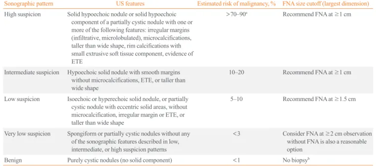
Table 1. Sonographic Patterns, Risk of Malignancy, and FNA Guidance for Thyroid Nodules in the 2015 American Thyroid Association Guidelines
Sonographic pattern US features Estimated risk of malignancy, % FNA size cutoff (largest dimension) High suspicion Solid hypoechoic nodule or solid hypoechoic
component of a partially cystic nodule with one or more of the following features: irregular margins (infiltrative, microlobulated), microcalcifications, taller than wide shape, rim calcifications with small extrusive soft tissue component, evidence of ETE
>70–90a Recommend FNA at ≥1 cm
Intermediate suspicion Hypoechoic solid nodule with smooth margins without microcalcifications, ETE, or taller than wide shape
10–20 Recommend FNA at ≥1 cm
Low suspicion Isoechoic or hyperechoic solid nodule, or partially cystic nodule with eccentric solid areas, without microcalcification, irregular margin or ETE, or taller than wide shape
5–10 Recommend FNA at ≥1.5 cm
Very low suspicion Spongiform or partially cystic nodules without any of the sonographic features described in low, intermediate, or high suspicion patterns
<3 Consider FNA at ≥2 cm observation without FNA is also a reasonable option
Benign Purely cystic nodules (no solid component) <1 No biopsyb
Adapted from Haugen et al., with permission from Mary Ann Liebert, Inc. [3].
FNA, fine needle aspiration; US, ultrasonography; ETE, extrathyroidal extension.
aThe estimate is derived from high volume centers, the overall risk of malignancy may be lower given the interobserver variability in sonography; bAspi- ration of the cyst may be considered for symptomatic or cosmetic drainage.
Thyroid Radiology also developed a new clinically feasible US risk-stratification system, the Korean Thyroid Imaging Report- ing and Data System (K-TIRADS), primarily based on the solid- ity and echogenicity of thyroid nodules by analyzing 2,000 nod- ules including 454 nodules that were pathologically proven as malignant [8,9]. Major differences in risk stratification by US between the K-TIRADS and ATA were: (1) isohyperechoic or partially cystic nodules with any suspicious features were classi- fied as intermediate suspicion (category 4), and (2) nodules of very low suspicion and in the benign group in the ATA guide- lines were combined and categorized as benign (category 2) in the K-TIRADS (Table 2, Fig. 1). Korean radiologists recom- mend FNA for category 5 nodules when the size is ≥1.0 cm;
however, the size criteria is lowered to >0.5 cm in the presence of extrathyroidal extensions, cervical lymph node or distant me- tastasis, trachea or recurrent laryngeal nerve invasion, and tumor progression.
What is the evidence for changing the size criteria for high suspicion nodules from >0.5 cm in the 2009 ATA guidelines to
≥1.0 cm in the 2015 ATA guidelines? In 2009, they referred to
a study showing that PTCs >0.5 cm had significantly higher lymph node metastasis and recurrence [10]. While in 2015, they described that PTMCs, the PTCs ≤1 cm size have disease-spe- cific mortality rates reported to be <1%, locoregional recur- rence rates of 2% to 6%, and distant recurrence rates of 1% to 2% based on Mazzaferri’s review [11]. They also insisted that these excellent outcomes are more related to the indolent nature of the disease rather than to the effectiveness of treatment. Fur- thermore, they suggested that a conservative approach of active surveillance management may be appropriate as an alternative to FNA in selected patients. However, FNA for US high suspi- cion nodules ≤1 cm size is appropriate considering that: (1) un- necessary long-term follow-up study for benign nodules with a high suspicion US pattern (approximately 20% to 40% of nod- ules with a high suspicion pattern) can be avoided; (2) a small percentage of patients with PTMC present with clinically sig- nificant regional or distant metastases; and (3) active surveil- lance for cytologically proven PTMC is also possible. For these reasons, the KTA guidelines will adopt the K-TIRADS recom- mendations to determine FNA indication instead of using the
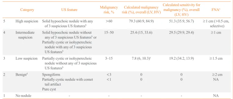
Table 2. Malignancy Risk Stratification according to K-TIRADS and FNA Indications
Category US feature Malignancy
risk, % Calculated malignancy risk (%), overall (LV, HV)
Calculated sensitivity for malignancy (%), overall
(LV, HV) FNAa
5 High suspicion Solid hypoechoic nodule with any of 3 suspicious US featuresb
>60 79.3 (60.9, 84.9) 51.3 (35.9, 56.7) ≥1 cm (>0.5 cm, selective) 4 Intermediate
suspicion Solid hypoechoic nodule without any of 3 suspicious US featuresb or Partially cystic or isohyperechoic
nodule with any of 3 suspicious US featuresb
15–50 25.4 (15, 33.6) 29.5 (29.9, 29.4) ≥1 cm
3 Low suspicion Partially cystic or isohyperechoic nodule without any of 3 suspicious US featuresb
3–15 7.8 (6, 10.3)c 19.2 (34.2, 13.9) ≥1.5 cm
2 Benignd Spongiform
Partially cystic nodule with comet tail artifact
Pure cyst
<3
<1 0
0 0
0 ≥2 cm
NA
1 No nodule - - - - NA
Adapted from Shin et al. [9]. LV and HV indicate low and high cancer volume data, respectively. Solid hypoechoic nodules include solid nodules with marked or mild hypoechogenicity.
K-TIRADS, Korean Thyroid Imaging Reporting and Data System; FNA, fine needle aspiration; US, ultrasonography; LV, low volume; HV, high vol- ume; NA, not applicable for FNA.
aFNA is indicated regardless of size and US feature of nodule in presence of poor prognostic factors including suspected lymph node metastasis by US or clinical evaluation, suspected extrathyroidal tumor extension, patients with diagnosed distant metastasis from thyroid cancer; bMicrocalcification, non- parallel orientation (taller-than-wide), spiculated/microlobulated margin; cMalignancy risk calculated from nodules excluding spongiform or partially cystic nodules with comet tail artifacts; dK-TIRADS 2 (benign category) includes partially cystic nodules with spongiform appearance or comet tail arti- facts which do not have any suspicious US feature.
ATA system.
ACTIVE SURVEILLANCE FOR PAPILLARY THYROID MICROCARCINOMA
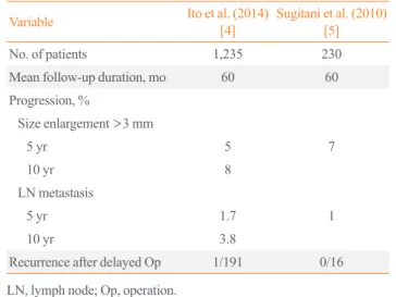
When a cytology result is diagnostic for thyroid malignancy, ac- tive surveillance as well as immediate surgery was recommend- ed in the 2015 ATA guidelines as a treatment option for patients with (1) very low risk of tumors clinically (without local inva- sion or metastasis) or cytologically (no evidence of aggressive disease), (2) high surgical risk because of comorbid conditions, or (3) a relatively short life expectancy. This recommendation is based on the results from two prospective clinical studies on ac- tive surveillance of patients with low risk PTMC from Japan started in the 1990s [4,5]. These studies enrolled 1,465 patients with biopsy-proven PTMCs that were not surgically removed and were followed for up to 15 years (average, 5 to 6 years).
Most patients showed stable tumor size after an average follow- up of 60 months whereas 5% to 7% of patients showed tumor enlargement (>3 mm) by US on 5-year follow-up, and 8%
showed this enlargement on 10-year follow-up. Additionally, 1% to 1.7% and 3.8% of patients at 5- and 10-year follow-up, respectively, showed evidence for lymph node metastases (Table 3). Furthermore, recurrence after delayed surgery was very rare (1/196 and 0/16) with an average of 6 years of follow-up. Inter- estingly, clinical progression (tumor growth or new lymph node metastasis) was related to patient age; younger patients (<40 years old) had a significantly higher progression rate (8.9%)
than patients >60 years old (1.6%) [4]. Citing these results, the 2015 ATA guidelines suggested that active surveillance could be a treatment option for PTMC patients instead of immediate sur- gery. Until now, there are no reliable clinical features (including molecular tests) that can differentiate PTMC in patients who develop progressive disease from indolent PTMC that does not cause significant disease [12-17]. Further studies are needed not only on the natural history of PTMC but also on the identifica- tion of markers indicating progressiveness or indolence in PTMC. The KTA will cautiously adopt active surveillance for selective PTMC patients as a treatment option, particularly for older patients.
Thyroid nodule
Partially cystic
Solid hypoechoic Pure cyst
Partially cystic with comet tail artifact
Spongiform
Benign (category 2) Intermediate
suspicion (category 4)
suspicionLow (category 3) suspicionHigh
(category 5)
Isohyperechoic
Any suspicious US featuresa Any suspicious US featuresa
Yes No Yes No
Fig. 1. Algorithm used in the Korean Thyroid Imaging Reporting and Data System for malignancy risk stratification based on solidity and echogenicity of thyroid nodules. Adapted from Shin et al. [9]. US, ultrasonography. aMicrocalcification, taller than wide shape, spiculated/
microlobulated margin.
Table 3. Results of Two Studies on Active Surveillance in Japan
Variable Ito et al. (2014)
[4] Sugitani et al. (2010) [5]
No. of patients 1,235 230
Mean follow-up duration, mo 60 60
Progression, %
Size enlargement >3 mm
5 yr 5 7
10 yr 8
LN metastasis
5 yr 1.7 1
10 yr 3.8
Recurrence after delayed Op 1/191 0/16
LN, lymph node; Op, operation.
SURGICAL EXTENT FOR
DIFFERENTIATED THYROID CANCER
Surgery is the most important component of treatment for DTC, and the extent of surgery has been debated for a long time. In previous guidelines including the 2009 ATA guidelines, total thyroidectomy had been endorsed as the primary initial surgical treatment for DTCs >1 cm regardless of locoregional or distant metastases based on study results showing that total thyroidec- tomy improved survival and decreased recurrence [18]. More- over, remnant ablation using radioactive iodine (RAI) which has been routinely performed to facilitate detection of recurrent or persistent disease during follow-up is much more efficient after total thyroidectomy. However, recent data have demonstrated that clinical outcomes are very similar following lobectomy or total thyroidectomy in patients with PTCs sized 1 to 2 cm and 2 to 4 cm [19-21]. Furthermore, RAI remnant ablation has not been proven to be completely efficient and improve outcomes in low to intermediate risk patients, so total thyroidectomy to facil- itate RAI therapy in these groups is needed in selective cases.
Surgical complications are another factor supporting lobecto- my instead of total thyroidectomy in the 2015 ATA guidelines.
Postoperative hypoparathyroidism and recurrent laryngeal nerve injury are well-known surgical complications after total thyroid- ectomy. Surgical complication rates naturally correlate with sur- gical volumes [22,23], although even in high volume surgeons (>100 thyroidectomies per year), complication rates were sig- nificantly higher after total thyroidectomy [24]. Based on this rationale, the 2015 ATA guidelines recommend (1) total thyroid- ectomy only for high risk patients with a tumor >4 cm and gross extrathyroidal extension (T4), clinical cervical nodes me- tastasis (cN1), or distant metastasis (M1); (2) either a bilateral or a unilateral thyroidectomy as an initial surgical treatment for DTC patients with 1 to 4 cm tumors without extrathyroidal ex- tension and without clinical node metastasis (cN0): lobectomy alone may be sufficient for low-risk DTC patients; and (3) lo- bectomy as an initial treatment for patients with <1 cm tumors without extrathyroidal extension and cN0. However, in actual clinical practice, 1 to 4 cm DTCs without extrathyroidal exten- sion and without clinical node metastasis (cN0) are so rare that total thyroidectomy would be necessary for patients with tumors in these ranges. Moreover, in Korea, surgical volume is much higher and postoperative complication rates are reported to be lower compared to other countries. The KTA guidelines flexibly accept the surgical extent for DTCs recommended by the ATA.
CONCLUSIONS
DTCs, especially PTMCs, have excellent prognosis with very low mortality, which makes it difficult to determine the most ef- ficient treatment options based on prospective, randomized con- trolled studies. Many of the published guidelines for the man- agement of thyroid nodules and DTCs were necessarily based on retrospective experiential reviews. In the 2015 ATA guide- lines, more conservative approaches to the diagnosis and treat- ment of DTCs were recommended because it was not certain if aggressive treatments were clearly beneficial rather than harm- ful for patients. The KTA will ‘flexibly’ and ‘selectively’ adopt the 2015 ATA guidelines such as active surveillance for PTMC patients and surgical extents for DTCs sized 1 to 4 cm; however, for FNA criteria according to US features, the K-TIRADS sys- tem will be used. Research to identify markers that can differen- tiate progressive from indolent PTMC as well as on the natural history of this disease should be continued in the future.
CONFLICTS OF INTEREST
No potential conflict of interest relevant to this article was re- ported.
ACKNOWLEDGMENTS
The revised KTA guidelines for the management of thyroid nodules and thyroid cancer will be published this year in the Journal of Korean Thyroid Association.
ORCID
Ka Hee Yi http://orcid.org/0000-0002-1999-9841
REFERENCES
1. Guth S, Theune U, Aberle J, Galach A, Bamberger CM. Very high prevalence of thyroid nodules detected by high frequen- cy (13 MHz) ultrasound examination. Eur J Clin Invest 2009;
39:699-706.
2. International Agency for Research on Cancer, World Health Organization. GLOBOCAN 2012: estimated cancer incidence, mortality and prevalence worldwide in 2012. Lyon: IARC;
c2016 [cited 2016 Aug 18]. Available from: http://globocan.
iarc.fr.
3. Haugen BR, Alexander EK, Bible KC, Doherty GM, Man-
del SJ, Nikiforov YE, et al. 2015 American Thyroid Associ- ation management guidelines for adult patients with thyroid nodules and differentiated thyroid cancer: the American Thyroid Association guidelines task force on thyroid nod- ules and differentiated thyroid cancer. Thyroid 2016;26:1- 133.
4. Ito Y, Miyauchi A, Kihara M, Higashiyama T, Kobayashi K, Miya A. Patient age is significantly related to the progres- sion of papillary microcarcinoma of the thyroid under obser- vation. Thyroid 2014;24:27-34.
5. Sugitani I, Toda K, Yamada K, Yamamoto N, Ikenaga M, Fujimoto Y. Three distinctly different kinds of papillary thy- roid microcarcinoma should be recognized: our treatment strategies and outcomes. World J Surg 2010;34:1222-31.
6. Moon WJ, Jung SL, Lee JH, Na DG, Baek JH, Lee YH, et al.
Benign and malignant thyroid nodules: US differentiation:
multicenter retrospective study. Radiology 2008;247:762-70.
7. Moon HJ, Kwak JY, Kim MJ, Son EJ, Kim EK. Can vascu- larity at power Doppler US help predict thyroid malignan- cy? Radiology 2010;255:260-9.
8. Na DG, Baek JH, Sung JY, Kim JH, Kim JK, Choi YJ, et al.
Thyroid imaging reporting and data system risk stratifica- tion of thyroid nodules: categorization based on solidity and echogenicity. Thyroid 2016;26:562-72.
9. Shin JH, Baek JH, Chung J, Ha EJ, Kim JH, Lee YH, et al.
Ultrasonography diagnosis and imaging-based management of thyroid nodules: revised Korean Society of Thyroid Radi- ology consensus statement and recommendations. Korean J Radiol 2016;17:370-95.
10. Noguchi S, Yamashita H, Uchino S, Watanabe S. Papillary microcarcinoma. World J Surg 2008;32:747-53.
11. Mazzaferri EL. Management of low-risk differentiated thy- roid cancer. Endocr Pract 2007;13:498-512.
12. Chow SM, Law SC, Chan JK, Au SK, Yau S, Lau WH. Pap- illary microcarcinoma of the thyroid: prognostic significance of lymph node metastasis and multifocality. Cancer 2003;98:
31-40.
13. Hay ID, Hutchinson ME, Gonzalez-Losada T, McIver B, Re- inalda ME, Grant CS, et al. Papillary thyroid microcarcino- ma: a study of 900 cases observed in a 60-year period. Sur-
gery 2008;144:980-7.
14. Roti E, degli Uberti EC, Bondanelli M, Braverman LE.
Thyroid papillary microcarcinoma: a descriptive and meta- analysis study. Eur J Endocrinol 2008;159:659-73.
15. Giordano D, Gradoni P, Oretti G, Molina E, Ferri T. Treat- ment and prognostic factors of papillary thyroid microcarci- noma. Clin Otolaryngol 2010;35:118-24.
16. Ito Y, Miyauchi A, Inoue H, Fukushima M, Kihara M, Hi- gashiyama T, et al. An observational trial for papillary thy- roid microcarcinoma in Japanese patients. World J Surg 2010;34:28-35.
17. Niemeier LA, Kuffner Akatsu H, Song C, Carty SE, Hodak SP, Yip L, et al. A combined molecular-pathologic score im- proves risk stratification of thyroid papillary microcarcino- ma. Cancer 2012;118:2069-77.
18. Bilimoria KY, Bentrem DJ, Ko CY, Stewart AK, Winchester DP, Talamonti MS, et al. Extent of surgery affects survival for papillary thyroid cancer. Ann Surg 2007;246:375-81.
19. Haigh PI, Urbach DR, Rotstein LE. Extent of thyroidectomy is not a major determinant of survival in low- or high-risk papillary thyroid cancer. Ann Surg Oncol 2005;12:81-9.
20. Nixon IJ, Ganly I, Patel SG, Palmer FL, Whitcher MM, Tut- tle RM, et al. Thyroid lobectomy for treatment of well differ- entiated intrathyroid malignancy. Surgery 2012;151:571-9.
21. Adam MA, Pura J, Gu L, Dinan MA, Tyler DS, Reed SD, et al. Extent of surgery for papillary thyroid cancer is not asso- ciated with survival: an analysis of 61,775 patients. Ann Surg 2014;260:601-5.
22. Kandil E, Noureldine SI, Abbas A, Tufano RP. The impact of surgical volume on patient outcomes following thyroid sur- gery. Surgery 2013;154:1346-52.
23. Duclos A, Peix JL, Colin C, Kraimps JL, Menegaux F, Pat- tou F, et al. Influence of experience on performance of indi- vidual surgeons in thyroid surgery: prospective cross sec- tional multicentre study. BMJ 2012;344:d8041.
24. Hauch A, Al-Qurayshi Z, Randolph G, Kandil E. Total thy- roidectomy is associated with increased risk of complications for low- and high-volume surgeons. Ann Surg Oncol 2014;
21:3844-52.