약학희지 제43권 제 2 호 198-^207(1999) Yakhak Hoeji Vol. 43. No. 2
CCI
4와 Lipopolysaccharide로 유도한 흰쥐 간 독성에 대한 YH439의 방어작용 : cytokines 및
nitric oxide 생성의 억제
김연숙 • 이종욱* • 김낙두*
서울대학교 약학대학, *유한양행 중앙연구소 (Received February 18. 1999)
YH439, a Hepatoprotective Agent, Suppresses Cytokines and Nitric Oxide Production in LPS-primed
Rats Administered with CCI 4
Youn Sook Kim, Jong Wook Lee* and Nak Doo Kim*
College of Pharmacy, Seoul N ational University, Seoul 151-742, Korea
^Central Research Institute, YuHan Co. LTD., Gunpo-Si, Kyonggi-Do 435-030, Korea
Abstract— The aim of the present investigation was to examine whether YH439, a hepatoprotective agent, exerts protective effect against hepatotoxicity and reduces the production of cytokines and NO in lipopolysaccharide (LPS)-primed rats with carbon tetrachloride (CCU). Administration of LPS fol
lowing a single dose of CCU injection resulted in remarkable elevations of the serum TNFa, IL-lp and IL- 6 level. The serum NO level was moderately elevated and severe liver damage was evidenced by in
creases in serum alanine aminotransferase (ALT) and sorbitol dehydrogenase (SDH) activities. YH439 decreased the levels of TTJF. IL-lp, IL-6. ALT, SDH as well as NO in the serum elevated by CCI4+LPS in a dose-dependent manner. Inducible nitric oxide synthase (iNOS) level was decreased in the liver of rats treated with YH439. The increased iNOS activity induced by LPS and interferon-y was significantly decreased in RAW 264.7 cells by YH439 treatment. YH439 increased the GSH level decreased by CCI4+ LPS and suppressed the ratio of GSSG/GSH. The reduction of hepatotoxicity by YH439 may be as
sociated with the decrease in the production of cytokines as well as suppression of iNOS protein in conjunction with an increase in the GSH level.
Keywords □ Tumor necrosis factor 2. interleukin-1 P, interleukin-6, nitric oxide, hepatoprotective agent, glutathione, inducible nitric oxide synthase.
CCI4는 간세포에서 CYP2E1 에 외해 CCl3 ■ (trichlo- romethyl free radical) 괴■ CCI3O2 • (peroxy radical) 로 환원 대사 되어 쿠퍼세포를 활성화시켜서 각중 cyto- kines 유리 를 촉진시 킨다. 느 은 ^ Lipopolysaccharide (LPS)에 의한 간득성외 기전은쿠퍼세포와호중구성 과 럽구가 관여하여 생성되는 TNF-a 등외 cytokines, eicosanoids, reactive oxygen species (R O S )^ 같은
본 논문에 관한 문의는 이 저자에게로 ( 전화■) 02-880-7840 ( 팩:스) 02-871-1795
매개체돌이 원인이 되는 것으로 알려져 있다.
LPS는 흰쥐의 간조직과 간세포에서 inducible n i
tric oxide synthase(iNOS)를 전사단계에서 유도하
며, ® iNOS에 의해 대량 생성된 N O는 자가면역 조직
득성외 병리에서 중요한 역할을 하며 심한 혈관 이완에
의한 septic shock 및 간 조직이나 간세포의 득성에 관
여한다.® Glutathione(GSH)은 th i이기를 가진 분자
로 세포를 산화제의 공격으로부터 보호하는데 매우 중
요하다® Edm und등에 의하면 CCU를 투여한 토끼를
LPS에 노출시켰을 때 생존율이 급격하게 감소되었으
YH439의 간 득성 방어작용
며 *® 또한 e c u 를 투여하고 LPS로 prim ing시킨 흰쥐 에서 ALT, AST, SDH 및 LDH 활성 이 상승적으로 증 가했다는 보고가 있다/^'*®
Isopropyl-2-( 1,3-dithioetane-2-ylidene)-2 [N-(4- methyl-2-thiazol-2-yl) carbamoyl] acetate (YH439) 는 합성 간장보호제로 개발되어 여러 화학적 물질로 유 발된 간득성 모델에서 효과가 있으며/®* 현재는 임상시 험 제2단계가 진행중이다. YH439는 CYP2E1 억제제이 며" " phase II 대사효소인 m EH 단백 발현과 GST 활 성을 증가 시킴이 보고되었다. 이 실험에서는 e c u 와 LPS를 병용 투여한 간염 모델을 이용하여 간 득성 메개 체로 알려져 있는 cytokines인 혈청TNF-a, IL-1, IL-6 및 N O에 미처는 YH439외 효과를 시험하였다. 또한 간 조직에서 GSH 함량 번화에 미치는 효과에 대해서도 검 토하였다.
실험방법
실험동물 - 5주령의 웅성 Sprague-Dawley 흰쥐를 대 한실 험 동물센터 에 서 구입 하여 서 울대 학교 약학대 학 실험동물 사육장에서 시 ^한 후 200~250 담이 되었을 때 실험에 사용하였다.
투여 및 시료채취 - YH439를 10, 25, 50, 100 mg/
kg외 용량으로 1일 1회, 3일간 투여후 마지막 투여2시 간째에 1.3 g/kg 용량의 e c u 를 0.25 m//100 g씩 복강 투여 하였다. CC14 투여 후 20시간째에 300 n ^ g 용 량외 LPS를 0.1 m//100 g씩 꼬리정맥에 정맥 주사하 였다. LPS 투여 후 4시간째에 채혈하여 3,500 rpm, 4°C에서 13분간 원심 분리하여 혈청을 얻었다/
간 무게 4배 용량외 50 mM Tris buffer(pH 7.4)틀 가하여 분쇄한 조직을 9,000 rpm 에서 30분, 105,000 g에서 1시간동안 초 원심 분리한 상등액인 cytos이을 분주하여 실험 시 까지 -70°C에 보관하였다.*®* 단백질 앙은 Bradford방법 으로 정 량하였다."®
혈청 A LT및 SDH활성 측정 - 혈청중의 ALT활성 은 영동제약의 측정k it를 이용하여 측정하였다.*"* 혈청 SDH활성은 Gerlach 및 Makoto의 방법에 따라, 4M fructose를 기질로 하여 366 nm 에서 2분간 롭광도 감 소를 측정하였다.^'®
혈청 TNF-a, IL -ip 및 IL-6 정량 - L 929 cell을 96-well microplate에 4xloV w ell이 되도록 가하고 37°C에서 24시간 배양하여 well에 부착되도록 하였다.
배지를 제거하고 혈청이 없는 D M EM 으로 용해한 1 H-g/m/ actinomycin D를 90 |i/씩 가하고, 혈청 시료를 10 씩 가하여 37°C에서 18시간 방치하였다, MTT assay로 L 929 cell외 세포득성을 측정하여 TNF-oc 수 준을 정량 하였다.*""" 혈청 IL-1P는 IL-1P 항체가 머 리 부착되 어 있는 ELISA k it를 사용하였다."® B 9.114 cell을 96-well microplate에 2~ 3x lOVwell이 되도 록 가하고, IL-6가 첨가되지 않은 R PM I 1640으로 희 석한 혈청 시료를 100 씩 가하여 37°C에서 68시간동 안 방치하였다. MTT assay로 세포증식을 측정하여 혈 청 IL-6 수준을 정 량 하였다.^ ®
혈청 nitrite/nitrate 정량 - 혈청 시료 4 1-1/를 NO analyzer(ANTEK model 745)에 주입하여 nitrite 및 nitrate 양을 sodium nitrite로부터 얻은 표준곡선으 로부터 농도를 환산하였다.®
iNOS 활성 측정 - RAW 264.7 cell에 0.5 [ig/ml LPS와 100 U/mZ IFN-y룰 가하여 37°C, 5% CO2 humidified incubator에서 6시간동안 배양하여 활성화 시켰다. Cytosol(단백질 100 나담에 해당)과 50 m M Tris buffer(pH 7.5), 100 nM L-arginine(200,000 dpm L-(*t: : )-arginine포함), 1 m M NADPH, 5 tetrahydrobiopterin 및 DM SO에 희석한 YH439CL0, 50 및 100 ^11^1)을 흔화하여 100 |4가 되도록 섞어서 37°C에서 25분간 shaking하여 반응시켰다. 이 반응액 에 Dowex 50W N a우 form column을 통과시 켜 얻은 상 등액 300 |ii에 Bray solution을 3 m /씩 넣어서 액체섬 광계수기로 방사눙을 측정하여 L-[^C]-citrulline 생성 량을 계산하였다.^'®
iNOS의 면역화학적 분석 - 간 cytos이을 Laemmli 방법®에 따라 Bio-Rad M ini Protean II를 이용하여 SDS-PAGE (Sodium dodesyl sulfate-polyacrylami- de gel electrophoresis)하였다. 전기 영동이 끝난 후 Davis의 방법 (1986) 에 따라 Western blot을 시행하였 다. 전이가 끝난 nitrocellulose지는 3% 탈지유 PBS용 액에 담가 4°C에서 하룻밤 방치하여 비특이성 결합을 방 지하고, iNOS의 일차항체인 rabbit anti-macropha
ge iNOS와 이차항체 인 AP conjugated goat anti
rabbit IgG, 기질인 BCIP/NBP액으로 면역화학적 분 석을 행하였다/^**
간조직내 GSH 및 GSSG 정량 - 0.6 M perchloric
add로 분쇄하여 얻은 상등액을 6.3 m M EDTA가 함유
된 0.125 M potassium phosphate buffer(pH 7.5)5.
10.2±1.4 471.4+122.9"*
9.0+2.7 1713.4+123.5*''"*'''
255.1±49Y>
203.6±46.5"*
108.0±29Y' 35.1±3.5"*
Control CCI4 (1.3 g/kg) LPS (300 |ig/kg) CCI4+LPS
+YH 439 (10 mg/kg) +YH 439 (25 mg/kg) + YH 439 (50 mg/kg) + YH 439 (100 mg/kg)
23.8±2.2 252.8+45.9'*''
21.5+2.9
1678.6±93.9"''"''^'
214.1±21.(f 180.8±27.T*>
102.9±18.3"">
39.4+3.0"*
R ats were pretreated w ith YH439 orally once a day for 3 consecutive days before intraperitoneal treatm ent of CCI4 and intravenous treatm ent of LPS. Blood samples were obtained 4 h after LPS injection. Values are represented as m ean± S.E. and percentage of decrease from CCI4+LPS. (n=8/group)
p<0.01. p<0.001 vs. control: "^<0.001 vs. CCI4:%<0.001 vs. L P S:%<0.001 vs. CCU+LPS.
희석한 시료 100 나/에 0.3 mM NADPH, 6 mM DTNB를 가하여 총부피가 1 m ; 이 되도록 하여 3(TC에 서 4분간 방치하였다. Glutathione reductase(26 U / m l) 30 롤 가하고 섞어 즉시 412 nm 에서 1분간 흡광 도 번화를 측정하였다. GSSG는 2-vinyl pyridine으로 환원형 GSH를 masking한 시료를 사용하였다.고
간조직내 glutathione reductase 활성 측정 - 간절편 을 50 mM Tris-0.154 M KCl homogenizing buffer (pH 7.4)로 분쇄하여 얻은 상등액을 사용하였다. 1 m M EDTA가 함유된 0.2 M potassium phosphate buffer(pH 7.5)에 3 m M DTNB, 2 mM NADPH를 넣 고 적당히 희석한 상등액(단백질 100 나담에 해당)을 가 하여 총부피가 1.9 m /이 되도록 하여 2~3분간 실온에 서 방치하였다. 20 mM GSSG 0,1 mZ을 가하여 412 nm 에서 1 분간 흡광도 번화를 측정했다. 상술한 medi
um 에 Glutathione reductase를 가하여 얻은 stan
dard curve로부터 농도틀 환산하였다/®
통계 처리 - 실험 결과는 m ean+ S.E.로 표시하였 으며 각 군간의 평균처는 unpaired Student's t-test 로 비교하였다.
실험결과
혈청 ALT 및 SDH활성에 대한 효과 - 흰쥐의 혈청 ALT활성이 CCLi를 투여하였을 때 대조군(23.8±2.2 SF U /L )의 10배 이상(252.8±46.0 SF U/L) 중가하 였으나 LPS에 의해서는 증가하지 않았다(21.5±2.9 SF U /L). 그러나 e c u 를 전투여하고 LPS를 처치하였 을 때는 70배(1678.6+93.9 SF U/L) 증가하였다. 이 러한 활성 중가는 10, 25, 50 및 100 mg/kg의 YH439
전투여에 의해서 각각 87, 89, 94 및 98% 억제되었다 (Table I) .
SD H 활성 역시 e c u 만 투여한 경우 대조 군(10.2+
1.4 U /L )외 46배(471.4±122.9 U/L) 중가하였으나 LPS투여 군은 대조 군과 같은 수준(9.0±2/7 U /L)이 었다. e c u 와 LPS를 병용 처리하였을 때는 대조군의 169배(1713.4±123.5 U /L) 중가하였다. YH439 10, 25, 50 및 100 mg/kg에서 각각 85, 88, 94 및 98% 억 제되 었다(Table I) .
혈청 TNF-OC, IL-lp 및 IL-6 수준에 대한 효과 - LPS투여 30분에 TNF-a외 수준은 755.6±214.4 pg/
m l 이었고 2시간 후에는 2436,6±73,7 pg/mZ로 최고
Fig. 1 -
T im e (h)
Time course of serum TNF-a concentrations in rats treated with CCI4+LPS. Rats were treated with CCI4 intraperitonealy and LPS intravenou
sly 20 h later. Blood samples were obtained 0.5. 1.
1.5. 2 and 4 hours after the LPS injection. TNF- ot concentrations were measured in L-929 fibro
blast cells by the lytic assay. Values are repre
sented as m ean±S.E.(n=8). **p<0.01, *** p<0.001 vs. control.
200 김연숙 - 이종욱 • 김낙두
Table I — Effects of YH 439 on serum alanine aminotransferase (ALT) and sorbitol dehydrogenase (SDH) ac- __________tivities in rats treated w ith CCI4. LPS. and CCU+LPS___________________________________________________
Treatment ALT (SF U/L) % Change SDH (U /L) % Change
00
%
15%12
% 6%
1%
00%
13%
11%
Wo
2%
2.9±2.9 ND
ND 51 ±24"^
80±rr*' 870±82"*"
2930±411^^ 1743±58^'
- 1412±47"'
483 ±53'* 1255±55^^
422 ±83"* 763±99^
109±19'^' 576±81 신
Control CCI4 (1.3 g/kg) LPS (300 Hg/kg) CCI4+LPS
+10 m g/kg YH 439 + 25 m g/kg YH 439 + 50 m g/kg YH 439 +100 mg/kg YH 439
Rats were pretreated w ith YH439 orally once a day for 3 consecutive days before m trapentoneal treatm ent of CCI4 and intravenous treatm ent of LPS. Blood samples were obtained 4h after LPS injection. Values are represented as m ean ± S.E. (n=8/group)
p<0.05, °오' p<0.01, p<0.001 vs. control; ^ p<0.001 vs. CCI4 and vs. L P S: p<0.001 vs. CCI4+LPS.
처에 도달하였다. 그 이후 감소하여 4시간 후에는 1249.2± 197.6 pg/m/로 감소하였다(Fig. 1). .
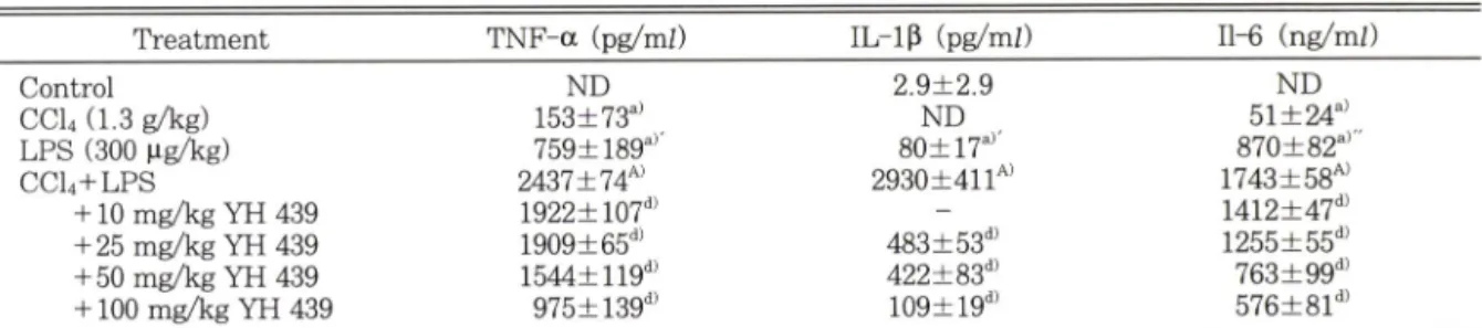
C C I 4 /LPS 투여 시 혈청 TNF-a가 가장 높았던 2시간에서 YH439의 효과률 측정하였을 때 YH439 전투여에 의해 농도 의존적으로 TNF-a 수준이 억제되었으며 10, 25, 50 및 100 mg/kg에서 각각 21, 22, 37및 60%가 억제 되 었다(Table II) .
LPS 투여 4시간후의 혈청 IL-1P 수준은 e c u 단득 투여에 의해서는 생성되지 않았고, CC1/LPS병용투여 에 외해서는 LPS단득 투여군(79.9± 16.7 pg/m O에 비해 37배 증가(2930.0±411.2 pg/m O하였다. C C l/
LPS 병용 투여군은 4시간에서 YH439의 전투여는 농 도 의존적으로 IL-1P 생성 억제효과가 있었다. 25, 50 및 100 mg/kg 투여군에서 C C l/L P S 투여 군에 비해 각각 84, 86 및 96% 억제하였다(Table II).
CCI4만 단득투여 하였을 때 IL~6가 191.0 ng/mZ까지 생성된 개체가 있었으나, LPS 단득 투여 군은 CC14 단 득 투여 군(51.1+23.5 ng/mO보다 평균 17배 더 많이 생성(870,2±81.7 ng/mO되었다. CCVLPS투여 군에 서의 혈청IL-6 수준(1743.3±57.9 ng/mO은 CCLj군의 34배, LPS군외 2배이였다. CCVLPS투여 군에서YH 439를 전투여 하였을 때 10, 25, 50 및 100 mg/kg투여 시 각각 19, 28, 56 및 67%를 억제하였다(Table II).
혈청 nitrite/nitrate 수준에 대한 효과 - CCI4 단득 투여군에서는 대조 군(48.3±8.1 비해 혈청 nitrite(N 0 2 ~)+nitrate(N0 3 -) 수준에 번화가 없었으 나(39.8±1.7 ^iM) LPS 투여 군은 대조군의 4.4배 (211.2±15.4 나M )이었다. CCL/LPS투여 군은 LPS 단득 투여군의 2배(409.2±14.9 m . p<0.001)가 생성 되었다. CC V LPS투여 군에서 YH439를 전투여 하였
Ca«(1.3o/Vo. i.p.) + LPS (300MO/Vg. I.v.)
Fig, 2 — Effects of YH439 on serum nitrite/nitrate concentrations in rats treated w ith CCI4, LPS. or CCI4+LPS. Rats were orally pretreated with YH 439 once a day for 3 consecutive days before in- traperitoneal treatment of CC14 and intravenous treatment of LPS. Blood samples were obtained 4h after LPS injection. Values are represented as mean±S.E. (n =8). ***p<0.001 vs control; *p<
0.05, ™p<0.001 vs. CCU+LPS.
을 때 10. 25. 50 및 100 mg/kg 투여시 각각 17, 15.
24 및 36% 억제하였다(Fig. 2).
1NOS 활성에 대한 효과 - LPS와 IFN-y로 활성화시 킨 RAW 264.7 세포외 cytosol에서외 iNOS활성은 3.2 [닛 CHL-citrulline pm ol/m in/m g protein 이 었다.
DM SO에 용해한 YH439를 첨가하여 측정하였을 때는 10, 50 및 100 에서 대조 군(3.2 pmol/min/mg pro- tein)에 비하여 각각 25. 41 및 40% 감소하였다. N^-ni- tro-L-arginine lOOl^iM을 첨가하였을 때는 86% 감소 (0.45+0.04 pmol/min/mg protein)하였다(Fig. 3).
iNOS단백 발현에 대한 효과 - 대조 군과 e c u 단득 투여 군에서는 iNOS가 발현되지 않았고. LPS단득 투
YH439의 간 득성 방어작용 201
Table II ~ Effect of YH 439 on serum levels of TNF-a IL-lp and IL-6 in rats treated w ith CCI4. LPS and CCI4+
LPS
Treatment TNF-a (pg/m/) IL - lp (pg/m /) 11-6 (ng/m /)
"55
Qv A vt
^
05
3 8 4 0 5
1 3 3
7»
1^
Y
to
1
1
^
±
±
±
±
±
±
±
■
^ 3 9 7 2 9 4 5 5 5 3 2 0
^ 7 1 7 4 9 9 5 9 2
1
1
1
202 김 연 숙 • 이중욱• 김낙두
100 N-L-Arg 100 LPS(1^g/ml)+IFN-r (100 U/ml)
JFig. 3 — Effects of YH439 on inducible nitric oxide syn- thase (iNOS) activities in RAW264.7 cell cytosol.
RAW264.7 cells were activated with combination of LPS and IFN-g for 6 hours. iNOS activities were measured as described in Materials and Methods. Values are represented as mean±S.E.
(n=6). V O .O l, ™p<0.001 vs. LPS+IFNPy.
여군의 간 세포에서는 단백질의 발현이 관찰되었다. 단 백발현 정도를 densitometry scanning 하였을 때 e c u /LPS 투여군외 간에서는 LPS단득 투여 군 (100%)외 1.5배 이상 진하게 iNOS단백질 수준이 유 도 발현되었다. CCiyLPS투여 군에서YH439를 전투여 한 간 세포에서는 10 mg/kg 투여시에는 감소효과가 없 었으나, 25 mg/kg 투여 군은 C O /L P S 투여 군에 비해 14%, 50 mg/kg 투여 군에서는 32%(p<0.05) 단백질 발 현이 감소되었다. 100 mg/kg을 투여하였을 때는 44%
(p<0.01) 억제되었다(Fig. 4).
GSH 및 GSSG수준에 대한 효과 - CC V LPS투여 군 (4.1+0.3 nmole/g liver)에 비해서 YH439 100 m g/kg을3일간 전투여한 후 CCL/LPS룰 투여한 군외 G SH 함량은(5.3+0.2 나mole/g liver)는 1.3배 중가 (p<0.005)하였다. GSSG수준은 YH439의 투여에 의해 감소하는 경향이 있었으나 유의성은 없었다. 그러나 총 G SH 에 대한 GSSG의 비율은 YH439 100 mg/kg의 투여로 32% 감소하였다(Fig. 5).
간조직내 Glutathione reductase 활성에 대한 효과 - 대조군의 간세포의 glutathione reductase 활성은 평균 247.6+7.5 mU/mg protein이었는데 YH439 100 mg/
kg을 3희 투여하고 24시간 후에 얻은 조직에서는 389.9
±3.8 mU/mg protein으로 증가(p<0.001)하였다. YH 439 100 mg/kg을 3일간 전투여한 후 CC1/LPS를 투여
Control CCM LPS 0 10 25 50 100 YH439 (rr^ ) CCU1.3 g/ko. i.p.) ♦ LPS<300
나g/kg, i.v.)
Fig. 4 — A) Immunoblot analysis • Effect of YH439 on the expression of iNOS protein in rats treated with e c u LPS or CCI4+LPS. Rats were pretreated with YH439 orally once a day for 3 consecutive days before intraperitoneal treatment of CCU and intravenous treatment of LPS. Liver sam
ples were obtained 4h after LPS injection. Lane 1, Control; 2. CCI4 (1.3 g/kg. i.p.): 3. LPS (300 ^lg/
kg. i.v.): 4. CCI4+LPS; 5, YH439 10 mg/kg+
CCI4+LPS:6. YH439 25 mg/kg+CCU+LPS: 7.
YH439 50 mg/kg+CCLrHLPS;8. YH439 100 mg/
kg+CCLi+LPS. B) Cumulative data obtained from 8 different experiments. The level of iNOS protein was quantified by densitometry, and ex
pressed relative to the levels in CC14+LPS treat
ed group. Each bar represents as mean+S.E.
(n =8). * Significantly different from CCI4+ LPS-treated rats. p<0.05, p<0.01.
하였을 때 Glutathione reductase외 활성 (376.0+27.7 mU/mg protein)은 YH439 비투여군(222.1±22.3 mU/mg protein)에 비헤서 glutathione reductase 활 성을 69.3% 증가시켰다(Fig. 6).
고 찰
본 연구에서는 간 득성 물질인 CC14와 과다한 염중반
응을 야기시키는 LPS를 병용투여한 간염 모델에서
YH439의 간 보호 효과에 대하여 연구하였다. CC14를
투여하고 20시간 뒤에 LPS를 투여하였을 때 CC14단득
처리에 비해 ALT는 6.6배. SDH는 3.6배 정도 중가하
였으며. LPS단득 처리에 비해서는 TNF-a는 3배, IL-
1P는 37배 및 IL-6는 2배. nitrite/nitrate는 2배.
YH439외 간 득성 방어작용 203
Control Y 100 , 0 1 00, YH439 (mg/kg)
C C l4 (1 -3 g/kg. i.p .) + L P S (3 0 0 >ig/kg, i.v.)
100,YH439 (mg/kg) 0 100 |YH439 (mg/kg)
CCI,(1.3
9/K
9, i.p ) ♦ LPS (300 M
9/kg, i.v.)
0 0 4(
1.3g/kg, i.p.) ♦ LPS (300 i.v.)
Fig. 5 — Effect of YH439 on the liver reduced glutathione (GSH) levels (a), oxidized glutathione (GSSG) levels (b) and oxidized glutathione (GSSG)/total glutathione (tGSH) (c) in rats treated with 40% PEG and CCLi+LPS. Rats were treated with YH439 (100 m g ^ ) orally once a day for 3 consecutive days. Liver samples were obtained 24 h after administration. Rats were pretreated with YH439 orally once a day for 3 consecutive days before intraperitoneal treatment of CC14 and intravenous treatment of LPS. Liver samples were obtained 4 h after LPS injection.
Values are represented as mean±S.E. (n=8). *p<0.05 vs. control 노<0.05, **p<0.01 vs. CCU+LPS.
iN O S발현은 1.5배 정도 증가하였다. 환원형 GSH도 단득 투여 시보다 더욱 감소하였다.
e c u 또는 LPS를 단득 투여 시보다 CCLi 및 LPS 를 병용 투여시, 간 득성 뿐만 아니라 염중 반응을 증폭 시키는 이유는 LPS로 활성화되는 쿠퍼세포가 RO S를 유리하여 CCI4로 인한 간 득성을 중가 시키기 때문이 다 따라서 CCU/LPS 간염 모델은 간득성을 특이 적으로 유발할 수 있는 좋은 실험 모델로 사용되어질 수 있을 것이다. YH439를 3일간 전처 리 하였을 때 용 량 의존적으로 CCLi/LPS로 증폭된 ALT와 SD H 의 활성을 억제하였다. IL-1P, TNF-a 및 IL-6등 cy- tokine은 염증 반응과 간 득성을 유발 하는 것으로 알
려저 있다. ^ YH439는 혈청 TNF-a가 최 대가 되는 LPS투여 후 2시간 째에 용량 외존적으로 억제하였으 며 따라서 LPS로 인한 TNF-a 유리를 면역 반응 초기 단계부터 억제 효과가 있는 것으로 사료된다. IL - lp 는 LPS 투여 4시간 후에 증폭되었으며 YH439는 용량 의존적으로 IL-IP 생성을 억제하였고 IL-6또한 용량 의존적으로 억제 되었다.
흰쥐 간 세포의 iN O S 발현을 면역 화학적으로 분석 한 결과 iN O S 단백 발현이 YH439투여로 완만하나 용량 외존적으로 감소되었으며 또한LPS로 활성화시킨 RAW 264.7 세포에서 iN O S외 활성을 억제하는 효과 가 있었다. 따라서 YH439는 효소활성을 직접 억제하는
cl$!l ^
/dloujrt}
_dAa s
i x
wo
50 5
1-
^
P
CI9>}16/9lt
} w
l_ 9SS9
CON Y 100 Y 0 Y 100
CCI,(1.3
9/kg,i.p.) + LPS(300 pg/kg. i.v.)
Fig. 6 — Effect of YH439 on liver glutathione reductase activities in rats treated with 40% PEG and CCI4+
LPS. Rats were pretreated with YH439 orally once a day for 3 consecutive days before in- traperitoneal treatment of CCI4 and intravenous treatment of LPS. liv er samples were obtained 4 h after LPS injection.Values are represented as mean±S.E. (n= 6^8 ). ***p<0.001 vs. control
<0.001 vs. CCI4+LPS.
작용뿐 아니라 전사단계에서 iN O S발현을 억제하는 효 과가 있는 것으로 보여지며 YH439에 외해 혈청 nitri- te 및 nitrate수준이 감소 된 것과 일치하는 결과이었 다. Endotoxin이나 TNF-a, IL-IP및 IFN-y 등의 염 중 cytokine둘은 간세포외 N OS를 중폭적으로 유도시 킨다.8■ "«>■ «> 또한 과량의 N O 는 저혈압을 유발하며 host cell인 간세포 자체의 득성을 유발하여 간득성 정 도와 상관관계가 있 다 ® 선택적인 iN O S억제제인 aminomethyHsothiourea(AE-ITU) 는 LPS를 투여 한 랫드에서 간 기능 저하룰 유외성 있게 억제하였으나 비선텍성 N OS활성 억제제인 N°-methyl-L-argi- nine(L-NMMA)은 혈청 ALT와 AST의 중가를 회복 시키지 못했다. 따라서 YH439는 ALT 및 AST활성을 억제 할뿐만 아니라 iN OS유도를 중폭시키는 작용을 갖는 TNF-a, IL-lp 및 IL-6외 생성을 억제하는 선텍 적인 iN OS 억제제로 작용하여 간질환에 개선효과가 있을 것으로 사료된다.« •훼* LPS는 체내의 monocyte, macrophage에서 superoxide등 free radical의 생성 을 중가 시켜서 lipid peroxidation과 세포득성을 유발 하는데 이때 체내에서는 GSH, glutathione peroxi
dase (GPX), superoxide dism utase(SOD) 및 ca- talase등이 득성에 대해 보호 작용을 한다. ^ TNF- a 와 IFN-y 으로 NOS를 활성화시킨 간세포에서
G SH고같과 GSSG증가가 관찰되고,"" GSH투여가 흰 쥐외 간 손상과 사망률을 억제한다는 보고가 있다/®
YH439를 단득 투여한 후 24시간 뒤에 간 조직의 총 G SH 양이 유의성 있게 중가하였으며 CC14와 LPS로 득성을 유발하였을때에 YH439 전투여는 환원형GSH 를 중가시키고 GSSG는 감소시켰다. Glutathione reductase(GR)활성도 YH439단득 투여 시나 또는 YH439를 전처 리하고e c u 와 LPS를 처 리하였을 때 모 두 유외성 있게 중가 시키는 것으로 보아 YH439는 항 산화효과가 있는 것으로 추정된다. Glutathione pe- roxidase활성은 각 군벌로 별차이를 보이지 않았으며 , C C V LPS투여 군에서만 유의성 있게 증가하였다.
이상의 결과로 보아 YH439는 염증 매개체인 TNF- 0C, IL-1P 및 IL~6와 같은cytokines의 생성을 억제하고 따라서 iN O S의 활성 및 유도를 억제하여 간 득성에 대 한 보호효과가 있을 것으로 사료된다. 또한 gluta
thione reductase활성을 중가하여 free radical의 득 성을 방어하는 간 조직의 G SH함량을 중가시켜 각종 화 학 득성 물질로 인한 간 득성에 보호효과틀 나타낼 것으 로 추정된다. GSH 중가 기전으로 Y~glutamyl-cy- stein synthetase(GCS)와 GSH synthase의 활성이 기여하는지에 대해 더 연구가 펄요하다.
감사의 말씀
본 연구는 유한 양행 중앙연구소 연구지원금으로 수 행되었으며 이에 감시■를 표합니다.
문 헌
1) Meeks, R., Harrison, R. J. and Bull, S. D. : Hepatotoxicology. CRC Press, Boc Raton, FL, 360 (1991).
2) Icho, T., Kojima, S., Shinohara, N., Kajiwara, Y., Kitabatake, K. and Kubota, K. : Protective ef
fects of tetrahydroneopteiin against free radical- induced injuiy. Biochem. Pharmacol. 45, 139 (1989).
3) Decker, T.. Lohmann-Matthes, M.L., Karck, U., Peters, T. and Decker, K. • Comparative study of cytotoxicity.tumor necrosis factor, and pros
taglandin release after stimulation of rat KupfF- er cells, murine Kupffer cells, and murine in
flammatory liver macrophages. /. Leukocyte Biol.
김연숙■이종욱• 김낙두
■
- m9 ■
^
M
§에iR씨3 31{ul£OJd OE n/ E}
0£
YH439외 간 득성 방어작용
45.
139 (1989).
4) Decker, K. : Signal paths and regualtion of su
peroxide, eicosanoid and cytokine formation in macrophages of rat liver. Biological Reactive In
termediates, vol. IV. New York. Plenum. 507 (1990).
5) Tiegs, G. and Wendel, A. : Leukotriene-medi- ated liver injury.
Biochem. Pharmacol.37. 2569 (1988).
6) Mitchell, J. A.. Kohlhaas. K. L. and Sorrentino, R. '■ Induction by endotoxin of nitric oxide syn
thase in the rat mesentery : lack of effect on ac
tion of vasoconstrictors.
Br. J. Pharmacol. 109,265 (1993).
7) Knowles. R. G.. Merrett, M.. Salter, M. and Moncada, S. ; Differential induction of brain, jung and liver nitric oxide synthase by en
dotoxin in the rat.
Biochem. J. 270.833 (1990).
8) Billiar, T. R., Curran. R. D., Stuehr, D. J., Fer
rari. F. K. and Simmons. R. L. : Evidence that the activation of Kupffer cells results in the pro
duction of L-arginine metabolites thar release cel
l-associated iron and inhibit hepatocyte proein synthesis.
Surgery 106,364 (1989).
9) Hothersall,
J . S.,Cunha, F. Q., Neild, G. H. and Norohna-Dutra, A. A. : Induction of nitric oxide synthesis in J774 cells lowers intracellular glu
tathione : effect of modulated glutathione redox status on nitric oxide synthase induction.
Biochem.J. 322.
477 (1997).
10) Farrar, W. E.. Jr., Eidson, M. and Kent. T. H. : Susceptibility of rabbits to pyrogenic and lethal effects of endotoxin after acute liver injury.
Proc.Soc. Exp. Biol. Med, 128.
711 (1968).
11) Chamulitrat, W.. Jordan, S. J. and Mason. R.
P. : Nitric oxide production during endotoxin shock in carbon tetrachloride-treated rats.
M o l Pharm. 46.391 (1994).
12) Chamulitrat, W., Blazka, M. E., Jordan. S. J., Luster, M. I. and Mason. R. P. : Tumor necrosis factor^ot and nitric oxide production in endotoxin- primed rats administered carbon tetrachloride.
Life Sciences 57,
2273 (1995).
13) Choi, E. Y.. Kim, S. G., Lee. J. W. '■ Suppression of rat hepatic cytochrome P450 2E1 expression
by isopropyl 2-( 1.3-dithioetane-2-ylidene)-2-[N- (4-methyl-thiavol-2-yl) carbamoyl]-acetate (YH 439), and experimental hepato protecta -nt : Pro
tective role against hepatic injury.
Biochem. Phar- rmcol. 52.1219 (1996).
14) Jeong. K. S. and Lee. I. J. : Transcriptional in hibition of cytochrome P450 2E1 by a synthetic compound. YH 439.
Arch. Biochem. Biophys. 326.137 (1996).
15) Oguchi, S.. lida. S.. Adachi, H.. Ohshima. H.
and Esumi, H. : Induction of Ca^^/calmodulin-de- pendent NO synthase in various organs of rats by Propionibacterium acnes and lipopolysaccha
ride treatment.
FEBS Letters 308.22 (1992).
16) Bradford. M. M. ■ A rapid and sensitive method for the quantification of protein dye biding.
A n a l Biochem. 72.248 (1976).
17) eitman, S. and Frankel, S. K. ; A colorimetric method for determination of serum glutamic ox
aloacetic and glutamic transaminase.
A m . J.Clin. Pathol. 28,
56 (1957).
18) Gerlach, U. : Sorbitol Dehydrogenase. Method of Enzymatic Analysis(III) 112 (1983).
19) Asada, M. : Sorbitol dehydrogenase and hepa
tocellular injury '■ an experimental and clinical study.
Gastroenterology 44.578 (1963).
20) Aggarwal, B. B., Kohr. W. J. and Hass, P. E. : Human tumor necrosis factor. /.
B iol Chem. 260.2345 (1985).
21) Fisch, H. and Gifford. G. E. : A photometric and plaque assay for macrophage mediated tu mor cell cytotoxicity,
h Im m unol Method. 57,311
(1983).
22) Sekiyama, K. D.. Yoshiba. M. and Thomson, A.
W. ; Circulating proinflammatory cytokines (IL- Ip, TNF-a, and IL-6) and IL-1 receptor anta
gonist (IL-lRa) in fulminant hepatic failure and acute hepatitis.
Clin. Exp. Immunol. 98,71 (1994).
23) Walley, K. R., Lukacs, N. W.. Standiford, T. J..
Strieter, R. M. and Kunkel, S. L. : Balance of inflammtory cytokines related to severity and mortality of murine sepsis.
Infec. Im m un. 64.4733 (1996).
24) Aarden, L. A.. Groot, E. R.. Schaap, O. L., and
Lansdorp, P. M. : Production of hybridoma grow
김연숙• 이종욱• 김낙두
th factor by human monocytes.
Eur./.
Immunol.17,
1411 (1987).
25) Garrick, J. B., Martins. Jr.. O. : The effect of LPS on cytokine synthesis and jung neutrophil influx after hepatic ischemia/reperfusion injury in the rat. /.
Surg. Res.68, 16 (1997).
26) Kitade, H., Sakitani, K . Inoue, K.. Masu, Y.
and N. Kawada ■ Interleukin-1 p markedly stimulates nitric oxide formation in the absence of other cytokines or lipopolysacchride in pri
mary cultured rat hepatocytes but not in Kupff- er cells.
Hepatology.23, 797 (1995).
27) Salter. M., Knowles. R. G. and Moncada, S. ■ Widespread tissue distribution, species distri
bution and changes in activity of Ca오느dependent and Ca오 '*'-independent nitric oxide synthases.
FEES Letters 391.
145 (1991).
28) Evans. T. J.. Strivens, E.. Carpenter, A. and Cohen, J. ■ DifFereces in cytokine response and induction of nitric oxide synthase in endotoxin- resistent and endotoxin-sensitive mice after in
travenous gram-negative infection.. }.
Immunol.150,
5033 (1993).
29) Laemmli. U. K. : Cleavage of structural proteins during assembly of the head of the bacteriophage T4. Nflfwre 227, 680 (1970).
30) Hom. G. J.. Grant. S. K . Wolfe. G.. Bach. T. J..
Macintyre, D. E. and Hutchinson. N. I. ; Lipo- polysaccharide-induced hypotension and vas
cular hyporeactivity in the rat : Tissue analysis of nitric oxide synthase mRNA and protein ex
pression in the presence and absence of dex
amethasone. N*^-monomethyl-L-arginine ofr in- domethacin. }.
Pharm. Exp. Ther,272. 452 (1995).
31) Griffith, O. W. ; Determination of glutathione disulfide using glatathione reductase and 2-vi- nylpyridine.
A n a l Biochem. 106,207 (1980).
32) Alcerboom. T. P. M. and Sies, H. : Assay of glu
tathione, glutathione disulfide and glutathione mixed disulfides in biological samples.
Methods Enzym ol 77.373 (1982).
33) Laskin, D. L. : Nonparenchymal cells and hepa- totoxicity.
Semin. Liver Dis. 10,293 (1990).
34) Mayer, A. M. S. and Spitzer. J. A. : Continous in
fusion of Escherichia coli endotoxin
in vivoprimes
in vitrosuperoxide anion release in rat po
lymorphonuclear leukocytes and Kupffer cells in a time-dependent manner.
Infect. Immum. 59,4590 (1991).
35) Kang, H. S., Kim, Y. H., Lee. C. S.. Lee, J. J., Choi. I. and Pyun, K. H. ; Suppression of in
terleukin-1 and turner necrosis factoi^a pro
duction by acanthoic acid. (-)-pimara-9(ll), 15- dien-19-oic acid, and its antifibrotic effects
in vivo.Cellular. Immunol. 170,
212 (1996).
36) Armendariz-Borunda, A.. Seyer, J. M., Postle- thwaite. A. E. and Kang. A. H. - Kupffer cells from carbon tetrachloride-injured rat livers pro
duce chemotactic factors for fibroblasts and mono- cytes: the role of tumor necrosis factoi^a.
Hepa- tobgif 14,895 (1991).
37) Beutler, B., Milsark. I. W. and Cerami. A. C. : Passive immunization against cachectin/tumor necrosis factor protects mice from lethal effect of endotoxin.
Science 229,869 (1985).
38) Exley, A. R , Cohen. J. and Buurman. W. • Mo
noclonal antibody to TNF in severe septic shock.
Lancet 335,
1275 (1990).
39) Whiting, J.. Rosenbluth. A., Narcisso. J. and Gol- lan, J. : Tumor necrosis factoi^a inhibits tan- rocolate uptake by hepatocytes- implications for the pathogenesis of endotoxin-induced cho
lestasis.
Gastroenterology 100,A811 (1991).
40) David A. G. and Andreas K. N. ; Cytokines, en
dotoxin. and glucocorticoids regulate the expres
sion of inducible nitric oxide synthase in hepa
tocytes.
Proc. Natl. Acad. Sci.(USA)
90,522 (1993).
41) Curran. R. D.. Billiar. T. R., Stuehr. D. J.. Hof
mann, K. and Simmons, R. L. : Hepatocytes produce nitrogen oxides from L-arginine in response to inflammatory products of Kupffer cells.
I Exp. Med. 170,1769 (1989).
42) Nathan. C. : Nitric oxide as a secretory product of mammalian cells.
FASEB]. 6, 3051 (1992).
43) Moncada, S. : The L-arginine : nitric oxide path
way.
Acta. Physiol Scand. 145,205 (1992).
44) Thiemermann. C., Ruetten, H., Wu. C. and Vane, J. R. : The multiple organ dysfunction syn
drome caused by endotoxin in the rat: at
tenuation of liver dysfunction by inhibitors of ni-
YH439의 간 득성 방어작용 207