621
©The Korean Society of Food Science and Technology
http://dx.doi.org/10.9721/KJFST.2012.44.5.621
독활(Aralia cordata Thunb) 추출물의 Nitric Oxide Synthesis 저해효과
강창호·구자룡·소재성*
인하대학교 생물공학과
Inhibitory Effects of Aralia cordata Thunb Extracts on Nitric Oxide Synthesis in RAW 264.7 Macrophage Cells
Chang-Ho Kang, Ja-Ryong Koo, and Jae-Seong So*
Department of Biological Engineering, Inha University
Abstract Assessment was made of the effects of Aralia cordata Thunb (DH) on the cell proliferation, inducible nitric oxide synthase (iNOS) mRNA gene expression and nitric oxide (NO) production in RAW 264.7 macrophage cells. For the screening of anti-inflammatory activities, ethanolic extracts of 55 species of traditional herbal medicines were examined for inhibitory effects, and it was confirmed that DH possessed inhibitory effects on NO production. As a result, DH significantly decreased the production of NO and iNOS gene expression at a concentration of 250µg/mL. The chloroform- soluble fractionates have the strongest No synthesis inhibitory effect. It is presumed that the inhibition of NO production in LPS-stimulated RAW 264.7 cells by DH components occurred via the modulation of iNOS and DH, and that the active compound from DH may be useful for therapeutic management of inflammatory-associate diseases.
Keywords: Aralia cordata Thunb, inducible nitric oxide synthase, RAW 264.7, nitric oxide
서 론
염증반응(inflammation)은 외부의 물리적인 상해 또는 미생물의 체내 감염으로부터 신체를 보호하기 위한 비특이적인 방어작용 이다. 이러한 염증반응은 체내의 면역기능을 강화시키고 보호하 는 기능을 하지만 불필요한 염증반응이 지속적으로 진행될 경우 세포나 조직의 손상이 진행되고 그로 인해 염증성 질병이 진행 되기 때문에 과도한 염증반응을 억제시키는 기능 또한 매우 중 요하다(1). 이원자 자유 라디칼인 Nitric oxide(NO)와 Prostaglandin 계열인 Prostaglandin E2(PGE2)는 염증반응의 대표적인 매개체이 다. NO는 생체 내에서 Nitric Oxide synthase(NOS) 라는 효소의 촉매 작용을 통해 L-arginine으로부터 생성되는 반응성이 강한 자 유 라디칼이다(2). NO는 생리적인 현상인 혈압조절과 신경전달 매개체로 작용하며, 혈액응고 면역기능 등의 역할을 하는 것으로 알려져 있다.
Mammalian cell에서 Type I, II, III의 NOS의 3가지 isoform이 존재하며, Type I인 neuronal NOS(nNOS)와 Type III인 endothe- lial NOS(eNOS)는 세포내에서 지속적으로 존재하기 때문에 con- stitutive NOS(cNOS)로 구분되나, Type II인 inducible NOS(iNOS) 는 cytokine과 LPS와 같은 특정 자극인자의 노출 후에 발현된다.
nNOS나 eNOS와는 달리 iNOS는 간 세포, 혈관 평활근 세포(3,4), 섬유 아세포(5) 또는 마우스의 대식 세포(6,7)와 같은 세포들에서
면역학적 자극이나 염증성 자극에 의하여 합성되고 이들 세포에 서는 다량의 NO를 생성한다.
NO가 필요이상으로 생성이 되면 염증 반응을 일으키고, 조직 의 파괴 및 면역 체계의 이상을 나타낸다고 보고 되었다. NO는 염증반응의 매개체로 수초가 지나면 nitrite(NO2), nitrate(NO3)등의 안정한 화합물로 존재하게 되며, 이 과정에서 형성되는 Peroxyni- triteanion(ONOO-)이 염증을 일으키게 되며(8,9) 세포 내 독소작용 을 야기하여 조직의 손상을 일으키는 것으로 알려져 있다. 또한 NO는 2형 동위효소(cyclooxygenase-2, COX-2)를 활성화시킴으로 써 염증성 물질인 PGE2의 생성을 증가시킨다(10). 따라서 염증성 질환의 치료를 위해 NO의 과도한 생성을 억제하는 것이 중요한 목표로 인식되고 있다. 현재 염증치료제로 이용되고 있는 비스테 로이드성 약물과 스테로이드성 약물 및 면역 억제제 등은 부작 용이 있어서 이러한 약물들의 단점들을 개선하여 증상에 대한 치 료제를 개발할 목적으로 천연물로부터 저해물질을 찾으려는 연 구가 많이 진행이 되고 있다.
독활(Aralia cordata Thunb)은 두릅나무과의 다년생 초본으로 멧두릅의 뿌리로 가을에 뿌리를 캐서 씻어 건조한 것이다. 10세 기 이전에는 독활의 별명을 강활이라 하였고, 강활과 독활의 기 원식물이 혼동되어 사실상 같은 약재를 사용한 것으로 알려져 있 으나 10세기 이후에는 강활과 독활을 구분하여 사용하였다. 한국 과 일본에서는 땃두릅의 뿌리를 독활로 쓰고 중국에서는 미나리 아재비과의 바디나물속의 식물의 뿌리를 우미독활로 땃두릅의 뿌 리를 구안독활로 쓰고 있다. 독활은 발한, 구풍, 외상, 자가면역 성질환 진통약으로 관절염, 감기, 부기, 두통, 치통, 신경통, 류마 티스, 피부가려움증 등에 쓰인다(11).
본 연구는 항염증효과를 나타내는 기능성 식품 및 의약품 소 재 개발을 위한 기초 연구로서 천연 식물 자원으로부터 NO 생 성 억제 활성 및 iNOS 발현의 억제효과를 규명하기 위해 문헌
*Corresponding author: Jae-Seong So, Department of Biological Engineering, Inha University, Incheon 402-751, Korea
Tel: 82-32-860-8666 Fax: 82-32-872-4046 E-mail: [email protected]
Received January 26, 2012; revised April 23, 2012;
accepted July 3, 2012
조사를 통해 항 염증 효과가 있는 55여 가지의 한약재를 선택한 후 NO에 대한 저해 활성을 조사하였으며, 가장 유용할 만한 효 과를 보인 독활을 선정하여 분리한 후 최종적으로 iNOS 발현 억 제효과와 세포독성을 확인하였다.
재료 및 방법
시료 및 시약
본 실험에 사용한 55종의 한약재는 서울경동시장 약재도매 상 가에서 원산지를 확인한 후 구입하였으며, 모든 원료는 건조 및 마쇄하여 사용하였다. 사용된 시약은 Sulfonic acid, phosphoric acid, N-1-napthylethylene-diamine dihydrochloride, Lipopolysaccha- rides(LPS), N-monomethyl-L-arginine(L-NMMA), 3-(4,5-dimethylth- iazol-2-yl)-2,5-diphenyltetrazolium bromide(MTT), Fetal bovine serum(FBS), penicillin-streptomycin, Trizol reagent, Hyaluronic acid(Sodium salt, from human umbilical cord), Hyaluronidase (Type I-S, from bovine testes), p-dimethylamino-benzaldehyde, potassium tetraborate, dimethyl sulfoxide(DMSO) 등은 Sigma사 (St. Louis, MO, USA) 제품을, Dulbeco’s Modified Eagle’s Medium (DMEM)은 Gibco사(Rockville, MD, USA) 제품을, deu- terated methanol(CD3OD)는 CIL사(Andover, MA, USA), potas- sium bromide(KBr)은 Aldrich사(Milan, Italy), silica gel-60은 Merk사 (Darmstadt, Germany)제품을, C18은 Alltech사(Deerfield, IL, USA) 제품을 구입하여 사용하였고, Maxim RT-PCR kit는 intron사(Seongnam, Korea) 제품을, 그 외의 시약과 용매는 일급 또는 특급을 사용하였다.
시료 추출물의 조제
건조 및 마쇄한 시료 50 g에 에탄올(95% EtOH, 시약용 1급)을 가하여 실온에서 24시간 동안 방치하고 여과하여, 다시 잔사에 에탄올을 가하여 침지한 후 여과하는 과정을 3회 반복하여 실행 하였다. 얻은 여액은 45oC 수욕상에서 진공회전 농축기(EYELA Ltd., Kyoto, Japan)로 감압 농축한 후 건조하여 냉장 보관하며 에 탄올 추출물 시료로 사용하였다. 또한 에탄올 추출물에서 70%
이상의 저해 활성을 나타낸 독활에 대해서는 먼저 증류수로 현 탁시킨 후 n-hexane, chloroform, ethyl acetate, n-butanol, water 순으로 용매 분획을 실시하였다.
세포 배양
Macrophage RAW264.7 세포주를 한국세포주은행(KCTC)에서 구입하여 사용하였다. 세포는 10% FBS, penicillin-streptomycin을 첨가한 DMEM 배지에서 37oC, 5% CO2에서 배양하였다.
NO 생성 저해 효과 측정
시료의 NO 생성 저해 활성은 Nitrite assay를 사용하여 측정하 였다(12). NO합성의 indicator로 사용되는 NO2 농도는 Griess reagent(1% Sulfonic acid, 0.1% N-1-napthylethylene-diamine dihydrochloride, 2.5% phosphoric acid)을 이용하여 배양배지에서 측정하였다. RAW264.7 세포를 96-well cell culture plate에 well당 2×10⁵ cells/mL가 되도록 분주한 후, LPS(1 µg/mL)와 추출물을 각 농도에 맞게 처리하여 20시간 동안 배양하였다. 세포배양액 상층 액 100 L를 동량의 Griess reagent와 혼합하여 실온에서 10분간 배양하여 546 nm에서 microplate reader(Bio-Rad Model 550, Korea)로 측정하였다. 모든 실험에서 fresh culture medium을 blank 로 사용하였다.
Cell viability측정
세포 생존성과 세포독성은 MTT assay(10)를 이용하여 96 well plate에서 수행하였다. MTT 용액을 20 µL를 첨가하여 4시간 동 안 반응시킨 후 세포에 DMSO 200 µL를 넣어 pipeting해준 다음 흡광도는 546 nm에서 microplate reader로 측정하였다.
Hyaluronidase 저해 활성 측정
Hyaluronidase(HAse)저해 활성은 Morgan-Elson 법(11)을 변형하 여 다음과 같이 측정하였다. Hyaluronic Acid(HA)가 HAse에 의 해 분해되어 생성되는 N-acetylglucosamine의 양을 DMAB 시약 으로 발색시킨 후 microplate reader를 이용하여 O.D.값을 측정 (595 nm)하였고, control에 대한 저해율를 구하였다. 우선 Microplate 에 0.1 M acetate buffer(pH 3.5)에 녹인 HAse 3 µL(in 23 units) 와 역시 0.1 M acetate buffer(pH 3.5, 5% DMSO)에 녹인 생약 시료 용액 6 µL를 각각의 well에 넣고 37oC에서 20분간 배양하 였다. 배양한 후 각 well에 HAse activator로 12.5 mM calcium chloride 6µL를 가한 후 다시 37oC에서 20분간 배양하였다. 배양 이 끝난 후 각각의 혼합용액에 기질로서 0.1 M acetate buffer(pH 3.5)에 녹인 HA(12 mg/mL) 15 µL를 넣고 37oC에서 40분간 배양 하였다. 효소반응을 중지 시키기 위해 0.4 N NaOH 6 µL 및 0.4 M potassium tetraborate 6µL를 반응 혼합물에 첨가하여 3분 동 안 수욕상에서 가열하였다. 가열한 반응용액을 완전히 냉각시킨 후 반응물에 발색제로써 DMAB 시약(ρ-dimetylaminobenaldehyde 0.4 g, acetic acid 35 mL 및 10 N HCl 용액 5 mL 혼합액) 180 µL을 가하여 37oC에서 20분간 배양한 다음 595 nm에서 흡광도 를 측정하였다. Hyaluronidase 활성 저해율은 다음 식에 의해 산 출하였다.
Inhibition (%)= ×100 Ac: A595 of the control As: A595 of the sample
Ac와 As는 각각 대조군과 실험군의 흡광도를 나타내며, 시료 대신 0.1 M acetate buffer(pH 3.5, 5% DMSO)를 첨가하여 처리 한 것을 대조군으로 하였다(13-15).
염증 유발 효소 발현 측정
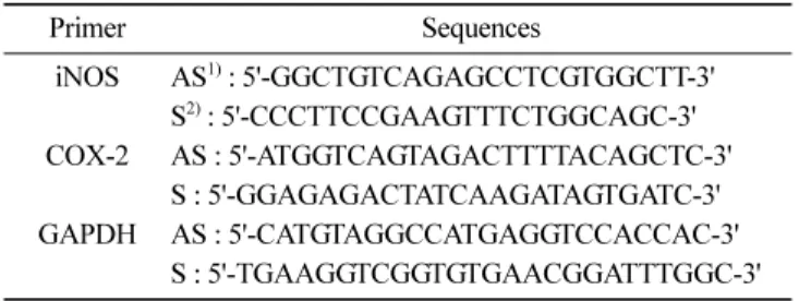
RT-PCR을 사용하여 독활 chloroform 추출물의 농도별 mRNA 상에서의 발현 정도를 확인하였다. 다음과 같은 조건으로 RT-PCR 을 실시하였다. cDNA 합성 45oC, 30분 initial denaturation 94oC, 5분 denaturation 94oC, 5분 annealing 50-55oC, 1분 elongation 72oC, 2분을 25 cycles한 다음, final elongation step을 72oC에서 5분간 수행하였다. RT-PCR의 생성물은 1% agarose gel을 사용하 여 100oC에서 30분간 전기영동하여 UV하에서 관찰하였다. 각 primer의 염기서열은 Table 1에 나타내었다(16-18).
활성물질의 추출 및 분획
독활 Ethanol extract 및 용매 분획
독활 분말 15 kg에 에탄올을 가하여 실온에서 24시간 방치 후 여과하여, 다시 잔사에 에탄올을 가하여 침지한 후 여과하는 과 정을 3회 반복 실행하여 얻은 여액을 감압 농축하여 추출물(AC) 로 조제하였다. 농축한 추출물(AC)을 증류수에 현탁시킨 후 hexane, chloroform, ethyl acetate, n-butanol 순으로 분획하여 감압 농축하 였다.
Ac–As ---Ac
독활 활성물질 분리정제 및 순도 확인
NO 저해 활성에 대해 높은 저해 효과를 보인 독활 에탄올 추 출물의 chloroform 분획(AC-C)에 대하여 분리를 실시하였다(Fig.
1). Silica gel이 충진된 column(30×600 mm)에 시료(AC-C)를 load- ing하고 chloroform-methanol을 이동상으로 하여 methanol의 농도 를 높여가며(chloroform-methanol=10:0, 9:1, 8:2, 7:3, 6:4, 4:6, 5:5, methanol) 용출하여 용출액의 극성도에 따라 19개의 획분으 로 분획한 다음 저해 활성을 측정하였다. 최대 저해활성을 나타 낸 획분(AC8-M)을 농축하여 C18(35-75µ)이 충진된 column(15×
300 mm)에 시료를 loading하고 methanol-water를 이동상으로 하 여 methanol의 농도를 높여가며(methanol-water=75:25, 85:15, 95:5, MeOH) 용출하여, 7개의 획분으로 분획한 다음 저해 활성 을 측정하였다(Fig. 1). 활성물질은 C18 column(Hypersil 5µ ODS, 4.6×250 mm, HP, Palo Alto, CA, USA)을 사용하여 순도를 확인 하였다. 활성물질의 분리/분취는 JAIGEL GS-310 column (20φ×500 mm)이 장착된 Recycling preparative HPLC(JAI LC- 908, Tokyo, Japan)를 사용하여 분석하였고, 단일성분은 Hypersil 5µ ODS column(reverse phase type, 4.6ⅹ250 mm)이 장착된 HPLC(Younglin, Anyang, Gyeonggi, Korea)와 LC-ESI-mass spec- trum (varian 1200L Quadruple LC/MS system, Palo Alto, CA, USA)을 사용하여 분석하였다.
통계분석
모든 실험은 3반복으로 측정하여 측정치를 평균값±표준편차로
나타내었으며, 실험결과의 통계적 유의성은 Student’s t-test에 의 해 시료간의 유의적 차이(p<0.05)를 검정하였다.
결과 및 고찰
Ethanol 추출물의 수율과 독활의 용매 분획
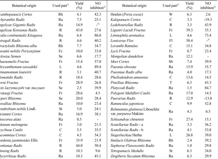
에탄올 추출물은 중량을 측정하기 위하여 동결 건조 후 각 시 료의 수율을 구하였다(Table 2). 강활, 초용담, 금은화 등은 각각 43, 44, 58%로 가장 높았으며, 산수유, 산사, 향부자, 여정자, 황 금, 희첨 등도 20% 이상의 높은 수율을 보였다. 독활은 6.4%의 상대적으로 낮은 수율을 보였다.
분리 시료로 선정된 독활 15 kg을 에탄올을 이용하여 대량 추 출한 결과 960 g의 추출물을 얻었으며, 최종 분리 시료인 chloro- form 분획물을 얻기 위하여 n-hexane, chloroform과 ethyl acetate 로 용매 분획한 결과 각각 723.84, 142.08, 24.96 g의 추출물을, n-butanol과 water 분획물은 각각 36.48, 32.64 g을 얻을 수 있었 으며, 142.08 g의 chloroform 분획물을 최종 활성물질 검색 시료 로 사용하였다.
NO 생성 저해 활성 검색
55종의 한약재 에탄올 추출물에 대하여 NO 생성 억제 활성을 검색한 결과 독활(DH), 길경(GG), 구절초(CJC) 등이 80% 이상의 높은 활성을 나타내었다(Table 2). Shin(19)의 연구결과에서도 독 활을 전탕 엑스분말로 NO 억제 효과를 관찰한 결과, 100 µg/mL 이상의 농도에서 NO 생산량이 유의하게 저하되는 것을 확인하 였다. 높은 활성을 나타낸 독활에 대해서는 n-hexane, chloroform, ethyl acetate, n-butanol, water 순으로 용매 분획하여 NO 저해 활 성을 조사하였다(Fig. 2). 그 결과, 250 µg/mL의 농도까지 Chloro- form 분획물에서 다른 분획물보다 높은 활성을 보였다(p<0.05).
500µg/mL의 농도에서는 세포 생존력이 ethyl acetate 분획물이 chloroform 분획물에 비해 현저하게 저하되어 최종 분리 시료로 chloroform 분획물을 선정하였다.
Hyaluronidase 저해 활성 측정
류머티스 관절염 등의 염증을 일으키게 하는 효소인 hyalu- Table 1. Primer sequences used for semi-quantitative RT-PCR
Primer Sequences
iNOS AS1) : 5'-GGCTGTCAGAGCCTCGTGGCTT-3' S2) : 5'-CCCTTCCGAAGTTTCTGGCAGC-3' COX-2 AS : 5'-ATGGTCAGTAGACTTTTACAGCTC-3'
S : 5'-GGAGAGACTATCAAGATAGTGATC-3' GAPDH AS : 5'-CATGTAGGCCATGAGGTCCACCAC-3'
S : 5'-TGAAGGTCGGTGTGAACGGATTTGGC-3'
1)AS: antisense
2)S: sense
Fig. 1. Purification of a compound for NO inhibition from Aralia cordata Thunb
ronidase의 저해에 의한 항염증 효과를 추정할 수 있는 hyalu- ronidase 저해 실험을 ethanol과 chloroform 분획에 대해서 측정하
였으며(Fig. 3), Chloroform 분획물에서 Ethanol 분획물보다 모든 농도에서 더 높은 저해 활성(p<0.05)을 보인 것을 확인할 수 있 Table 2. List of traditional medicinal herbs with Nitric Oxide inhibition
Botanical origin Used part1) Yield (%)
NO
inhibiton2) Botanical origin Used part1) Yield (%)
NO inhibiton2)
Acanthopanacis Cortex Rh 4.1 42.3 Hoelen (Poria cocus) W 6.3 2.6
Achyranthis Radix Ra 7.5 25.1 Kalopanacis Cortex C 3.3 -19.3
Angelicae Gigantis Radix Ra 14.9 -3) Ledebouriellae Radix R 3.3 43.9 Angelicae Koreanae Radix R 43.0 27.6 Ligustri Lucidi Fructus Fr 39.3 35.1 Aralia continentalis Kitagawa Ra 6.4 86.0 Limnophila aromatica L 4.6 73.4
Astragali Radix R 4.6 40.2 Lonicerae Flos Fl 58.4 -3)
Atractylodis Rhizoma alba Rh 7.7 34.7 Loranthi Ramulus C 15.1 34.8 Aurantii nobilis Pericarpium Fc 10.0 33.0 Lycii Fructus Fr 8.7 23.4
Celosiae Semen Se 6.6 17.7 Mongolian dandelion Ra 12.1 -
Chaenomelis Fructus Fr 15.4 57.0 Mori Cortex Sb 7.4 39.9
Chrysanthemum zawadskii L 4.6 89.4 Paeonia obovata Ra 15.9 35.7
Cinnamomum loureirii R 3.1 40.7 Paeoniae Radix alba Ra 4.0 17.3
Clematidis Radix R 18.4 28.6 Phellodendron amurense C 13.6 14.5
Corni Fructus Fr 28.9 26.8 Pinelliae Rhizoma T 6.3 43.3
Coix lacryma-jobi var. ma-yuen Se 2.5 39.9 Platycodi Radix Ra 1.5 84.7 Crataegi Fructus Fr 28.6 4.5 Polygoni Multiflori Caulis Ra 17.0 14.5
Cyperi Rhizoma Se 20.0 28.4 Puerariae Radix R 12.9 -13.0
Davalliae Rhizoma Ra 10.0 23.4 Ranunculus japonicus C 9.9 52.8
Dendrobium nobile Lindl. St 5.0 24.1 Rehmannia glutinosa Liboschitz
var. purpurea Makino Ra 4.3 6.5
Dictamni Cortex Ra 16.9 38.1
Dioscorea alata Ra 0.5 - Schisandrae chinensis Fr 27.4 15.1
Solanum melongena Fr 3.0 21.5 Scutellariae Radix - a Ra 3.3 36.2
Erycibeae Caulis C 5.5 35.5 Scutellariae Radix - b Ra 4.1 53.8
Eucommiae Cortex C 4.5 54.3 Siegesbeckiae Herba L 26.8 39.0
Gardenia jasminoides Ellis Fr 35.9 21.9 Smilacis Glabrae Rhizoma Rh 2.4 39.2 Gentianae Radix R 44.0 50.4 Sophorae Flavescentis Radix Ra 1.0 29.8
Ginseng Radix R 10.3 9.6 Tetrapanacis Medulla St 6.3 34.0
Glycyrrhizae Radix Ra 10.3 45.1 Zingiberis Siccatum Rhizoma Ra 6.3 29.9
1)C: cortex, Fl: flower, Fc: fruit cortex, Fr: fruit, L: leaf, R: root, Ra: radix, Rh: rhizome, Sb: stem bark, Se: seed, St: stem, T: tuber, W: wolf
2) NO inhibitory effect was represented as % against control 0.1 M acetate buffer (pH 3.5, 5% DMSO): Inhibitory effect (%)={(Ac-As)/Ac}100±SD
.3)-: No effect
Fig. 2. Inhibition of NO production by DH solution in LPS-induced RAW264.7 cells. All values are mean±SD. Values not sharing the same letter are significantly different, p<0.05.
었다. Cha(20)의 연구결과에서 생약재를 통한 hyaluronidase 저해 효과 실험결과에서 대다수의 생약재들이 1000 µg/mL 이상의 농 도를 사용하여도 저해효과를 나타내지 않는 것으로 조사되었다.
Hyaluronidase 활성을 억제하는 기능을 갖는 물질들에는 flavonoids, tannins, spice의 curcumines, 감초의 glycyrrhizin 등이 알려져 있고, 이들 성분들과 일부 식물 추출물들이 함염증제로써 사용되고 있 다(21).
염증 유발 효소 발현 측정
독활 Chloroform 분획물의 NO 합성 저해효과가 유전자의 조 절에 의한 것인지 확인해 보기 위하여 RT-PCR을 통해 확인해 보 았다. iNOS는 평소에는 세포 내에 존재하지 않으나 일단 유도되 면 장시간 동안 다량의 NO를 생성하며, 생성된 NO는 병리적인 혈관확장, 세포독성, 조직손상 등과 같은 생체에 유해한 작용을 나타낸다. 그리고 염증상태에서 iNOS에 의해 생성된 NO는 혈관 투과성, 부종 등의 염증반응을 촉진시킬 뿐만 아니라 염증매개체 의 생합성을 촉진하여 염증을 심화시키는 것으로 알려져 있다 (22-23). 다수의 염증 억제 약물들의 작용기전은 prostagladin 합성
억제를 나타내며 이는 COX-2의 생성 및 활성저해에 의한 것이 다. 따라서 COX-2에 의한 prostagladin의 합성은 염증반응을 매 개하는 것으로 여겨진다. 염증발현 유도물질인 LPS 처리군과 억 제효과가 우수한 L-NMMA와의 비교해 보았을 때 iNOS와 COX- 2 gene의 발현이 억제되는 것을 확인할 수 있었다(Fig. 4).
독활 활성물질 분리정제 및 순도 확인
최종 시료인 독활 chloroform(AC-C)을 silica gel 60 column chromatography을 이용하여 chloroform-methanol을 이동상으로 하 여 methanol의 농도를 높여가며(chloroform-methanol=10:0, 9:1, 8:2, 7:3, 6:4, 4:6, 5:5, methanol) 용출하였다. 용출액의 극성도에 따라 19개의 획분으로 분획하여 저해 활성을 측정한 결과, 80%
의 높은 활성을 나타내는 획분(AC8)을 확인하였으며 계속적인 분리를 진행하였다(Fig. 5). 활성분획(AC8)을 chloroform과 meth- anol을 이용하여 분리한 다음 methanol에 의해 녹은 fraction을 C18
column chromatography를 이용해 water-methanol을 이동상으로 하 여 methanol의 농도를 높여가며(water-methanol=75:25, 85:15, 95:5, MeOH)용출하여, 7개의 분획을 얻은 후 각각 농축하여 활 성을 측정한 결과, 모든 분획에서 cytotoxicity를 나타내지 않았으 며, 5번째 분획(AC8-MⅤ)에서 80% 이상의 높은 활성을 나타내 었다(Fig. 6). 최종 분리한 AC8-MⅤ의 순도 확인을 위해 95%
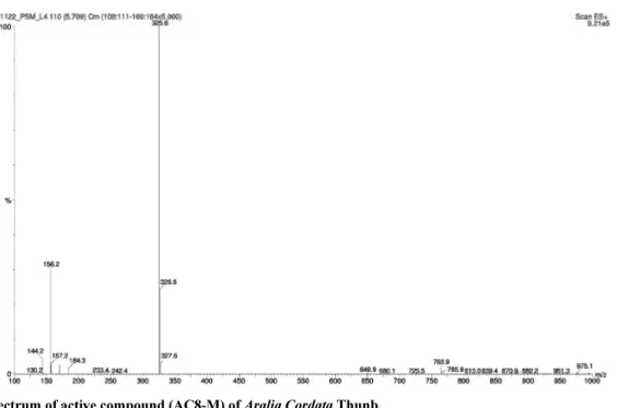
methanol isocratic 조건으로 analytical HPLC를 실시한 결과 단일 peak를 보였으며, 단일물질인지 확인해 보기 위해 활성 물질(AC8- MV)의 LC-ESI-mass spectrum을 통해 분자량을 측정하였으며, 순 수한 단일물질로 분리되었음을 확인할 수 있었다(Fig. 7).
Fig. 3. Hyaluronidase inhibition of EtOH extract and CHCl3 Fr.
with different concentrations. All values are mean±SD. Values not sharing the same letter are significantly different, p<0.05.
Fig. 5. Inhibition of each active fractionates from DH-CHCl3 fraction based on the first silica gel column chromatography on NO production in LPS-induced RAW264.7 cells. All values are mean±SD. *p<0.05; **p<0.01;***p<0.001 compared with AC8 group.
Fig. 4. Inhibition by the chloroform soluble fractionate (DH) on iNOS and COX-2 mRNA expression in LPS-induced RAW 264.7 macrophage. 1; Control, 2; LPS (10µg/mL), 3; L-NMMA (10 µM), 4; L-NMMA (10µM)+LPS (10 µg/mL), 5; CHCl3 (250µg/mL), 6;
CHCl3 (250µg/mL)+LPS (10 µg/mL), 7; CHCl3 (125µg/mL)+LPS (10µg/mL)
요 약
항염증효과가 있는 기능성 식품 및 의약품 소재의 개발을 위 하여 천연 식물 자원으로부터 NOS 저해 활성 물질을 분리하고 그 이화학적인 특성에 대해 알아보기 위해 항염증 효과가 있다 고 알려져 있는 58가지의 생약재에서 NO 저해효과를 확인해 본 결과 독활에서 80% 이상의 높은 저해활성을 가진 것을 알 수 있 었다. 독활 생약재 에탄올 추출물에서 n-hexane, chloroform, ethyl acetate, n-butanol, water 순으로 용매 분획을 실시한 후 NO 생성 저해 활성을 측정한 결과, chloroform 분획에서 가장 높은 저해 활성을 보여 최종 분리 시료로 선정하였으며, open column chro- matography(silica gel, C18)를 이용하여 최종적인 활성 물질(AC8- MV)을 분리할 수 있었다. 분리한 활성 물질(AC8-MV)의 순도를
확인하기 위하여 analytical HPLC와 LC-ESI-mass spectrum를 분 석한 결과 순수한 단일 물질로 분리 되었음을 확인할 수 있었으 며, 차후 nuclear magnetic resonance spectrometer(NMR)를 통해 구조분석을 실시할 것이다. 또한 AC8-MV와 NO저해효과가 유의 적으로 차이가 없던 AC8-MVI 활성물질에 대한 순도 및 구조분 석을 연구할 것이다. 이를 통해 독활로부터 분리한 활성물질(AC8- MV)이 NO inhibitor 로서 일반 항염증 약물 및 기능성 식품 소 재로 실용화될 수 있는 가능성을 시사하였다.
감사의 글
본 연구는 인하대학교의 지원에 의해 이루어진 연구이며 지원 에 감사드립니다.
Fig. 6. Inhibition of each fractionate isolated from AC8 fraction based on the second C18 column chromatography on NO production in LPS-induced RAW264.7 cells. All values are mean±SD. Values not sharing the same letter are significantly different, p<0.05.
Fig. 7. ESI-MS spectrum of active compound (AC8-M) of Aralia Cordata Thunb.
문 헌
1. Stuehr DJ, Cho HJ, Kwon NS, Weise M, Nathan CF. Purification and characterization of the cytokine-induced macrophage nitric oxide synthase: an FAD- and FMN-containing flavoprotein. P.
Natl. Acad. Sci. USA 88: 7773-7777 (1991)
2. Lee HJ, Kim NY, Jang MK, Son HJ, Sohn DH, Lee SH, Ryu JH.
A sesquiterpene, dehydrocostus lactone, inhibits the expression of inducible nitric oxide sysnthase and TNF-alpha in LPS-activated macrophage. Planta Med. 65: 104-108 (1999)
3. Busse R, Mlsch A. Induction of nitric oxide synthase by cytok- ines in vascular smooth muscle cells. FEBS Lett. 275: 87-90 (1990)
4. Bravo MJ, Nakayama K. The role of attention in different visual- search tasks. Percept. Psychophys. 51: 465-472 (1992)
5. Werner-Felmayer G, Werner ER, Fuchs D, Hausen A, Reibnegger G, Wachter H. Tetrahydrobiopterin-dependent formation of nitrite and nitrate in murine fibroblasts. J. Exp. Med. 172: 1599-1607 (1990)
6. Lancaster JR, Hibbs JB. EPR demonstration of iron-nitrosyl com- plex formation by cytotoxic activated macrophages. P. Natl.
Acad. Sci. USA 87: 1223-1227 (1990)
7. Stuehr DJ, Marletta MA. Mammalian nitrate biosynthesis: Mouse macrophages produce nitrite and nitrate in response to Escheri- chia coli lipopolysaccharide. P. Natl. Acad. Sci. USA 82: 7738- 7742 (1985)
8. DeRojas-Walker T, Tamir S, Ji H, Wishnok JS, Tannenbaum SR.
Nitric oxide induces oxidative damage in addition to deamination in macrophage DNA. Chem. Res. Toxicol. 8: 473-477 (1995) 9. Szab C, Zingarelli B, O’Connor M, Salzman AL. DNA strand
breakage, activation of poly (ADP-ribose) synthetase, and cellular energy depletion are involved in the cytotoxicity of macrophages and smooth muscle cells exposed to peroxynitrite. P. Natl. Acad.
Sci. USA 93: 1753-1758 (1996)
10. Weisz A, Cicatiello I, Esumi H. Regulation of the mouse induc- ible-type nitric oxide synthase gene promoter by interferon, bacte- rial lipopolysaccharide, and NG-monomethyl-L-arginine. J. Biol.
Chem. 316: 209-215 (1996)
11. Padwad Y, Ganju L, Jain M, Chanda S, Karan D, Banerjee PK, Sawhney RC. Effect of leaf extract of seabuckthorn on lipopolysaccharide induced inflammatory response in murine macrophages. Int. Immunopharmacol. 6: 46-52 (2006)
12. Reissig JL, Strominger JL, Leloir LF. A modified colorimetric method for the estimation of N-acetylamino sugars. J. Biol.
Chem. 217: 959-966 (1955)
13. Golberg RL, Huff JP, Lenz ME, Glickman P, Katz R, Thonar EJ- MA. Elevated plasma levels of hyaluronate in patients with osteoarthritis and rheumatoid arthritis. Arth. Rheum. 34: 799-807 (1991)
14. Kakehi K, Kinoshita M, Yasueda S. Hyaluronic acid: Separation and biological implications. J. Chromatogr. B 797: 347-355 (2003)
15. Yui N, Okano T, Sakurai Y. Inflammation responsive degradation of crosslinked hyaluronic acid gel. J. Control Release 22: 105- 116 (1992)
16. López AS, Alegre E, Díaz A, Mugueta C, González A. Early- response gene signaling is induced by angiogenic oligosaccha- rides of hyaluronan in endothelial cells. Inhibition by non-angio- genic, high-molecular-weight hyaluronan. Immunol. Lett. 106:
163-171 (2006)
17. Sakamoto K, Yonoki Y, Kubota Y, Kuwagata M, Saito M, Naka- hara T, Ishii K. Inducible nitric oxide synthase inhibitors abol- ished histological protection by late ischemic preconditioning in rat retina. Exp. Eye Res. 82: 512-518 (2006)
18. Hara MR, Cascio MB, Sawa A. GAPDH as a sensor of NO stress. Biochim. Biophys. Acta 1762: 502-509 (2006)
19. Shin MS. Comparative studies on the effects of Aralia continen- talis root and Angelica pubescens root. PhD thesis, University of Kyungwon, Seongnam, Korea (2006)
20. Cha BC, Lee EH, Cho JY. Glutathione S-transferase activity and hyaluronidase inhibitory effect of medicinal plants. Korean J.
Physiol. Pharmacol. 35: 184-188 (2004)
21. Kushwah A, Amma MK, Sareen KN. Effect of some anti-inflam- matory agents on lysosomal & testicular hyaluronidases. Indian J.
Exp. Biol. 16: 222-224 (1978)
22. Santos-Gomes PC, Seabra RM, Andrade PB, Fernandes-Ferreira M. Determination of phenolic antioxidant compounds produced by calli and cell suspensions of sage (Salvia officinalis L.). J.
Plant Physiol. 160: 1025-1032 (2003)
23. Tesuka Y, Irikawa S, Kaneko T, Banskota AH, Nagaoka T, Xiong Q, Hase K, Kadota S. Screening of Chinese herval drug extracts for inhibitory activity on nitric oxide production and identification of an active compound of Zanthoxylum bugeanum. J. Ethnophar- macol. 77: 209-217 (2001)