A Formulated Korean Red Ginseng Extract Inhibited Nitric Oxide Production through Akt- and Mitogen Activated Protein Kinase-dependent Heme
Oxygenase-1 Upregulation in Lipoteichoic Acid-stimulated Microglial Cells
Ji Eun Shin1, Kyungmin Lee1, Ji-Hee Kim2, Iskander Madhi1 and YoungHee Kim1,2*
1Department of Molecular Biology, College of Natural Sciences, Pusan National University, Busan 46241, Korea
2BK21Plus Research Group for Longevity and Marine Biotechnology, Pusan National University, Busan 46241, Korea Received December 7, 2018 /Revised January 8, 2019 /Accepted January 10, 2019
Korean red ginseng made from steaming and drying fresh ginseng has long been used as a traditional herbal medicine due to its effects on the immune, endocrine, and central nerve systems and its anti-in- flammatory activity. In this study, we investigated the molecular mechanism responsible for the an- ti-inflammatory effects of a formulated Korean red ginseng extract (RGE) in response to lipoteichoic acid (LTA), a cell wall component of gram-positive bacteria. RGE inhibited LTA-induced nitric oxide (NO) secretion and inducible nitric oxide synthase (iNOS) expression in BV-2 microglial cells, without affecting cell viability. RGE also inhibited nuclear translocation of nuclear factor kappa B (NF-κB) p65 and degradation of IκB-α. In addition, RGE increased the expression of heme oxygenase-1 (HO-1) in a dose-dependent manner, and the inhibitory effect of RGE on iNOS expression was abrogated by small interfering RNA-mediated knockdown of HO-1. Moreover, RGE induced nuclear translocation of nu- clear factor E2-related factor 2 (Nrf2), a transcription factor that regulates HO-1 expression. Furthermore, the phosphoinositide-3-kinase (PI-3K) inhibitor and mitogen-activated protein kinase (MAPK) in- hibitors suppressed RGE-mediated expression of HO-1, and RGE enhanced the phosphorylation of Akt, extracellular signal-regulated kinases (ERKs), p38, and c-JUN N-terminal kinases (JNKs). These results suggested that RGE suppressed the production of NO, a proinflammatory mediator, by induc- ing HO-1 expression via PI-3K/Akt- and MAPK-dependent signaling in LTA-stimulated microglia. The findings indicate that RGE could be used for the treatment of neuroinflammation induced by gram- positive bacteria and that it may have therapeutic potential for various neuroinflammation-associated disorders.
Key words : Heme oxygenase-1, Korean red ginseng, neuroinflammation, nitric oxide, Nrf2
*Corresponding author
*Tel : +82-51-510-2526, Fax : +82-51-513-9258
*E-mail : [email protected]
This is an Open-Access article distributed under the terms of the Creative Commons Attribution Non-Commercial License (http://creativecommons.org/licenses/by-nc/3.0) which permits unrestricted non-commercial use, distribution, and reproduction in any medium, provided the original work is properly cited.
Journal of Life Science 2019 Vol. 29. No. 4. 402~409 DOI : https://doi.org/10.5352/JLS.2019.29.4.402
Introduction
Gram-positive bacterial infections of the central nervous system (CNS) cause bacterial meningitis, encephalomyelitis, brain abscess or sepsis [26]. Bacterial invasion induces a rap- id inflammatory response, which is mediated by the brain’s innate immune cells such as microglia. Lipoteichoic acid (LTA) is a cell wall component of Gram-positive bacteria such as Staphylococcus aureus [13]. Several studies have dem- onstrated that LTA may bind to target cells through Toll-like receptors-2 (TLR-2) which plays a critical role in LTA-in- duced microglial activation [31]. Signaling via TLR-2 is
mediated by different adaptor proteins, including MyD88, which promotes downstream signaling via mitogen-acti- vated protein kinases (MAPKs), and nuclear factor-kappaB (NF-κB) leading to the expression of pro-inflammatory mole- cules [23]. In Gram-positive infection, microglia produce considerable amounts of nitric oxide (NO) through ex- pression of inducible nitric oxide synthase (iNOS). Large amounts of NO function to restore CNS homeostasis by clearing pathogens and infected cells. However, deregulated or chronic activation of microglial cells can induce too many pro-inflammatory molecules including NO, leading to neu- ronal cell death and brain injury [24]. Moreover, the neuro- inflammatory responses of the CNS are well-known features of various neurodegenerative diseases, such as Alzheimer’s disease (AD) and Parkinson’s disease. Therefore control of microglial activation and subsequent suppression of the pro- duction of neurotoxic pro-inflammatory molecules could provide a potential therapeutic approach for the treatment of neurodegenerative diseases as well as meningitis [38].
Heme oxygenase-1 (HO-1) is an inducible enzyme that catalyzes the oxidation of cellular heme into carbon mon- oxide (CO), biliverdin, and free iron [30]. HO-1 and its enzy- matic by-products provide a host defense mechanism that can protect the body against oxidative injury and also con- tributes to the anti-inflammatory activity of cells and tissues [30]. In activated macrophages, HO-1 expression or CO treat- ment inhibits the production of the pro-inflammatory media- tors such as NO, prostaglandin E2 (PGE2), and inflammatory cytokines [28]. Moreover, a large number of remedial agents have been reported to induce HO-1 expression and exert their anti-inflammatory effects through HO-1 induction.
The root of Korean Panax ginseng C.A. Meyer has been used as a traditional herbal medicine not only in Korea but also in Asia and Western countries. It has been reported that ginseng and ginseng saponins have a wide range of pharma- cological activities including anti-tumorigenic, immune- stimulating, antistress and antioxidant effects [3, 5, 7, 9, 33, 39]. It is especially well established that ginseng ameliorates inflammatory responses [14, 29, 41]. Red ginseng is made by steaming and drying the fresh ginseng. The pharmaco- logical efficacy of Korean red ginseng is known to be en- hanced by these processes mostly due to the changes in the characteristics of the constituent ginsenosides [11]. In the present study, we investigated the molecular mechanism re- sponsible for the anti-inflammatory effects of a formulated Korean red ginseng extract (RGE) in LTA-stimulated microglia. We elucidated that RGE inhibits LTA-induced iNOS expression via upregulation of HO-1.
Materials and Methods
Materials
Lipoteichoic acid from Staphylococcus aureus, and other re- agents not referred were purchased from Sigma (St. Louis, MO, USA). A formulated Korean red ginseng extract (Han- samin Gold) was purchased from Nonghyup Red Ginseng (Seoul, Korea). HO-1 siRNA, and antibodies for iNOS, HO-1, NF-κB p65, inhibitor of kappa B-alpha (IkB-a), histone deace- tylase 3 (HDAC3), ERK, JNK, p38, Akt, α-tubulin, and b-actin were purchased from Santa Cruz Biotechnology (Santa Cruz, CA, USA). Antibody against for phosphorylated ERK (p- ERK), p-JNK, p-p38, p-Akt were purchased from Cell Signal- ing Technology (Beverly, MA, USA). Antibody against for TATA-binding protein (TBP) was purchased from Cloud- clone corp (Katy, Texas, USA). Cell culture medium, DMEM,
and fetal bovine serum (FBS) were purchased from Gibco BRL (now part of Invitrogen Corporation, Carlsbad, CA, USA). X-treme GENE siRNA Transfection Reagent were pur- chased from Roche (Indianapolis, IN, USA).
Cell culture
Mouse BV-2 microglial cells were grown in DMEM me- dium supplemented with 5% heat inactivated fetal bovine serum (FBS) and 0.1% penicillin-streptomycin (BioSource International, Camarillo, CA, USA) at 37℃ in a humidified atmosphere of 5% CO2 and 95% air.
Cell viability assay (MTT assay)
The cytotoxicity of RG was assessed using the micro- culture tetrazolium (MTT)-based colorimetric assay. The re- maining cells after Griess reaction were used for MTT assay.
MTT was added to each well (final concentration is 62.5 μg/
ml). After incubation for 3 hr at 37℃ in 5% CO2, the super- natant was removed and the formazan crystals produced in viable cells were solubilized with dimethylsulfoxide (DMSO). The absorbance of each well was then read at 570 nm using a microplate reader (Bio-Rad, Hercules, CA, USA).
Measurement of nitrite concentration
NO synthesis in cell cultures was measured by a micro- plate assay method. To measure nitrite, 100 ml aliquots were removed from conditioned medium and incubated with an equal volume of the Griess reagent (1% sulfanilamide/0.1%
N-(1-naphthyl)-ethylenediamine dihydrochloride/2.5% H3PO4) at room temperature for 10 min. Nitrite concentration was determined by measuring the absorbance at 540 nm with a microplate spectrophotometer (Bio-Rad, Hercules, CA, USA). The sodium nitrite was used as a standard.
Transient transfection with siRNA
Transfection of cells with siRNA was performed using the X-treme GENE siRNA Transfection Reagent (Roche Applied Science), according to the manufacturer’s instructions.
Commercially available human HO-1 and Nrf-2 specific siRNAs (Santa Cruz, Heidelberg, Germany) and negative control siRNAs (Santa Cruz) were used for transfection. In brief, X-treme GENE siRNA Transfection Reagent (10 μl) was added to 100 μl serum-free medium containing 2 μg of each siRNA oligo, and was incubated for 20 min at room temper- ature. Gene silencing was measured after 48 hr by Western blotting.
A B
C
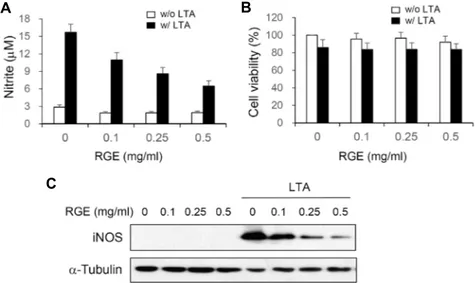
Fig. 1. RGE inhibits NO production and iNOS expression in LTA-stimulated microglial cells. BV-2 cells were treated with different concentrations of RGE for 3 hr and then incubated with or without LTA (10 mg/ml) for 20 hr. Nitrite content was measured using the Griess reaction (A). Cell viabilities were determined by MTT assay (B). Each bar represents the mean ± S.E. from 3 independent experiments. **p<0.01 vs the LTA-treated group. The expression of iNOS and α-tubulin were detected by Western blot using specific antibodies (C).
Preparation of nuclear extracts
Nuclear extracts were prepared as described previously [2]. Cells were washed with PBS, resuspended in ice-cold isotonic buffer A [10 mM HEPES (pH7.9) 1.5 mM MgCl2, 10 mM KCl, 0.5 mM dithiothreitol (DTT) and protease in- hibitor cocktail], and incubated at 4℃ for 10 min. Cells were centrifuged at 15,000× g for 1 min and pellet was re- suspended in ice-cold buffer B [20 mM HEPES (pH 7.9), 420 mM NaCl, 1.5 mM MgCl2, 0.2 mM EDTA, 25% glycerol, 0.5 mM DTT and protease inhibitor cocktail] followed by in- cubation at 4℃ for 30 min with occasional vortexing. The resulting suspension was centrifuged at 15,000x g for 5 min, and the supernatant was stored at -20℃.
Western blot analysis
Cells were harvested in ice-cold lysis buffer (1% Triton X-100 and 1% deoxycholate in PBS). The protein content of the cell lysates was determined using Bradford reagent (Bio-Rad; Hercules, CA, USA). The proteins in each sample were resolved by 10% SDS-polyacrylamide gel electropho- resis (SDS-PAGE), transferred to a polyvinylidene difluoride (PVDF) membrane, and incubated with the appropriate antibodies. The proteins were visualized using an enhanced chemiluminescence detection system (Amersham Biosciences, Piscataway, NJ, USA) with horseradish peroxidase-con- jugated anti-rabbit or anti-mouse secondary antibodies.
Anti-a-tubulin or anti-b-actin antibodies were used as load-
ing control for cytosolic protein and anti-HDAC3 or anti-TBP antibodies were used as loading control for nuclear protein.
Statistical analysis
All results were expressed as the mean ± SE (standard error). Each experiment was repeated at least three times.
Statistical analysis was performed by using SPSS software to determine significant differences. We used one-way anal- ysis of variance (ANOVA) followed by Tukey’s post hoc test for comparison of three or more groups. A value of p<0.05 was considered statistically significant.
Results
RGE suppresses LTA-inducted neuroinflammatory molecules
To investigate whether RGE could abrogate LTA-medi- ated neuroinflammation, we examined the effects of RGE on NO production expression of in BV-2 microglial cells.
Stimulation of BV-2 microglial cells with LTA increased NO synthesis and iNOS expression, whereas RGE pretreatment significantly attenuated the LTA-induced NO synthesis and iNOS expression in a dose-dependent manner (Fig. 1A, Fig.
1C). To determine the effect of RGE on cell viability, BV-2 microglial cells were treated with various concentration of RGE in the absence or presence of LTA. While LTA induced a little toxicity on BV-2 cells, RGE at concentrations up to
Fig. 2. RGE inhibits LTA-induced NF-κB activation. BV-2 cells were treated with various concentrations of RGE for 3 h and followed by LTA (10 μg/ml) treatment for 1 hr.
NF-κB p65 level in nuclear and cytosolic fraction was assessed by Western blotting. The level of IκB-α in cyto- solic extracts were also analyzed by Western blotting.
A
B
C
(hr)
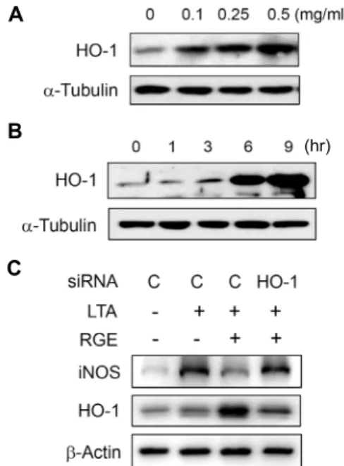
Fig. 3. RGE-induced HO-1 suppresses iNOS expression. BV-2 cells were cultured with increasing concentrations of RGE for 6 hr (A) or with 0.5 mg/ml of RGE for the indicated times (B). HO-1 expression was determined by Western blot. (C) The si-Control or si-HO-1 transfected cells were treated with RGE (0.5 mg/ml) for 3 hr, then stimulated with LTA (10 μg/ml) for 20 hr. The ex- pression iNOS or HO-1 were determined by Western blotting.
0.5 mg/ml induced no cytotoxicity (Fig. 1B). These results suggest that RGE suppresses NO release in LTA-stimulated microglial cells by inhibiting iNOS expression level, and these effects are not due to cytotoxicity.
RGE suppresses LTA-induced activation of NF-κB NF-kB is known to mediate the expression of iNOS gene in response to LTA. To determine the effects of RGE on NF-kB activity, we examined nuclear translocation of NF-kB by Western blotting. As shown in Fig. 2, nuclear level of NF-kB p65 was markedly increased and cytosolic level of NF-kB p65 was significantly decreased by LTA treatment.
However, RGE pretreatment reduced nuclear level of NF-kB p65 in a dose-dependent manner, at the same time RGE in- creased cytosolic level of NF-kB p65. In accord with this re- sult, RGE inhibited LTA-induced degradation of IkB-a in a dose-dependent manner. These results suggest that RGE suppresses LTA-induced nuclear translocation of NF-kB via blocking of IkB-a degradation.
RGE reduces iNOS expression through induction of HO-1
To investigate whether RGE induces HO-1 expression in microglia, BV-2 cells were incubated with various concen- trations of RGE. The HO-1 protein level was significantly increased after 6 hr by RGE in a dose-dependent manner (Fig. 3A, Fig. 3B). To elucidate that RGE-induced HO-1 sup- presses the expression of iNOS, we applied an HO-1 small interfering (si) RNA system to knock down HO-1 function.
BV-2 cells were transfected with HO-1 siRNA or control siRNA , and treated with LTA in the absence or presence
of RGE. As shown in Fig. 3C, a decrease of HO-1 blocked RGE-mediated suppression of LTA-stimulated iNOS ex- pression, whereas transfection with control siRNA showed no effect. These results suggest that HO-1 expression is up-regulated by RGE and involved in RGE-induced anti-in- flammatory activity.
RGE-induced HO-1 expression is mediated by Nrf2 Since NF-E2-related factor 2 (Nrf2) is known to regulate the expression of HO-1, we investigated whether RGE in- duces nuclear accumulation of Nrf2, which is critical to its transcriptional activity, in BV-2 cells. As shown in Fig. 4, nuclear level of Nrf2 was increased by RGE in a dose-de- pendent manner and reached peak at 3 hr. These results sug- gest that RGE activates Nrf2, which in turn induces HO-1 expression.
PI-3K/Akt and MAPKs mediates RGE-induced HO- 1 expression
To elucidate the molecular target of RGE in further up- stream signaling pathway of HO-1 expression, we examined the effect of pharmaceutical protein kinase inhibitors of
A
B (hr)
Fig. 4. Effects of RGE on nuclear translocation of Nrf2. BV-2 cells were incubated with indicated concentrations of RGE for 3 hr (A) or with 0.5 mg/ml of RGE for the indicated times (B) and then the levels of Nrf2 in nuclear extracts were analyzed by Western blotting.
A
B
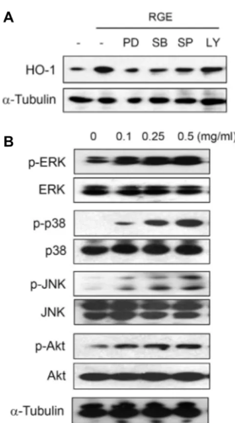
Fig. 5. Involvement of Akt and MAPKs in RGE-mediated ex- pression of HO-1. (A) BV-2 cells were incubated with PD98059 (10 μM), SB203580 (10 μM), SP600125 (10 μM), and LY294002 (10 μM), for 30 min and then treated with RGE (0.5 mg/ml) for 6 hr. (B) Cells were treated with various concentrations of RGE for 30 min. Equal amounts of cytosolic extract were subsequently analyzed by Western blotting with specific antibodies.
MAPKs, PD 98059 (ERK inhibitor), SB203580 (p38 kinase in- hibitor), SP600125 (JNK inhibitor), and PI-3K/Akt inhibitor, LY294002. As shown in Fig. 5A, RGE-induced HO-1 expre- ssion was significantly inhibited by 3 MAPK inhibitors and slightly inhibited by LY294002. Moreover, RGE induced the phosphorylation of 3 MAPKs and Akt (Fig. 5B). These results indicate that PI-3K/Akt and MAPK signalings occur up- stream of RGE-mediated HO-1 expression.
Discussion
In present study, we investigated the anti-inflammatory effects of RGE in LTA-stimulated microglial cells. We used BV-2 microglial cells because there is a close resemblance between BV-2 microglial cells and primary microglia in terms of the inflammatory signaling pathways and BV-2 mi- croglial cells are an appropriate model for the activation of microglia in vitro. We found that GRE pretreatment sig- nificantly inhibited the production of NO and expression of iNOS in response to LTA without affecting cell viability (Fig.
1). NO is released from activated astrocytes and microglia, and neurons are remarkably sensitive to NO-induced cell death [4, 40]. High amounts of NO produced by iNOS from activated microglial cells are believed to play a critical role in the pathogenesis of various inflammation-related diseases.
So suppression of iNOS expression in microglial cells could represent an attractive target to treat various neuroinfla- mmatory diseases [8, 37]. Thus our results suggest that RGE could be a useful remedial agent for Gram-positive bac- teria-mediated inflammatory diseases such as pneumonia, meningitis, and sepsis. Red ginseng and several ginseno- sides are reported to ameliorate neuroinflammation in re-
sponse to LPS [15, 16, 19-22, 35]. Our study showed that red ginseng also attenuates LTA-induced neuroinflammatory response. Since LTA is known to provoke inflammatory re- sponse through TLR-2 [31], red ginseng might suppress TLR-2 signaling as well as TLR-4.
A major transcriptional regulator of iNOS genes is NF-κB, which is also a key regulator of a variety of genes involved in immune and inflammatory responses [1]. Inappropriate regulation of NF-κB is directly involved in a wide range of human disorders, including a variety of cancers, neuro- degenerative diseases, sepsis, and numerous other inflam- matory conditions [6, 10, 32]. Therefore, the development of a drug that controls NF-κB is a promising strategy for the treatment of inflammatory disease [10]. In resting cells, NF-κ B dimers remain in the cytosol bound to IkB that block their nuclear import. In response to stimulation, IKK complex phosphorylate IkBs which in turn are rapidly ubiquitinated and degraded by 26S proteasome complex. Consequently,
released NF-κB dimers translocate to the nucleus and stim- ulate target genes expression [10]. Our study showed that RGE significantly inhibited NF-κB translocation into the nu- cleus (Fig. 3). In addition, RGE suppressed the LTA-stimu- lated degradation of IkB-a. Therefore these results suggest that RGE inhibits NF-κB activity through the suppression of degradation of IkB-a.
HO-1 is known to exhibit anti-inflammatory effects by at- tenuating production of pro-inflammatory mediators [12], and then thought to be used as a potential therapeutic agent for treating inflammatory diseases. We found that RGE sig- nificantly increased HO-1 expression in microglial cells (Fig.
3A and B). Since HO-1 expression is reported to be increased by chemical-mediated oxidative stress [17], we elucidated if RGE induced HO-1 expression by cell damage. RGE did not decrease cell viability when we conducted MTT assay. So HO-1 might be specifically induced by RGE, not by cell damage. Furthermore, the blocking of HO-1 expression via siRNA markedly attenuated the inhibitory effects of RGE on LTA-induced iNOS expression (Fig. 3C). These data suggest that RGE inhibit TLR2-mediated iNOS expression via HO-1 induction.
Nrf2 is a transcription factor which plays a central role for inducible expression of HO-1 [34]. In normal conditions, Nrf2 is arrested in the cytoplasm by binding with Kelchlike ECH-associated protein 1 (Keap1) and degraded by the ubiq- uitin-dependent proteasome. Under activation, Nrf2 is dis- sociated from Keap1, translocates to the nucleus, and binds antioxidant response elements (AREs) located in the pro- moter regions of HO-1 [25]. In this study, nuclear trans- location of Nrf2 was significantly enhanced by treatment with RGE (Fig. 4). This result suggests that RGE up-regulates HO-1 expression through Nrf2 activation.
Several studies reported that Nrf2 activation and HO-1 expression are regulated by PI-3K and MAPKs such as ERK, JNK, and p38 in response to various stimuli [18, 27].
However, these signaling pathways depend on the type of cells and stimuli in terms of their contribution to HO-1 expression. In this study, RGE-mediated HO-1 expression was suppressed by the inhibitors of PI-3K/Akt, ERK, JNK, and p38 (Fig. 5), and RGE induced the phosphorylation of these kinases. These results suggest that RGE induce HO-1 expression via the activation of PI-3K/Akt, ERK, JNK, and p38. Nrf2 was reported to be phosphorylated at multiple sites by MAPKs to facilitate its nuclear translocation [36].
Therefore, PI-3K/Akt and MAPKs could up-regulate HO-1
through direct phosphorylation of Nrf2 or indirect mecha- nisms.
In conclusion, we demonstrated that RGE inhibited NO release and iNOS expression in LTA-stimulated microglial cells, and these effects are mediated by PI-3K/Akt and ERK, JNK, p38-dependent HO-1 induction. Our findings suggest that RGE could be used for the treatment of Gram-positive bacteria-induced neuroinflammation and may have ther- apeutic potential for various neuroinflammation-associated disorders.
Acknowledgement
This research was supported by a 2-Year Research Grant of Pusan National University.
References
1. Ahn, K. S. and Aggarwal, B. B. 2005. Transcription factor NF-κB: A sensor for smoke and stress signals. Ann. N. Y.
Acad. Sci. 1056, 218-233.
2. Andrews, N. C. and Faller, D. V. 1991. A rapid micro- preparation technique for extraction of DNA-binding pro- teins from limiting numbers of mammalian cells. Nucleic Acids Res. 19, 2499.
3. Attele, A. S., Wu, J. A. and Yuan, C. S. 1999. Ginseng phar- macology: Multiple constituents and multiple actions.
Biochem. Pharmacol. 58, 1685-1693.
4. Bal-Price, A. and Brown, G. C. 2001. Inflammatory neuro- degeneration mediated by nitric oxide from activated glia- inhibiting neuronal respiration, causing glutamate release and excitotoxicity. J. Neurosci. 21, 6480-6491.
5. Bhattacharya, S. K. and Mitra, S. K. 1991. Anxiolytic activity of Panax ginseng roots: An experimental study. J. Ethnophar- macol. 34, 87-92.
6. Blackwell, T. S., Blackwell, T. R., Holden, E. P., Christman, B. W. and Christman, J. W. 1996. In vivo antioxidant treat- ment suppresses nuclear factor-kappa B activation and neu- trophilic lung inflammation. J. Immunol. 157, 1630-1637.
7. Block, K. I. and Mead, M. N. 2003. Immune system effects of echinacea, ginseng, and astragalus: A review. Integr.
Cancer Ther. 2, 247-267.
8. Doherty, G. H. 2011. Nitric oxide in neurodegeneration:
Potential benefits of non-steroidal anti-inflammatories. Neu- rosci. Bull. 27, 366-382.
9. Gillis, C. N. 1997. Panax ginseng pharmacology: A nitric ox- ide link? Biochem. Pharmacol. 54, 1-8.
10. Giridharan, S. and Srinivasan, M. 2018. Mechanisms of NF- kappaB p65 and strategies for therapeutic manipulation. J.
Inflamm. Res. 11, 407-419.
11. He, M., Huang, X., Liu, S., Guo, C., Xie, Y., Meijer, A. H.
and Wang, M. 2018. The difference between white and red
ginseng: Variations in ginsenosides and immunomodulation.
Planta Med. 84, 845-854.
12. Jazwa, A. and Cuadrado, A. 2010. Targeting heme oxygen- ase-1 for neuroprotection and neuroinflammation in neuro- degenerative diseases. Curr. Drug Targets 11, 1517-1531.
13. Jiang-Shieh, Y. F., Yeh, K. Y., Wei, I. H., Chang, C. Y., Chien, H. F., Tsai, R. Y., Chang, M. L., Lee, A. W., Pai, M. H. and Wu, C. H. 2005. Responses of microglia in vitro to the gram-positive bacterial component, lipoteichoic acid. J.
Neurosci. Res. 82, 515-524.
14. Jin, Y., Kotakadi, V. S., Ying, L., Hofseth, A. B., Cui, X., Wood, P. A., Windust, A., Matesic, L. E., Pena, E. A., Chiuzan, C., Singh, N. P., Nagarkatti, M., Nagarkatti, P. S., Wargovich, M. J. and Hofseth, L. J. 2008. American ginseng suppresses inflammation and DNA damage associated with mouse colitis. Carcinogenesis 29, 2351-2359.
15. Kang, A., Hao, H., Zheng, X., Liang, Y., Xie, Y., Xie, T., Dai, C., Zhao, Q., Wu, X., Xie, L. and Wang, G. 2011. Peripheral anti-inflammatory effects explain the ginsenosides paradox between poor brain distribution and anti-depression efficacy.
J. Neuroinflammation 8, 100.
16. Kang, A., Xie, T., Zhu, D., Shan, J., Di, L. and Zheng, X. 2017.
Suppressive effect of ginsenoside Rg3 against lipopoly- saccharide-induced depression-like behavior and neuro- inflammation in mice. J. Agric. Food Chem. 65, 6861-6869.
17. Keyse, S. M. and Tyrrell, R. M. 1989. Heme oxygenase is the major 32-kDa stress protein induced in human skin fi- broblasts by UVA radiation, hydrogen peroxide, and so- dium arsenite. Proc. Natl. Acad. Sci. USA. 86, 99-103.
18. Kim, J. H., Park, G. Y., Bang, S. Y., Park, S. Y., Bae, S. K.
and Kim, Y. 2014. Crocin suppresses LPS-stimulated ex- pression of inducible nitric oxide synthase by upregulation of heme oxygenase-1 via calcium/calmodulin-dependent protein kinase 4. Mediators Inflamm. 2014, 728709.
19. Lee, J. S., Song, J. H., Sohn, N. W. and Shin, J. W. 2013.
Inhibitory effects of ginsenoside Rb1 on neuroinflammation following systemic lipopolysaccharide treatment in mice.
Phytother. Res. 27, 1270-1276.
20. Lee, K. W., Jung, S. Y., Choi, S. M. and Yang, E. J. 2012.
Effects of ginsenoside re on LPS-induced inflammatory me- diators in BV2 microglial cells. BMC Complement. Altern.
Med. 12, 196.
21. Lee, Y. Y., Park, J. S., Lee, E. J., Lee, S. Y., Kim, D. H., Kang, J. L. and Kim, H. S. 2015. Anti-inflammatory mechanism of ginseng saponin metabolite Rh3 in lipopolysaccharide- stimulated microglia: Critical role of 5'-adenosine mono- phosphate-activated protein kinase signaling pathway. J.
Agric. Food Chem. 63, 3472-3480.
22. Lin, W. M., Zhang, Y. M., Moldzio, R. and Rausch, W. D.
2007. Ginsenoside Rd attenuates neuroinflammation of dop- aminergic cells in culture. J. Neural Transm. Suppl. 72, 105-112.
23. Liu, Y., Yin, H., Zhao, M. and Lu, Q. 2014. TLR2 and TLR4 in autoimmune diseases: A comprehensive review. Clin. Rev.
Allergy Immunol. 47, 136-147.
24. Lull, M. E. and Block, M. L. 2010. Microglial activation and
chronic neurodegeneration. Neurotherapeutics 7, 354-365.
25. Motohashi, H., Katsuoka, F., Engel, J. D. and Yamamoto, M.
2004. Small Maf proteins serve as transcriptional cofactors for keratinocyte differentiation in the Keap1-Nrf2 regulatory pathway. Proc. Natl. Acad. Sci. USA. 101, 6379-6384.
26. Neher, J. J. and Brown, G. C. 2007. Neurodegeneration in models of Gram-positive bacterial infections of the central nervous system. Biochem. Soc. Trans. 35, 1166-1167.
27. Nemmiche, S., Chabane-Sari, D., Kadri, M. and Guiraud, P. 2012. Cadmium-induced apoptosis in the BJAB human B cell line: Involvement of PKC/ERK1/2/JNK signaling pathways in HO-1 expression. Toxicology 300, 103-111.
28. Otterbein, L. E., Bach, F. H., Alam, J., Soares, M., Tao Lu, H., Wysk, M., Davis, R. J., Flavell, R. A. and Choi, A. M.
2000. Carbon monoxide has anti-inflammatory effects in- volving the mitogen-activated protein kinase pathway. Nat.
Med. 6, 422-428.
29. Park, J. S., Shin, J. A., Jung, J. S., Hyun, J. W., Van Le, T.
K., Kim, D. H., Park, E. M. and Kim, H. S. 2012. Anti-in- flammatory mechanism of compound K in activated micro- glia and its neuroprotective effect on experimental stroke in mice. J. Pharmacol. Exp. Ther. 341, 59-67.
30. Ryter, S. W. and Choi, A. M. 2010. Heme oxygenase-1/car- bon monoxide: Novel therapeutic strategies in critical care medicine. Curr. Drug Targets 11, 1485-1494.
31. Schwandner, R., Dziarski, R., Wesche, H., Rothe, M. and Kirschning, C. J. 1999. Peptidoglycan- and lipoteichoic acid- induced cell activation is mediated by toll-like receptor 2.
J. Biol. Chem. 274, 17406-17409.
32. Segain, J. P., Raingeard, de la Bletiere, D., Bourreille, A., Leray, V., Gervois, N., Rosales, C., Ferrier, L., Bonnet, C., Blottiere, H. M. and Galmiche, J. P. 2000. Butyrate inhibits inflammatory responses through NFkappaB inhibition: Im- plications for crohn's disease. Gut 47, 397-403.
33. Shin, H. R., Kim, J. Y., Yun, T. K., Morgan, G. and Vainio, H. 2000. The cancer-preventive potential of Panax ginseng:
A review of human and experimental evidence. Cancer Causes Control 11, 565-576.
34. Srisook, K., Kim, C. and Cha, Y. N. 2005. Molecular mecha- nisms involved in enhancing HO-1 expression: De-repre- ssion by heme and activation by Nrf2, the "one-two" punch.
Antioxid. Redox Signal. 7, 1674-1687.
35. Sun, X. C., Ren, X. F., Chen, L., Gao, X. Q., Xie, J. X. and Chen, W. F. 2016. Glucocorticoid receptor is involved in the neuroprotective effect of ginsenoside Rg1 against inflamma- tion-induced dopaminergic neuronal degeneration in sub- stantia nigra. J. Steroid Biochem Mol. Biol. 155, 94-103.
36. Sun, Z., Huang, Z. and Zhang, D. D. 2009. Phosphorylation of Nrf2 at multiple sites by MAP kinases has a limited con- tribution in modulating the Nrf2-dependent antioxidant response. PLoS One 4, e6588.
37. Tieu, K., Ischiropoulos, H. and Przedborski, S. 2003. Nitric oxide and reactive oxygen species in parkinson's disease.
IUBMB Life 55, 329-335.
38. Van Eldik, L. J., Thompson, W. L., Ralay Ranaivo, H., Behanna, H. A. and Martin Watterson, D. 2007. Glia proin-
초록:홍삼추출액은 lipoteichoic acid로 자극된 소교세포에서 Akt 및 MAPK 의존적으로 heme oxygenase-1 발현을 유도함으로써 NO 생성을 억제함
신지은1․이경민1․김지희2․이스칸더 마디1․김영희1,2*
(1부산대학교 자연과학대학 분자생물학과, 2BK21플러스 장수해양바이오사업단)
생삼을 쪄서 건조시킨 홍삼은 전통적으로 사용되고 있는 약재로서 면역, 내분비 및 중추신경계 작용을 증진시 키며 염증을 억제하는 효과가 있는 것으로 알려져 있다. 본 연구에서는 그람 양성균의 세포벽 성분인 lipoteichoic acid (LTA)에 의한 염증반응에 홍삼추출액(RGE)이 항염증 효과를 가지는지 관찰하고 그 작용 기전을 연구하였다.
BV-2 소교세포에서 RGE는 세포에 독성을 유도하지 않으면서 LTA로 인한 nitric oxide (NO)의 생성과 inducible nitric oxide synthase (iNOS) 발현을 억제하였으며, NF-kB p65의 핵으로의 이동과 IkB-a의 분해 또한 억제하였다.
한편, RGE는 농도의존적으로 heme oxygenase-1 (HO-1)의 발현을 유도하였으며, HO-1 siRNA를 처리했을 때는 RGE가 iNOS의 발현을 억제하지 못하였다. RGE는 HO-1의 발현에 관여하는 전사인자인 nuclear factor E2-related factor 2 (Nrf2)를 핵으로 이동을 촉진시켰다. 또한 RGE에 의한 HO-1의 발현은 phosphatidylinositol-3-kinase (PI-3K) 및 MAPK 억제제에 의해 감소되었으며, RGE가 Akt와 ERK, p38, JNK의 인산화를 유도하였다. 이상의 결과를 종합해보면, RGE는 PI-3K/Akt 및 ERK, p38, JNK 신호전달과정을 통해 HO-1의 발현을 유도함으로써 NO 와 같은 염증매개물질의 생성을 억제한다는 것을 알 수 있다. 그러므로 홍삼추출액은 그람 양성균에 의한 신경염 증과 염증관련 신경계 질환의 치료제로서 사용될 수 있을 것이라 사료된다.
flammatory cytokine upregulation as a therapeutic target for neurodegenerative diseases: Function-based and target-based discovery approaches. Int. Rev. Neurobiol. 82, 277-296.
39. Wakabayashi, C., Murakami, K., Hasegawa, H., Murata, J.
and Saiki, I. 1998. An intestinal bacterial metabolite of gin- seng protopanaxadiol saponins has the ability to induce apoptosis in tumor cells. Biochem. Biophys. Res. Commun. 246, 725-730.
40. Wei, T., Chen, C., Hou, J., Xin, W. and Mori, A. 2000. Nitric oxide induces oxidative stress and apoptosis in neuronal
cells. Biochim. Biophys. Acta 1498, 72-79.
41. Yang, Y., Yang, W. S., Yu, T., Sung, G. H., Park, K. W., Yoon, K., Son, Y. J., Hwang, H., Kwak, Y. S., Lee, C. M., Rhee, M.
H., Kim, J. H. and Cho, J. Y. 2014. ATF-2/CREB/IRF-3-tar- geted anti-inflammatory activity of Korean red ginseng wa- ter extract. J. Ethnopharmacol. 154, 218-228.