Extract from Prunus mume Sieb. et Zucc. Fruit Prevents LPS-induced Homotypic Aggregation of Monocytic THP-1 Cells via Suppression of Nitric Oxide
Production and NF-κB Activation
Hye-Rim Lee1†, Youngsook Park2†, Hyun Jeong Kim1, Aram Lee2, Jihea Choi2, Jaeho Pyee1, Heonyong Park1 and Jongmin Kim2*
1Department of Molecular Biology & Institute of Nanosensor and Biotechnology, Dankook University, 126, Jukjeon-dong, Suji-gu, Yongin-si, Gyeonggi-do 448-701, Korea
2Department of Life Systems, Sookmyung Women’s University, 52 Hyochangwon-gil, Yongsan-gu, Seoul 140-742, Korea Received May 13, 2015 /Revised July 3, 2015 /Accepted July 8, 2015
Homotypic cell adhesion (homotypic aggregation) in activated monocytes plays a central role in phys- iological and pathological processes including inflammatory responses, differentiation and migration.
The extract of the Prunus mume Sieb. et Zucc. fruit (Maesil) has potential benefits to human health;
such as anti-viral, anti-microbial, and anti-cancer activities. Indeed, Maesil extract may modulate in- flammatory responses via interference with homotypic aggregation in monocytes. In the present study, the molecular mechanisms underpinning the therapeutic efficacy of Maesil extract in inflammatory diseases were investigated. It was found that Maesil extract inhibited homotypic aggregation in lip- opolysaccharide (LPS)-activated monocytes. This was mediated by reduction of nitric oxide (NO) pro- duction, partly via inhibition of inducible nitric oxide synthase (iNOS) expression in LPS-activated THP-1 cells. It was confirmed that NO inhibition is a key mechanism in Maesil induced blockade of monocyte aggregation through identification of reversal of this inhibitory effect by the NO-producing agent S-nitroso-N-acetyl penicillamine (SNAP). In addition, Maesil extract significantly attenuated LPS-induced IκB-α phosphorylation and NF-κB translocation into the nucleus. In conclusion, Maesil extract exerts anti-inflammatory effects via inhibition of homotypic aggregation of LPS-activated mon- ocytes through mechanisms involving the suppression of NO production and NF-κB activity, suggest- ing Maesil extract as a potential therapeutic candidate for the prevention and treatment of chronic in- flammatory diseases.
Key words : Homotypic aggregation, inflammationnitric oxide, maesil extract, NF-κB
†Authors contributed equally.
*Corresponding author
*Tel : +82-2-710-9553, Fax : +82-2-2077-7322
*E-mail : [email protected]
This is an Open-Access article distributed under the terms of the Creative Commons Attribution Non-Commercial License (http://creativecommons.org/licenses/by-nc/3.0) which permits unrestricted non-commercial use, distribution, and reproduction in any medium, provided the original work is properly cited.
Journal of Life Science 2015 Vol. 25. No. 7. 801~809 DOI : http://dx.doi.org/10.5352/JLS.2015.25.7.801
Introduction
The fruit of Prunus mume Sieb. et Zucc. (Korean name, Maesil) belongs to the Rosaceae family and there are several varieties of Maesil in Korea, including Cheonmae and Cheongchuk. It has long been used as a traditional remedy and health food in Korea, China, Vietnam, and Japan [6, 21, 26, 34]. To date, a number of phytochemical, pharmaco- logical, biological, and clinical studies on Maesil have been reported [5, 7, 13, 16, 17]. Many chemical constituents have been isolated from Maesil extract including volatile com-
pounds [27], hydroxycinnamic acid derivatives [26], citric acid derivative (mumefural) [7], and triterpenoids [16].
Maesil extract has been reported to show anticancer [1, 13, 30], antiviral [38], anti-microbial [29], anti-oxidant [37], and cardiovascular protective [35] effects. Maesil extract has been used pharmaceutically in folk medicine for antitussive, ex- pectoration, antiemetic, antidiarrheal, anthelmintic, and anti- pyretic actions [26, 37]. Despite the known beneficial effects of Maesil extract in chronic inflammatory diseases such as cancer and cardiovascular disease, there are very few reports concerning its antiinflammatory effects and the detailed mechanisms involved. The present study sought to de- termine a potential application for Maesil extract in phytop- therapy through identification of the anti-inflammatory re- sponses elicited upon exposure of immune cells to multiple components of Maesil extract. Homotypic cell adhesion (homotypic aggregation) of activated monocytes is essential for physiological processes including immune response, dif- ferentiation, and monocyte migration. It is also intimately
involved in the pathogenesis of chronic inflammatory dis- eases such as atherosclerosis [25]. Under chronic inflamma- tion, proinflammatory stimuli, such as lipopolysaccharide (LPS), activate monocytes and induce the expression of cell adhesion molecules (CAMs) and the production of nitric ox- ide (NO) [9, 18, 36]. As a result, activated monocytes move to target tissues and induce heterotypic/homotypic cell ad- hesion [11], indicating that CAMs and NO are key regulators of the inflammatory processes that control cell-cell adhesion [18, 32, 36]. During this process, increased expression of lym- phocyte function-associated antigen-1 (LFA-1) is correlated with the induction of homotypic aggregation of monocytes [22, 24]. In addition, integrin β2, and intercellular adhesion molecule-1 (ICAM-1) have also been shown to be involved in homo- and heterotypic cell adhesion [31]. During in- flammatory processes, the expression of inducible nitric ox- ide synthase (iNOS) is significantly induced by various proinflammatory stimuli such as LPS, interleukin-1β, and tu- mor necrosis factor, and large amounts of NO are produced [3]. Excessive NO is associated with cytotoxicity and pro-in- flammatory activity [10]. Nuclear factor (NF)-κB is consid- ered as one of the most important factors in the immune response. Activation of NF-κB in response to pro-in- flammatory stimuli is initiated by the rapid phosphorylation of IκB-α, through activation of IκB-α kinase. Phosphorylation of IκB-α results in its proteolytic degradation by ubiquitina- tion [15], thus liberating the NF-κB complex and enabling translocation to the nucleus with subsequent activation of pro-inflammatory genes such as iNOS [15]. In addition, mi- togen-activated protein kinase (MAPK) signaling is consid- ered to play important roles in cell-cell adhesion [31, 33] and has been implicated in the regulation of iNOS expression [4, 5, 20]. In this study, the underlying mechanisms by which Maesil extract dampens the inflammatory response were investigated. It was found that Maesil extract inhibits the production of NO and NF-κB activation in LPS-stimulated monocytes, resulting in reduction of homotypic aggregation.
These data support the proposal that Maesil extract has po- tential as a functional food material, prompting the need to expand research efforts in this area.
Materials and Methods
Preparation of Prunus mume fruit (Maesil) extract Maesil extract was obtained from Institute of Native Genetic Resources Inc. (Yongin, Korea). Briefly, Maesil fruits of two different cultivars, Cheonmae and Cheongchuk
(Aojiku) were obtained from local farmers in Suncheon and fruits without seeds were extracted through a series of proc- esses, including freeze drying and grinding into powder fol- lowed by mixing and shaking in distilled water at 100°C for 2 hr or in 20%, 40% or 100% (v/v) ethanol for 20 hr.
The extracts were filtered through a Whatman filter paper (GE Healthcare UK Limited, Buckinghamshire, U.K.), con- centrated with rotary evaporator, and finally freeze dried, weighed, and stored in a refrigerator at -20°C until use.
Cell culture
THP-1 cells (obtained from Korean Cell Line Bank-KCLB No 40202) isolated from human monocytic leukemia were cultured in RPMI-1640 medium (Wel GENE Inc.) containing 10% fetal bovine serum (Wel GENE Inc.) and antibiotics (100 U/ml penicillin/streptomycin). The culture was maintained at 37℃ in a humidified 5% CO2 atmosphere. For stimulation experiments, cells were incubated with LPS in the presence or absence of Maesil extract for the indicated time periods.
Cell viability assay
Cytotoxicity was measured using the WST-1 assay kit (Daeil LabService, Seoul, Korea) according to the manu- facturer’s instructions. THP-1 cells (1×10⁵/ml) were treated with various concentrations of Maesil extract for 24 hr, fol- lowed by 1 hr incubation with WST-1 at 37°C and 5% CO2. The absorbance was measured at 450 nm using an ELISA plate reader (Bio-Rad, Model 550). The cell viability was cal- culated as relative absorbance compared to control.
Homotypic aggregation assay
THP-1 cells were plated in 6-well plates. The cells were treated 1 μg/ml LPS (Sigma–Aldrich) along with each ex- tract at the indicated concentrations for 24 hr. The homotypic aggregation of THP-1 cells was observed by microscope (Zeiss Autoplan 2) at 10X magnification or quantified by counting the number of adhesive cells.
Western blotting
After cell activation for the indicated time periods, THP-1 cells were washed twice in ice-cold PBS and then lysed by a 30 minute rocking incubation in RIPA buffer (50 mM Tris, pH 7.5, 150 mM NaCl, 1% NP-40, 0.5% sodium deoxy- cholate, 0.1% SDS, and 1 mM phenylmethylsulfonyl fluo- ride). Nuclear and cytoplasmic extracts were prepared using Nuclear Complex Co-IP Kit (Active Motif) according to man-
ufacturer's instructions. Cell debris was removed by cen- trifugation and total protein concentration of the soluble cell lysate was measured using the BCA assay (Intron). Equal amounts of protein (10~25 μg) were subjected to sodium do- decyl sulfate-polyacrylamide gel electrophoresis, and then transferred to a polyvinylidene difluoride membrane (Milli- pore, USA) at 300 mA for 1 hr. The membrane was blocked with 5% bovine serum albumin (BSA) and probed with anti- bodies specific to LFA-1, Integrin β2, ICAM-1 (Abcam), Tubulin, p-IκB-α, NF-κB p65, GAPDH (Cell Signaling Tech- nology), Lamin A/C (Santa Cruz Biotech). After washing, the membranes were incubated with HRPconjugated secon- dary antibodies (Cell Signaling Technology) and immunor- eactivity was detected using enhanced chemiluminescence (Amersham, USA).
Measurement of NO production
Intracellular NO content was monitored via the fluo- rescence intensity of DAF-2DA (Calbiochem). Cells were pre-incubated with DMSO or Maesil extract and were wash- ed with HEPES buffer (5 mM HEPES, 140 mM NaCl, 5 mM KCl, 2 mM CaCl2, 1 mM MgCl2, 5 mM Glucose, pH7.4) and treated with LPS. Following this, cells were incubated at 37°C in 5% CO2 with DAF-2DA for 30 min, harvested, and lysed by sonication. The fluorescence of collected super- natants was measured using a spectrofluorophotometer (RF 5301PC Shimadzu) at excitation and emission of 495 and 515 nm (slit 10 nm), respectively. NO content was calculated via fluorescence intensity [19].
Results and Discussion
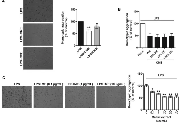
Maesil extract inhibits homotypic aggregation of LPS-activated THP-1 cells
Cell-cell adhesion plays central roles in the inflammatory response and migration of monocytes. A homotypic ag- gregation assay is a useful method to identify the efficacy and molecular mechanisms of an extract or the active con- stituents of a medicinal plant against inflammation.
Accordingly, the effects of Maesil extract on homotypic ag- gregation of LPS-activated THP-1 cells were investigated, with focus on the following: 1) different cultivars of Maesil;
2) different extraction methods (hot water or ethanol); and 3) different concentrations of Maesil extract. Two different Maesil varieties, namely Cheonmae and Cheongchuk (Aojiku), were compared. Hot water extract from Cheonmae (CME)
substantially inhibited homotypic aggregation of LPS-acti- vated THP1 cells; Hot water extract from Cheongchuk (CCE) also exhibited inhibitory activity, but to a much lesser extent than Cheonmae hot water extract (Fig. 1A). These data sug- gest that Cheonmae hot water extract (referred to as Maesil extract from here onwards), and to some extent Cheongchuk hot water extract, may confer potent anti-inflammatory ac- tivity against pathological processes in activated monocytes.
Maesil extract has traditionally been consumed as a sauce, a syrup, and as juice, with extraction methods employing hot water. However, it is also consumed as a liquor, which is extracted using alcohol. Thus, in the present study, Cheonmae Maesil was also extracted using water, 20%, 40%, or 100% ethanol and these preparations were investigated for their effect on homotypic aggregation of LPS activated THP-1 cells. As shown Fig. 1B, all of these extracts showed a similar inhibitory effect on homotypic aggregation, indicat- ing that the anti-inflammatory properties of Maesil are evi- dent regardless of the solvent used for extraction. Finally, the effect of various concentrations of Maesil (Cheonmae hot water) extract on homotypic aggregation of LPS-activated THP-1 cells was determined. Incubation of THP-1 cells with Maesil extract resulted in dose-dependent inhibition of LPS-induced homotypic aggregation. Indeed, aggregation was inhibited up to 50% when concentrations of 1,040 μ g/ml Maesil extract were used (Fig. 1C), suggesting that a concentration of 10 μg/ml Maesil (Cheonmae hot water) ex- tract is sufficient to inhibit LPS-activated homotypic ag- gregation of THP-1 cells.
In vitro cytotoxic effect of Maesil extract against THP-1 cells
Next, it was investigated whether or not Maesil extract is capable of eliciting a cytotoxic effect on THP-1 cells. To assess this, THP-1 cells were treated with Maesil extract (0.1200 μg/ml) for 24 hr and then an alteration of LPS-in- duced cytotoxicity was analyzed. It was found that Maesil extract at concentrations of 0.110 μg/ml appeared non-cyto- toxic, but Maesil extract at concentrations of 100-200 μg/ml was, if minimal, cytotoxic in THP-1 cells (Fig. 2). Therefore, to exclude an experimental noise by Maesil extract-induced cytotoxicity, further experiments were conducted with Maesil extract at less than noncytotoxic concentrations (≤
10 μg/ml). In addition, it is worthy of note that a range of concentrations (≤10 μg/ml) of Maesil extract employed in this study are likely to be physiologically and clinically rele-
A
B
C
Fig. 1. Inhibition of LPS-activated THP-1 cell homotypic aggregation by Maesil extract. THP-1 cells were incubated with 1 μg/ml LPS along with Cheonmae hot water extract (CME) or Cheongchuk hot water extract (CCE) (A) or Cheonmae ethanol extracts (EE) (0%-WE, 20%, 40% and 100%) (B), various concentrations of Maesil (Cheonmae hot water) extract (ME, 0.1-40 μg/ml) (C) for 24 hr. Homotypic aggregation of LPS activated THP-1 cells was observed under the microscope and quantified by counting the adhesive cells. Images are representative of at least 3 different observations. Bar graphs show mean ± S.E.
(n=3). *p<0.05. **p<0.01.
Fig. 2. Effect of Maesil extract on THP-1 cell viability. THP-1 cells were treated with various doses of Maesil extract for 24 hr and cell cytotoxicity was measured by the WST-1 assay. Data were plotted as bar graphs (mean
± S.E., n=3). *p<0.05.
vant in THP-1 cells. A recent report has shown that MK615, an extract of the Prunus mume, is able to relieve severe symp- toms of patients with cancer when 6.5 g of MK615 was ad- ministered twice per day [12]. Based on this and other data, the effective concentrations (≤10 μg/ml) of Maesil extract
used in this study are comparable to doses administrated in clinical and in vivo studies [12, 23]. It is also noteworthy that an advantage of using whole extracts seems likely to appear unexpected efficacy caused by unknown compounds in the whole extracts. Many studies have shown that such synergistic effects from unknown compounds in whole ex- tracts occasionally render the crude extract more efficacious when comparing to a single compound isolated at the equiv- alent dose [8, 18].
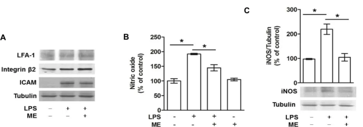
Maesil extract markedly inhibits the production of nitric oxide via suppression of iNOS expression in LPS-activated THP-1 cells, but not integrin beta 2 family members and ICAM-1
Given that homotypic aggregation of activated monocytes is regulated by NO production and CAM expression [18, 32, 36], it is plausible that Maesil extract regulates these me- diators, thereby crucially modifying homotypic aggregation of monocytes. Previous studies have shown that the integrin
A B
C
Fig. 3. Effect of Maesil extract on protein expressions of inducible nitric oxide synthase (iNOS) and cell adhesion molecules (CAMs) and production of nitric oxide in LPS activated THP-1 cells. THP-1 cells were incubated for 24 hr with 1 μg/ml LPS alone or in the presence of 10 μg/ml Maesil extract (ME). Protein levels of LFA-1, Integrin β2, ICAM-1 (A), iNOS (C) and tubulin were determined. Cells were treated with LPS alone or in the presence of 10 μg/ml ME extract and NO was measured (B). Data are plotted as bar graphs (mean ± S.E., n=3). *p<0.01.
beta 2 family members including LFA-1, and integrin β2, as well as ICAM-1 are key molecules in the regulation of monocyte adhesion [22, 31]. Therefore, the effects of Maesil extract on the expression of LFA-1, integrin β2, and ICAM-1 in LPS activated THP-1 cells were investigated. THP1 cells were treated with 1 μg/ml LPS, alone or in the presence of 10 μg/ml Maesil extract for 24 hr and subjected to west- ern blot analysis. The protein levels of the three CAMs were increased by LPS treatment, whereas Maesil extract had no effect on the protein expression of any of the adhesion mole- cules examined (Fig. 3A). These findings suggest that Maesil extract at the low concentrations (10 μg/ml) regulates homo- typic aggregation of activated monocytes through, if any, other CAMs than examined in this study. Accordingly, fur- ther experiments will be needed to identify cell adhesion molecules specifically involved in this event. To characterize underlying molecular mechanisms for the inhibition of ho- motypic aggregation of LPS-activated monocytes by ex- posure to Maesil extract, it was assessed whether Maesil ex- tract regulates the production of NO in these cells. It is known that NO plays an important role in inflammatory responses via mediation of monocyte activation and cell ad- hesion [32, 36]. Many studies have shown that monocytes produce low levels of NO, but treatment with LPS induces high levels of NO in these cells [3, 10]. Consistent with pre- vious studies, NO production was found to be remarkably elevated in THP-1 cells upon treatment with LPS. This LPSinduced NO production was shown to be substantially diminished in the presence of Maesil extract, whereas Maesil extract alone had no effect on the basal NO level (Fig. 3B).
Given that enhanced production of NO in LPSactivated
monocytes requires the induction of iNOS protein [3], the effect of Maesil extract on the expression level of iNOS was explored in these cells. Treatment of monocytes with LPS appeared to markedly enhance the expression of iNOS; an effect that was completely abrogated by concurrent treat- ment with Maesil extract (Fig. 3C). These data suggest that Maesil extract inhibits excessive NO production and damp- ens inflammatory pathways in LPS activated monocytes.
However, appropriate NO production plays a crucial role in physiological functions [2, 28] and thus a fine balance in NO production is fundamental to maintaining homeostasis in monocytes. In addition, phenolic compounds and terpe- noids, which are known chemical constituents of Maesil ex- tract, seems likely to have beneficial effects on chronic in- flammatory diseases associated with excessive NO pro- duction [14]. It is therefore assumed that phenolic com- pounds and terpenoids may be key active constituents of Maesil extract that plays important roles in the regulation of NO production in inflammatory conditions. Detailed mo- lecular mechanisms remain to be identified.
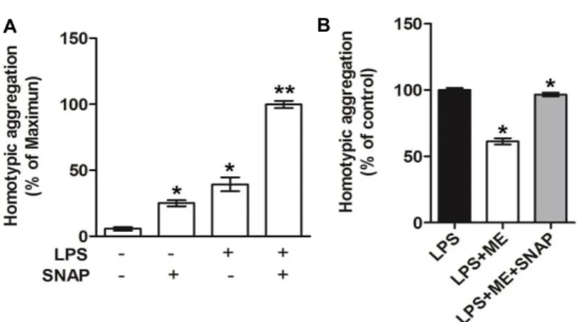
Maesil extract inhibits homotypic aggregation via in- hibition of nitric oxide production in LPS-activated THP-1 cells
To confirm whether inhibition of NO production by Maesil extract is a crucial factor for homotypic aggregation, a reversal effect of an NO-donor called S-nitroso-N-acetyl penicillamine (SNAP) on homotypic aggregation in THP-1 cells was examined. As expected, basal homotypic ag- gregation of monocytes was increased upon treatment with SNAP; co-treatment of THP-1 cells with LPS and SNAP was
A B
Fig. 4. Effect of Maesil extract and the NO-producing agent S-nitroso-N-acetyl penicillamine (SNAP) on homotypic aggregation in LPS activated THP- 1 cells. THP-1 cells were incubated for 24 hr with 1 μg/ml LPS alone or in the presence of 200 mM SNAP (A), 10 μg/ml Maesil extract, or both Maesil extract and 200 mM SNAP (B). Homotypic aggregation of LPS activated THP-1 cells was ob- served under the microscope and quantified by counting the adhesive cells. Images are repre- sentative of at least 3 different observations. Bar graphs show mean ± S.E. (n=3). *p<0.05. **p<0.01.
A B
Fig. 5. The inhibitory effect Maesil extract on LPS-induced IκB-α phosphorylation in LPS activated THP-1 cells. Serum-depleted THP-1 cells were treated with 1 μg/ml LPS along with Maesil extract for various periods of time. Western blotting was performed with p-IκB-α (A) and NF-κB p65 antibodies in LPS activated THP-1 cytosolic and nuclear extracts (B). Each band was quantified by densitometry. Bar graphs show mean ± S.E. (n=3), *p<0.05. **p<0.001.
shown to considerably enhance homotypic aggregation (Fig.
4A). In addition, the inhibitory effect of Maesil extract on LPS induced homotypic aggregation of monocytes was abro- gated by treatment of THP 1 cells with SNAP (Fig. 4B). These data suggest that Maesil extract-induced blockade of in- flammatory responses such as an inhibition of homotypic aggregation occurs through the regulation of NO production.
Attenuation of LPS-induced IκB-α phosphorylation and NF-κB translocation into the nucleus by Maesil ex- tract
The participation of NO signaling in homotypic cell adhe-
sion is under tight regulation, involving several intercellular signaling pathways. To determine the signal transduction pathways underlying the inhibition of LPS-induced homo- typic aggregation by Maesil extract, several key signaling molecules in the cell-cell adhesion pathway [18, 31] were studied. LPS-induced phosphorylation of p38 MAPK, ERK, and JNK was not altered by Maesil extract at a concentration of 10 ug/ml (Data not shown), whereas IκB-α phosphor- ylation was dramatically reduced by the co-treatment with Maesil extract for 60 min (Fig. 5A). Phosphorylation of IκB-α leads to its degradation and this event releases NF-κB from the NF-κB/IκB-α complex in the cytoplasm, thus allowing
translocation to the nucleus and subsequent promotion of pro-inflammatory gene expression, including iNOS. To be sure, the effects of Maesil extract on NF-κB translocation into the nucleus in LPS activated THP-1 cells were investigated.
It was found that the nuclear fraction of NF-κB was in- creased by LPS. To the contrary, Maesil extract significantly attenuated the NF-κB translocation into the nucleus in LPS-stimulated monocytes (Fig. 5B). These data suggest that Maesil extract inhibits the NF-κB pathway through in- hibition of LPS-induced phosphorylation of IκB-α and NF-κB translocation into the nucleus [15, 18]. However, further studies will be required to elucidate whether inhibition of the NF-κB pathway is directly involved in Maesil extract mediated inhibition of LPS-induced THP-1 cell homotypic aggregation via NO regulation. In conclusion, the present study provides an evidence for the following mechanisms:
(1) Maesil extract inhibits homotypic aggregation of LPS-ac- tivated THP-1 cells; (2) Maesil extract reduces the production of NO in LPS activated-THP-1cells and this event is medi- ated by the suppression of iNOS expression; and (3) Maesil extract inhibits LPS induced phosphorylation of IκB-α and NF-κB translocation into the nucleus. Taken together, there is strong evidence to suggest that Maesil extract could be utilized as a phytotherapeutic agent for the prevention of inflammatory diseases.
Acknowledgments
I thank Dr. Danielle McLean for critical reading of the manuscript and thoughtful discussions. This Research was supported by the Sookmyung Women's University Research Grants (1-1503-0049).
References
1. Adachi, M., Suzuki, Y., Mizuta, T., Osawa, T., Adachi, T., Osaka, K., Suzuki, K., Shiojima, K., Arai, Y., Masuda, K., Uchiyama, M., Oyamada, T. and Clerici, M. 2007. The
"Prunus mume Sieb. et Zucc" (Ume) is a rich natural source of novel anti-cancer substance. Int. J. Food Prop. 10, 375-384.
2. Beckman, J. S. and Koppenol, W. H. 1996. Nitric oxide, su- peroxide, and peroxynitrite: the good, the bad, and ugly.
Am. J. Physiol. 271, 1424-1437.
3. Bogdan, C. 2001. Nitric oxide and the immune response.
Nat. Immunol. 2, 907-916.
4. Chan, E. D. and Riches, D. W. 1998. Potential role of the JNK/SAPK signal transduction pathway in the induction
of iNOS by TNF-alpha. Biochem. Biophys. Res. Commun. 253, 790-796.
5. Chen, C., Chen, Y. H. and Lin, W. W. 1999. Involvement of p38 mitogen-activated protein kinase in lipopoly- saccharide-induced iNOS and COX-2 expression in J774 macrophages. Immunology 97, 124-129.
6. Choi, S. Y., Chung, M. J. and Sung, N. J. 2002. Volatile N-ni- trosamine inhibition after intake Korean green tea and Maesil (Prunus mume SIEB. et ZUCC.) extracts with an amine-rich diet in subjects ingesting nitrate. Food Chem.
Toxicol. 40, 949-957.
7. Chuda, Y., Ono, H., Ohnishi-Kameyama, M., Matsumoto, K., Nagata, T. and Kikuchi, Y. 1999. Mumefural, citric acid derivative improving blood fluidity from fruit-juice concen- trate of Japanese apricot (Prunus mume Sieb. et Zucc). J.
Agric. Food Chem. 47, 828-831.
8. Chung, K. F., Dent, G., McCusker, M., Guinot, P., Page, C.
P. and Barnes, P. J. 1987. Effect of a ginkgolide mixture (BN 52063) in antagonising skin and platelet responses to platelet activating factor in man. Lancet 1, 248-251.
9. Forslund, T., Nilsson, H. M. and Sundqvist, T. 2000. Nitric oxide regulates the aggregation of stimulated human neu- trophils. Biochem. Biophys. Res. Commun. 274, 482-487.
10. Guzik, T. J., Korbut, R. and Adamek-Guzik, T. 2003. Nitric oxide and superoxide in inflammation and immune regu- lation. J. Physiol. Pharmacol. 54, 469-487.
11. Hong, S., Kim, S. H., Rhee, M. H., Kim, A. R., Jung, J. H., Chun, T., Yoo, E. S. and Cho, J. Y. 2003. In vitro anti-in- flammatory and pro-aggregative effects of a lipid com- pound, petrocortyne A, from marine sponges. Naunyn Schmiedebergs Arch. Pharmacol. 368, 448-456.
12. Hoshino, T., Takagi, H., Naganuma, A., Koitabashi, E., Uehara, S., Sakamoto, N., Kudo, T., Sato, K. and Kakizaki, S. 2013. Advanced hepatocellular carcinoma responds to MK615, a compound extract from the Japanese apricot
"Prunus mume". World J. Hepatol. 5, 596-600.
13. Jeong, J. T., Moon, J. H., Park, K. H. and Shin, C. S. 2006.
Isolation and characterization of a new compound from Prunus mume fruit that inhibits cancer cells. J. Agric. Food Chem. 54, 2123-2128.
14. Jiang, F. and Dusting, G. J. 2003. Natural phenolic com- pounds as cardiovascular therapeutics: potential role of their antiinflammatory effects. Curr. Vasc. Pharmacol. 1, 135- 156.
15. Karin, M. and Delhase, M. 2000. The I kappa B kinase (IKK) and NF-kappa B: key elements of proinflammatory signal- ling. Semin. Immunol. 12, 85-98.
16. Kawahara, K., Hashiguchi, T., Masuda, K., Saniabadi, A. R., Kikuchi, K., Tancharoen, S., Ito, T., Miura, N., Morimoto, Y., Biswas, K. K., Nawa, Y., Meng, X., Oyama, Y., Takenou- chi, K., Shrestha, B., Sameshima, H., Shimizu, T., Adachi, T., Adachi, M. and Maruyama, I. 2009. Mechanism of HMGB1 release inhibition from RAW264.7 cells by oleanolic acid in Prunus mume Sieb. et Zucc. Int. J. Mol. Med. 23, 615-620.
17. Kim, D. O., Jeong, S. W. and Lee, C. Y. 2003. Antioxidant
capacity of phenolic phytochemicals from various cultivars of plums. Food Chem. 81, 321-326.
18. Kim, H. J., McLean, D., Pyee, J., Kim, J. and Park, H. 2014.
Extract from Acanthopanax senticosus prevents LPS-induced monocytic cell adhesion via suppression of LFA-1 and Mac-1. Can. J. Physiol. Pharmacol. 92, 278-284.
19. Kim, J., Park, J., Choi, S., Chi, S. G., Mowbray, A. L., Jo, H. and Park, H. 2008. X-linked inhibitor of apoptosis protein is an important regulator of vascular endothelial growth fac- tor-dependent bovine aortic endothelial cell survival. Circ.
Res. 102, 896-904.
20. Kim, Y. H., Lee, S. H., Lee, J. Y., Choi, S. W., Park, J. W.
and Kwon, T. K. 2004. Triptolide inhibits murine-inducible nitric oxide synthase expression by down-regulating lip- opolysaccharide-induced activity of nuclear factor-kappa B and c-Jun NH2-terminal kinase. Eur. J. Pharmacol. 494, 1-9.
21. Ko, B. S., Kim, da, S., Kang, S., Ryuk, J. A. and Park, S.
2013. Prunus mume and Lithospermum erythrorhizon extracts synergistically prevent visceral adiposity by iImproving en- ergy metabolism through potentiating hypothalamic leptin and insulin signalling in ovariectomized rats. Evid. Based Complement Alternat. Med. 2013, 750986.
22. Lee, G. K., Jung, K. C., Park, W. S., Kook, M. C., Park, C.
S., Sohn, H. W., Bae, Y. M., Song, H. G. and Park, S. H.
1999. LFA-1- and ICAM-1-dependent homotypic aggrega- tion of human thymocytes induced by JL1 engagement. Mol.
Cells 9, 662-667.
23. Matsushita, S., Tada, K. I., Kawahara, K. I., Kawai, K., Hashiguchi, T., Maruyama, I. and Kanekura, T. 2010.
Advanced malignant melanoma responds to Prunus mume Sieb. Et Zucc (Ume) extract: Case report and in vitro study.
Exp. Ther. Med. 1, 569-574.
24. Mentzer, S. J., Faller, D. V. and Burakoff, S. J. 1986.
Interferon-gamma induction of LFA-1-mediated homotypic adhesion of human monocytes. J. Immunol. 137, 108-113.
25. Mina-Osorio, P., Shapiro, L. H. and Ortega, E. 2006. CD13 in cell adhesion: aminopeptidase N (CD13) mediates homo- typic aggregation of monocytic cells. J. Leukoc. Biol. 79, 719-730.
26. Mitani, T., Horinishi, A., Kishida, K., Kawabata, T., Yano, F., Mimura, H., Inaba, N., Yamanishi, H., Oe, T., Negoro, K., Mori, H., Miyake, Y., Hosoda, A., Tanaka, Y., Mori, M.
and Ozaki, Y. 2013. Phenolics profile of mume, Japanese apricot (Prunus mume Sieb. et Zucc.) fruit. Biosci. Biotechnol.
Biochem. 77, 1623-1627.
27. Miyazawa, M., Shirakawa, N., Utsunomiya, H., Inada, K.
and Yamada, T. 2009. Comparision of the volatile compo- nents of unripe and ripe Japanese apricot (Prunus mume Sieb. et Zucc.). Nat. Prod. Res. 23, 1567-1571.
28. Nguyen, T., Brunson, D., Crespi, C. L., Penman, B. W., Wishnok, J. S. and Tannenbaum, S. R. 1992. DNA damage
and mutation in human cells exposed to nitric oxide in vitro.
Proc. Natl. Acad. Sci. USA 89, 3030-3034.
29. Otsuka, T., Tsukamoto, T., Tanaka, H., Inada, K., Utsuno- miya, H., Mizoshita, T., Kumagai, T., Katsuyama, T., Miki, K. and Tatematsu, M. 2005. Suppressive effects of fruit-juice concentrate of Prunus mume Sieb. et Zucc. (Japanese apricot, Ume) on Helicobacter pylori-induced glandular stomach le- sions in Mongolian gerbils. Asian Pac. J. Cancer Prev. 6, 337-341.
30. Park, C., Jin, C. Y., Kim, G. Y., Jeong, Y. K., Kim, W. J.
and Choi, Y. H. 2011. Induction of apoptosis by ethanol ex- tract of Prunus mume in U937 human leukemia cells through activation of caspases. Oncol. Rep. 26, 987-993.
31. Park, H., Park, S. G., Lee, J. W., Kim, T., Kim, G., Ko, Y.
G. and Kim, S. 2002. Monocyte cell adhesion induced by a human aminoacyl-tRNA synthetase-associated factor, p43:
identification of the related adhesion molecules and signal pathways. J. Leukoc. Biol. 71, 223-230.
32. Peng, H. B., Spiecker, M. and Liao, J. K. 1998. Inducible ni- tric oxide: an autoregulatory feedback inhibitor of vascular inflammation. J. Immunol. 161, 1970-1976.
33. Pillinger, M. H., Feoktistov, A. S., Capodici, C., Solitar, B., Levy, J., Oei, T. T. and Philips, M. R. 1996. Mitogen-acti- vated protein kinase in neutrophils and enucleate neu- trophil cytoplasts: evidence for regulation of cell-cell adhesion. J. Biol. Chem. 271, 12049-12056.
34. Sun, L., Yang, W., Zhang, Q., Cheng, T., Pan, H., Xu, Z., Zhang, J. and Chen, C. 2013. Genome-wide characterization and linkage mapping of simple sequence repeats in mei (Prunus mume Sieb. et Zucc.). PLoS ONE 8, e59562.
35. Utsunomiya, H., Takekoshi, S., Gato, N., Utatsu, H., Motley, E. D., Eguchi, K., Fitzgerald, T. G., Mifune, M., Frank, G.
D. and Eguchi, S. 2002. Fruit-juice concentrate of Asian plum inhibits growth signals of vascular smooth muscle cells induced by angiotensin II. Life Sci. 72, 659-667.
36. Webb, J. L., Polak, J. M. and Evans, T. J. 2001. Effect of adhe- sion on inducible nitric oxide synthase (iNOS) production in purified human neutrophils. Clin. Exp. Neuroimmunol.
123, 42-48.
37. Yan, X. T., Lee, S. H., Li, W., Sun, Y. N., Yang, S. Y., Jang, H. D. and Kim, Y. H. 2014. Evaluation of the antioxidant and anti-osteoporosis activities of chemical constituents of the fruits of Prunus mume. Food Chem. 156, 408-415.
38. Yingsakmongkon, S., Miyamoto, D., Sriwilaijaroen, N., Fujita, K., Matsumoto, K., Jampangern, W., Hiramatsu, H., Guo, C. T., Sawada, T., Takahashi, T., Hidari, K., Suzuki, T., Ito, M., Ito, Y. and Suzuki, Y. 2008. In vitro inhibition of human influenza A virus infection by fruit-juice concen- trate of Japanese plum (Prunus mume SIEB. et ZUCC). Biol.
Pharm. Bull. 31, 511-515.
초록:매실 추출물의 산화질소 생성과 NF-κB 활성 조절을 통한 LPS유도성 THP-1 세포 동형성 응집 의 억제 효과
이혜림1†․박영숙2†․김현정1․이아람2․최지혜2․피재호1․박헌용1․김종민2*
(1단국대학교 분자생물학과, 2숙명여자대학교 생명시스템학과)
활성화된 단핵구의 동형성 세포 부착(동형성 응집)은 염증반응, 분화, 이동과 같은 생리학적, 병리학적 과정에 서 중요한 역할을 한다. 매실 추출물은 항바이러스, 항균, 항암작용과 같은 효과를 보인다고 알려 져있다. 따라서, 매실 추출물은 단핵구의 동형성 응집 억제를 통해 염증반응을 조절할 가능성을 가진다. 본 연구에서는, 염증성 질환에서 매실 추출물의 치료효능을 뒷받침할 수 있는 분자적 기전을 조사하였다. 매실 추출물이 지질다당질 (LPS)로 활성화된 단핵구의 동형성 응집을 억제함을 확인하였다. 이러한 효과는 LPS로 활성화된 THP-1 세포의 iNOS 단백질 발현 억제를 통해, 산화질소(NO) 생산의 감소로 조절되는 것을 발견하였다. 또한 NO 생성물질인 SNAP 처리 실험을 통해 단핵구 동형성 응집을 억제하는데 매실에 의한 NO 억제가 필수적인 기작임을 확인하였 다. 게다가, 매실 추출물은 LPS로 유도된 IκB-α 의 인산화와 NF-κB의 핵내로의 이동을 현저하게 감소시키는 것을 확인하였다. 매실 추출물은 NO생성과 NF-κB 활성 억제를 통해 LPS로 활성화된 단핵구의 동형성 응집을 저해하 고 이를 통해 항염증 효과를 유도할 수 있다는 결론으로부터 만성 염증성 질환의 치료와 예방에 매실 추출물의 효능을 제시하고자 한다.