A Thesis
For The Degree of Doctor of Veterinary Medicine
Expression of Constitutive Endothelial and
Inducible Nitric Oxide Synthase in the Sciatic
nerve of Lewis Rats with Experimental
Autoimmune Neuritis
Graduate School, Cheju National University
Department of Veterinary Medicine
Yongduk Lee
Expression of Constitutive Endothelial and Inducible
Nitric Oxide Synthase in the Sciatic Nerve of Lewis
Rats with Experimental Autoimmune Neuritis
Yongduk Lee
(Supervised by Professor Taekyun Shin)
A thesis submitted in partial fulfillment of the requirement for the degree of Doctor of Veterinary Medicine
2001. 10.
This thesis has been examined and approved.
Thesis director, Cha-Soo Lee, Prof. of Veterinary Medicine
Du-Sik Lee, Prof. of Veterinary Medicine
Sang-Mok Lee, Prof. of Veterinary Medicine
Hee-Seok Kim, Prof. of Veterinary Medicine
Tae-Kyun Shin, Prof. of Veterinary Medicine
2001. 12.
Department of Veterinary Medicine
GRADUATE SCHOOL
Abstract
Expression of Constitutive Endothelial and
Inducible Nitric Oxide Synthase in the Sciatic
nerve of Lewis Rats with Experimental
Autoimmune Neuritis
Advised by Professor Taekyun Shin
Yongduk Lee
Department of Veterinary Medicine
Graduate School, Cheju National University, Jeju 690-756, Korea
Abstract
This study examined the expression of constitutive endothelial nitric oxide synthase (eNOS) and inducible NOS (iNOS) in the sciatic nerve of Lewis rats with experimental autoimmune neuritis (EAN). Western blot analysis showed that both eNOS and iNOS expression in the sciatic nerves of rats increased significantly during the peak stage of EAN, but declined thereafter. Only minimal amounts of these enzymes were identified in
normal rat sciatic nerves. Immunohistochemical studies showed that eNOS was increased in vascular endothelial cells and Schwann cells, but not in inflammatory cells, during the peak stage of EAN. However, iNOS was found mainly in inflammatory macrophages in sciatic nerve EAN lesions. These findings suggest that, depending on the stage of peripheral nervous system autoimmune disease, the increased expression of both eNOS and iNOS might be involved in either the production of detrimental effects during the induction stage of EAN or in the recovery from EAN paralysis. ... Keywords: autoimmune neuritis, nitric oxide synthase, Schwann cells
CONTENTS
1. Introduction--- 1
2. Materials and Methods--- 4
3. Results--- 10
4. Discussion--- 25
5. Conclusion--- 28
Ⅰ. Introduction
Nitric oxide (NO) is a short-lived free radical with biological
functions in the nervous, cardiovascular, and immune systems (Moncada et
al., 1991; Bredt and Snyder, 1992). NO is synthesized from L-arginine by
activation of the enzyme nitric oxide synthase (NOS). NOS exists in two forms: (1) a constitutive, Ca2+-dependent form that is rapidly activated by agonists that elevate intracellular free Ca2+, for example, neuronal NOS (nNOS) and endothelial NOS (eNOS); and (2) a Ca2+-independent inducible form (iNOS) (Moncada et al., 1991; Xie and Nathan, 1994). Neuronal NOS is constitutively expressed in discrete neuronal cells, whereas endothelial NOS is present in endothelial cells of blood vessels. Inducible NOS is expressed in various cell types when activated (Xie et al., 1992). NOS is known to play both beneficial and detrimental roles in injury of the nervous system through the generation of NO. There is general agreement that excess production of NO, mainly by iNOS, causes tissue damage, whereas constitutive nNOS and eNOS function in normal physiological events, such as regulation of the microcirculation, synaptic plasticity, and neuroprotective processes (Iadecola, 1993; Dawson and Dawson, 1996). In the rat model of autoimmune central nervous system disease known as experimental
autoimmune encephalomyelitis (EAE), increased expression of the three forms of NOS was observed in EAE- affected spinal cords, and it was postulated that iNOS was involved in macrophage apoptosis in EAE lesions (Kim et al., 2000). Whether constitutive endothelial isoforms of NOS contribute to autoimmune damage in the peripheral nervous system (PNS)
has not been established.
Experimental autoimmune neuritis (EAN) is an autoimmune disease model of human peripheral demyelinating disease that is mediated by CD4+ T cells (Fujioka et al., 2000; Matsumoto, 2000). EAN lesions in susceptible animals are characterized by the infiltration of T cells and macrophages during the peak stage of the disease (Fujioka et al., 2000). Some Schwann cells undergo apoptosis during EAN (Conti et al., 1998). There is a
general agreement that inflammatory mediators, including pro-inflammatory cytokines from macrophages and T cells, mediate the paralysis (Hartung et
al., 1992; Kieseier et al., 2000). Furthermore, in our previous report on the
prototype autoimmune disease model EAE, we showed that the majority of host cells survive, including neurons and glial cells, whereas invading inflammatory cells are eliminated via the activation of apoptosis-related molecules (Moon et al., 2000). If this is also the case in EAN,
death-related molecules might be activated in the inflammatory cells of EAN lesions. Little is known about the expression of NOS and cpp32, which is an important death-related enzyme, in this process (Wintergerst et
al., 2000).
Many investigators have focused on the detrimental effect of NOS in central nervous system disease, and have determined that the iNOS
inhibitor aminoguanidine effectively ameliorates the paralysis of rat EAE (Zhao et al., 1996; Shin et al., 2000). However, it is known that EAN is not effectively ameliorated by the administration of either
NG-L-monomethyl-arginine (L-NMMA), a competitive inhibitor of NOS, or aminoguanidine, a selective inhibitor of cytokine-inducible NOS (Zielasek et
al., 1995). In fact, EAN is aggravated by treatment with NOS inhibitors,
suggesting that NOS is beneficial in PNS physiology (Ruuls et al., 1996). This discrepancy may originate from a different role or pattern of NOS expression in the PNS. Although NOS is known to be involved in PNS diseases, little is known of the expression pattern of constitutive eNOS in PNS autoimmune neuritis.
This study examined changes in the expression of constitutive eNOS and inducible NOS (iNOS) over the course of EAN by Western blot
analysis, and elucidated the function of NOS in host and inflammatory cells by immunohistochemistry.
Ⅱ. Materials and Methods
1. Animals
Lewis rats were obtained from Harlan (Sprague Dawley Inc., Indianapolis, IN) and bred in our animal facility. Female rats weighing 160-200 g, aged 7-12 weeks were used in the experiments.
2. Monoclonal antibodies and polyclonal antisera
The following monoclonal antibodies (mAb) were used in this study: W3/25 (anti-CD4); OX8 (anti-CD8); R73 (anti-TCR); ED1 to label
macrophages; and OX42 (anti-complement receptor type 3), which also labels macrophages. These mAb were purchased from Serotec (Blackthorn, Bicester, Bucks, UK). Monoclonal anti-â actin antibody was purchased from Sigma (St. Louis, MO). Rabbit anti-iNOS, rabbit anti-eNOS, and mouse monoclonal anti-eNOS antibodies were obtained from Transduction Laboratories (Lexington, KY). Rabbit anti-S100 antisera was purchased from Dako (Copenhagen, Denmark).
3. EAN induction
Each rat was injected into the hind footpads bilaterally with an emulsion containing equal parts of fresh P2 peptides in phosphate buffer (g/mL) and complete Freunds adjuvant (CFA; Mycobacterium tuberculosis H37Ra, 5 mg/mL; Difco). Some rats were also given intraperitoneal injections of Bordetella pertussis toxin (2 g/ea) (Sigma Chemical Co., St. Louis, MO). After the injections, rats were observed daily for clinical signs of EAN.
The progress of EAN was divided into seven clinical stages: Grade (G) 0, no signs; G1, floppy tail; G2, mild paraparesis; G3, severe
paraparesis; G4, tetraparesis; G5, moribund condition or death; and R0, the recovery stage (Matsumoto, 2000). Control rats were immunized with CFA only. Five rats were sacrificed under ether anesthesia at various stages of EAN.
4. Tissue sampling
In this study, tissue sampling was performed on days 12 -15 and on day 21 post-immunization (PI), during the peak and recovery stages of EAN, respectively. Five rats in each group were sacrificed under ether anesthesia. The sciatic nerves were removed and frozen at -70℃ for protein analysis. Pieces of the sciatic nerves were either snap frozen in pre-chilled isopentane using liquid nitrogen or processed for paraffin
embedding after fixation in 4% paraformaldehyde in phosphate-buffered saline (PBS, pH 7.4).
5. Western blot analysis
Frozen sciatic nerves and spinal cords were thawed at room
temperature, minced, lysed in a buffer consisting of 40 mM Tris-HCl, pH 7.4, 120 mM NaCl, and 0.1% Nonidet P-40 (polyoxyethylene [9] p-t-octyl phenol) containing the protease inhibitors, leupeptin (0.5 g/ml), PMSF (1 mM), and aprotinin (5 g/ml), and then homogenized.
Samples were electrophoresed under denaturing conditions on 7.5% SDS-polyacrylamide gels, and the separated proteins were transferred to PROTRANⓇ nitrocellulose transfer membrane (Schleicher and Schuell, Keene, NH), as previously described (Ahn et al., 2001). eNOS and iNOS were detected using mouse monoclonal anti-eNOS and rabbit anti-iNOS antisera diluted 1:5000 with TBS-T, respectively (Transduction
Laboratories, Lexington, KY). The reaction was visualized by labeling with horseradish peroxidase-conjugated goat anti-rabbit IgG secondary antibody (Vector, Burlingame, CA). Visualization was achieved using Amersham ECL reagents (Arlington Heights, IL). The immunoblot membranes were re-probed with a monoclonal antibody to beta actin (Sigma) using the methods described above. The immunoblot signals were quantitated using a densitometer (M GS-700 imaging Densitometer, Bio-Rad, CA). The relative expressions of eNOS and iNOS were calculated, and the value for the
control was arbitrarily defined as 1.
6. Immunohistochemistry
Deparaffinized sections of the sciatic nerves were treated with 0.3% hydrogen peroxide in methyl alcohol for 30 min to block endogenous peroxidase. Frozen sections to be used for R73 immunostaining were treated with ethyl ether for 10 minutes (Ohmori et al., 1992). After three washes with PBS, the sections were exposed to 10% normal goat serum, and then incubated with primary antisera including mouse monoclonal anti-eNOS antibodies, rabbit anti-iNOS antisera (diluted 1:200)
(Transduction Laboratories, Lexington, KY), and rabbit anti-S100 antisera (Dako, Copenhagen, Denmark) for 1 hour at RT. ED1 antibodies were used to identify macrophages in paraffin sections. Rabbit anti-S100
antiserum was used to label Schwann cells (Mata et al., 1990). After three washes, the appropriate biotinylated secondary antibody and the
avidin-biotin-peroxidase complex (ABC) from the Elite kit (Vector,
Burlingame, CA) were added sequentially. Peroxidase was developed with diaminobenzidine (DAB)-hydrogen peroxidase solution (0.001%
3,3'-diaminbenzidine and 0.01% hydrogen peroxidase in 0.05M Tris buffer). The sections were counterstained with hematoxylin before being mounted.
To co-localize eNOS and S100 in the same section, S100 was first immunostained using the single-staining protocol described above, and the color was developed using the aminoethyl carbazole (AEC) substrate kit
(Zymed, San Francisco, CA). After photographing the S100-immunostained lesions in the sciatic nerve, the slide was bleached in ethanol. The slide was then immunostained using the primary eNOS antisera in the same manner, and the color was developed using the AEC substrate kit (Zymed). The co-localization of both antigens in a single cell was readily apparent.
7. Terminal deoxynucleotidyl transferase (TdT)-mediated dUTP nick end-labeling (TUNEL)
DNA fragmentation was detected by in situ nick end-labeling, as recommended by the manufacturer (Intergen, Purchase, N.Y.). In brief, the paraffin sections were deparaffinized, rehydrated, and washed in PBS. The sections were treated with 0.02% proteinase K (Sigma, St. Louis, MO) for 15 minutes at room temperature, blocked with 3% hydrogen peroxide in PBS for 10 minutes and washed with PBS. Tissues were incubated with equilibration buffer for 5 minutes and allowed to react with TdT enzyme for 60 minutes at 37 C. The reaction was stopped by incubation in stop buffer for 15 minutes. Finally, sections were allowed to react with
peroxidase-labeled anti-digoxigenin antibody for 30 minutes. Positive cells were visualized using diaminobenzidine substrate counterstained with hematoxylin.
For TUNEL and iNOS double-labeling, slides previously processed for TUNEL were subsequently immunostained with polyclonal iNOS
by incubating the sections in Sigma Fast TM nitroblue
tetrazolium/5-bromo-4-chloro-3-indolyl phosphate (NBT/BCIP) substrate tablet solution (Sigma).
8. Statistical analysis
Statistical comparisons among groups were made using the
Student-Newman-Keuls Multiple Comparisons test. Differences with
Ⅲ. Results
1. Clinical observation of EAN
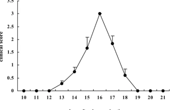
The clinical course of EAN is shown in Fig. 1. EAN rats
immunized with P2 peptides developed floppy tails (G1) on days 9-10
PI, and showed progressive hindlimb paralysis (G2 or G3) on days
12-15 PI. All of the rats subsequently recovered (Fig. 1)
Fig. 1. Clinical course of experimental autoimmune neuritis (EAN) in Lewis rats. EAN was induced by immunization with 100 μg of P2
peptides emulsified with complete adjuvant into bilateral hind footpads. Each rat was treated with 50 ng of pertussis toxin on the day of
immunization. The results are expressed as mean clinical scores ± S.E. (n = 5). 0 0.5 1 1.5 2 2.5 3 3.5 10 11 12 13 14 15 16 17 18 19 20 21
days after immunization
2. Histological findings of sciatic nerves
Histological examination revealed that a large number of
inflammatory cells infiltrated the area around the sciatic nerves of
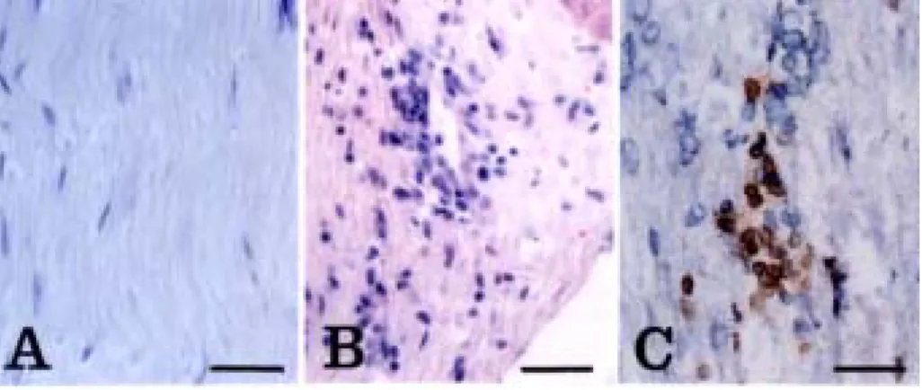
rats with EAN during the peak stage (day 13 PI) (Fig.2B). In the
normal control rats (Fig. 2A), no infiltrating cells were detected in
the sciatic nerves. Inflammatory cells in the EAN lesions consisted of
R73+/ TCRαβ+ T cells (Fig.2C) and ED1+ macrophages (Fig. 6B).
Fig. 2. Histology of EAN-affected sciatic nerves. Some inflammatory cells were found in the sciatic nerve of EAN-affected rats (day 13 PI) (B), whereas normal rats were devoid of inflammatory cells in the sciatic nerve (A). The inflammatory cells consisted mainly of R73+/ TCR+ T cells (C). A and B, Hematoxylin-eosin staining. C, immunostaining with R73 mAb. C is a frozen section. A, Normal rat; B and C, EAN-affected rats (day 13 PI). Counterstained with hematoxylin. Scale bar = 30 μm.
3. Western blot analysis of eNOS and iNOS in EAN
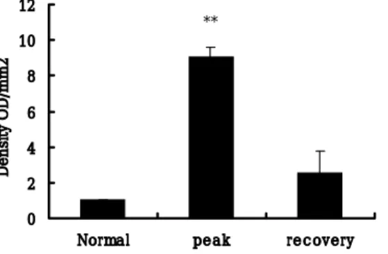
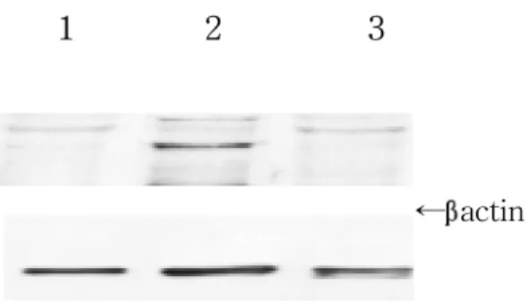
Constitutive eNOS expression in the sciatic nerves was significantly increased during the peak stage (day 13 PI, G3) of EAN (Fig. 3, lane 2) (p < 0.01). Its levels declined during the recovery stage of EAN (day 21 PI, R0) (Fig. 3, lane 3).
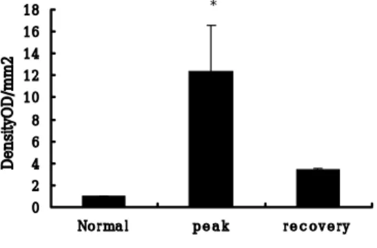
The expression of iNOS in the sciatic nerves of rats with EAN was also increased compared to the normal control (p < 0.01) (Fig. 4).
1 2 3
←eNOS
←β actin
Fig. 3. A, Representative Western blot analysis of endothelial NOS in the sciatic nerve shows a strong band in the paralytic stage (lane 2, G3, day 13 PI), and a weaker band in the recovery stage (lane 3, RO, day 21 PI) of EAN rats compared with normal controls (lane 1). The molecular mass of eNOS is 140 kDa.
Fig. 3. B, The quantification of eNOS after Western blotting and normalization for β-actin content showed a great increase of eNOS at the peak stage of EAN
compared to the control group, followed by a decline in the recovery stage. The immunoblot was quantitated by densitometry, and the relative value of the control was arbitrarily defined as 1. Data points represent the mean +/- SEM; n = 3 rats/group. 0 2 4 6 8 10 12
Normal peak recovery
Density OD/mm2
1 2 3
←iNOS←βactin
Fig. 4. A, Western blot analysis of iNOS in the sciatic nerve during the normal (lane 1), paralytic (lane 2, G3, day 13 PI), and recovery stages (lane 3, RO, day 21 PI) of EAN rats. The molecular mass of iNOS is 130 kDa.
Fig. 4. B, Quantitation of sciatic nerve iNOS expression over the course of EAN. The immunoblot was quantitated by densitometry, and the relative value of the control was arbitrarily defined as 1. Data points represent the mean +/- SEM; n = 3 rats/group.
0 2 4 6 8 10 12 14 16 18 No r ma l p e a k r e c o v e r y DensityOD/mm2 *
4. Immunohistochemical localization of eNOS and iNOS in EAN
In EAN-affected rats, eNOS (Fig. 5) was intensely stained in vascular endothelial cells (Fig. 5B), in some axons that were encased by Schwann cells (Fig. 5D), and in Schwann cells of the sciatic nerve (Figs. 5C and 5D). In contrast, eNOS was weakly stained in some vascular endothelial cells in the sciatic nerves of normal control rats (Fig. 5A). Furthermore, both eNOS (Fig. 5E) and S100 (Fig. 5F) were co-localized in Schwann cells within the EAN lesions. Few inflammatory cells were positive for eNOS (Table 1).
Contrary to the expression pattern of eNOS in EAN, iNOS
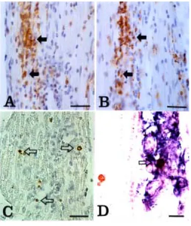
immunoreactivity was mainly localized to inflammatory cells (Fig. 6A) that were stained positively for ED1 (Fig. 6B) in the EAN lesions.
This finding suggests that the increased expression of constitutive eNOS in axons and some Schwann cells, which recover their function after recovery from paralysis, mediates a beneficial role in the process of PNS inflammation, whereas inflammatory cells expressing iNOS are eliminated via apoptosis during the recovery stage of EAN.
Fig. 5. Immunostaining of eNOS in the sciatic nerve of normal rats (A) and rats with EAN (G3, day 13 PI) (B-D). eNOS (A) was weakly stained in the vessels (arrow) of the sciatic nerve of normal rats. The expression of eNOS was enhanced in vessels (B, arrow) and some
Schwann cells (C, arrowheads) in EAN lesions. In a higher magnification of C. eNOS-positive axons encased by Schwann cells(D, arrows) were found. Arrowheads indicate same cells in C and D. Furthermore, eNOS (E) co-localized with S100 (F) in each Schwann cell of the EAN lesions. Arrows indicate the same group of Schwann cells. A-D: DAB substrate. E and F: AEC substrate. A D: counterstained with hematoxylin. A-C, E, F: Scale bar = 30 m. D: Scale bar = 10μm.
Table 1. Immunohistochemical analysis of endothelial nitric oxide synthase in the sciatic nerve of rats with EAN and control rats.
a
The sciatic nerves were examined at the peak (grade 3: day 14 post-immunization) and recovery (day 21 post-immunization) stages of EAN
b Increased intensity of eNOS immunoreactivity in some cells. c
ND = Inflammatory cells including macrophages and lymphocytes were not detected in the sciatic nerves of rats.
Cell type
Normal
EAN
aPeakstage
Recovery
Vascular endothelial cells
+
+++
b++
Perineurium
+
++
+
Schwann cells
+
+++
b++
Axis cylinder
+
+
+
5. Correlation between apoptosis and the expression of iNOS in inflammatory cells
The relationship between apoptosis and iNOS expression was examined in EAN-affected rat sciatic nerves, because the majority of inflammatory cells were eliminated by apoptosis, leading to spontaneous recovery from paralysis. Some TUNEL-positive cells (Fig. 6C)were scattered throughout the sciatic nerve of rats with EAN, as previously shown (Weishaupt et al., 2000). In a double labeling experiment in this study, a TUNEL-positive cell (Fig. 6D, brown color) in a perivascular lesion was shown to be iNOS-positive (Fig. 6D, blue color). This suggests that some iNOS-positive inflammatory cells undergo apoptosis through the activation of iNOS in EAN lesions.
Fig. 6. Detection of apoptosis and iNOS expression in the sciatic nerve of rats with EAN (G3, day 13 PI). iNOS was mainly immunostained in perivascular inflammatory cells (A) that were positive for ED1 (B). TUNEL-reactive cells were scattered throughout the EAN lesions. Arrows indicate TUNEL-positive cells (C). Double labeling for iNOS and TUNEL in the sciatic nerve of EAN rats revealed that TUNEL-positive cells (brown color, arrowhead) were also positive for iNOS (blue) in EAN lesions. However, not all iNOS-positive cells were positive for TUNEL, or vice-versa. A-C: Counterstained with hematoxylin. AB, Scale bar = 30μ m. D, Scale bar = 10 μm.
Ⅳ. Discussion
This is the first report to show that constitutive eNOS and iNOS are differentially expressed in EAN lesions of Lewis rats. We also showed that constitutive eNOS was mainly expressed in neural elements, probably restoring axonal function. However, iNOS, which is expressed in
macrophages, mediates the elimination of macrophages. In any case, either cNOS or iNOS participates in restoring PNS function after injury. This result is in part consistent with a previous report that three forms of NOS are over-expressed after sciatic nerve ligature (Gonzalez-Hernandez and Rustioni, 1999).
It is generally agreed that Schwann cells stimulated with tumor necrosis factor (TNF)- and interferon (IFN)-ã in in vitro systems generate NO via the activation of iNOS (Gold et al., 1996, Nagano et al., 2001). Gold et al. (1996) confirmed that stimulated Schwann cells play a potential immunoregulatory role through generation of NO, which suppresses T cell proliferation and ultimately induces T cell death. Nagano et al. (2001) further verified that TNF- and IFN-ã exert a synergistic inhibitory action on immortalized Schwann cell viability, an effect that is accompanied by iNOS induction and ceremide accumulation. These findings suggest that stimulated Schwann cells generate NO, which may induce T cell or Schwann cell death through the accumulation of ceramide.
In this study, we found that vessels and Schwann cells had
increased eNOS expression, but not iNOS expression, during the peak stage of EAN. These findings suggest that constitutive eNOS increases in
Schwann cells in response to inflammatory stimuli in sciatic nerves in autoimmune disease models (Kieseier et al., 2000). The increased
expression of eNOS, in combination with iNOS from macrophages, might be involved in the recovery of the peripheral nervous system from
autoimmune disease models, because NO from Schwann cells suppresses T cell proliferation (Gold et al., 1996). However, it is important to note that the data of previous studies of the expression of NOS, particularly iNOS, were obtained from Schwann cells grown in culture. In this study, we found that eNOS, but not iNOS, was expressed in the Schwann cells of sciatic nerves of rats with EAN, suggesting that Schwann cells do not express iNOS in vivo. We do not exclude the limited possibility that eNOS is involved in PNS tissue injury processes, such as Schwann cell apoptosis (Weishaupt et al., 2001), because eNOS has been shown to be associated with cardiocyte apoptosis in certain experimental models (Kawaguchi et al., 1997). The precise mechanism of eNOS expression needs further study.
Recently, many investigators have focused on the beneficial effect of iNOS during the pathogenesis of autoimmune encephalomyelitis. This is because NOS II knockout mice are more severely affected by EAE
(Sahrbacher et al., 1998), and EAE-resistant rats become susceptible after inhibition of iNOS (OBrien et al., 1999). Similar findings were also recognized in EAN (Ruuls et al., 1996), although different NOS inhibitors were used. It is postulated that constitutive eNOS expression in axons and Schwann cells increases at the time of sciatic injury in EAN. The
consequent increase in both eNOS and iNOS and possibly the generation of NO may accelerate normal physiological events, such as regulation of microcirculation, synaptic plasticity, and neuro-protective processes
(Iadecola, 1993; Dawson and Dawson, 1996). In this study, some macrophages were shown to undergo apoptosis in EAN-affected sciatic nerves, and these cells have been shown to play a beneficial role in recovery from EAN (Kiefer et al., 2001).
Ⅴ. Conclusion
EAN is a CD4+ T cell-mediated disease model. This disease is characterized by the infiltration of T cells and macrophages that are ultimately eliminated through apoptosis. The inflammatory process is paralleled by the expression of three forms of nitric oxide synthase in EAN-affected lesions. This study illustrated that an increased expression of constitutive eNOS may be a compensatory response against PNS
inflammation. Thus, eNOS is involved in the activation of axons, vessels, and adjacent Schwann cells. Macrophages expressing iNOS play a role in transmitting signals to host tissues, and the level of iNOS expression is inversely related to the elimination of T cells and macrophages in EAN lesions by either paracrine or autocrine signals.
Considering all of the above findings, the parallel between EAN paralysis and NOS expression in EAN implies that while NOS may be beneficial, it functions in either a beneficial or detrimental role depending on the stage of inflammation, the capacity to generate NO, or the cell type.
Ⅵ. References
Ahn, M., Kang, J., Lee, Y., Riu, K., Kim, Y., Jee, Y., Matsumoto, Y., Shin, T., 2001. Pertussis toxin-induced hyperacute autoimmune encephalomyelitis in Lewis rats is correlated with increased expression of inducible nitric oxide synthase and tumor necrosis factor alpha. Neurosci. Lett. 308(1), 41-44.
Bredt, D.S., Snyder, S.H., 1992. Nitric oxide, a novel neuronal messenger. Neuron 8, 3-11.
Conti, G., Scarpini, E., Rostami, A., Livraghi, S., Baron, P.L., Pleasure, D., Scarlato, G., 1998. Schwann cell undergoes apoptosis during experimental allergic neuritis (EAN). J. Neurol. Sci. 161(1), 29-35.
Dawson, V.L., Dawson, T.M., 1996. Nitric oxide in neuronal degeneration. Proc. Soc. Exp. Biol. Med. 211(1), 33-40.
Fujioka, T., Purev, N., Kremlev, S.G., Ventura, E.S., Rostami, A., 2000. Flow cytometric analysis of infiltrating cells in the peripheral nerves in experimental allergic neuritis. J. Neuroimmunol. 108, 181-191.
Hartung, H.P., Jung, S., Stoll, G., Zielasek, J., Schmidt, B., Archelos, J.J., Toyka, K.V., 1992. Inflammatory mediators in demyelinating disorders of the CNS and PNS. J. Neuroimmunol. 40(2-3), 197-210
Iadecola, C., 1993. Regulation of the cerebral microcirculation during neural activity: is nitric oxide the missing link? Trends Neurosci. 16(6), 206-214.
Gold, R., Zielasek, J., Kiefer, R., Toyka, K.V., Hartung, H.P., 1996 Secretion of nitrite by Schwann cells and its effect on T-cell activation in vitro. Cell. Immunol.168(1), 69-77.
Gonzalez-Hernandez, T., Rustioni, A., 1999. Expression of three forms of nitric oxide synthase in peripheral nerve regeneration. J. Neurosci. 55(2), 198-207.
Kawaguchi, H., Shin, W.S., Wang, Y., Inukai, M., Kato, M., Matsuo-Okai, Y., Sakamoto, A., Uehara, Y., Kaneda, Y., Toyo-oka, T., 1997. In vivo gene transfection of human endothelial cell nitric oxide synthase in
cardiomyocytes causes apoptosis-like cell death. Identification using Sendai virus-coated liposomes. Circulation 95(10), 2441-2447.
Kiefer, R., Kieseier, B.C., Stoll, G., Hartung, H., 2001. The role of macrophages in immune-mediated damage to the peripheral nervous system. Prog. Neurobiol. 64(2), 109-127.
Kieseier, B.C., Krivacic, K., Jung, S., Pischel, H., Toyka, K.V., Ransohoff, R.M., Hartung, H.P., 2000. Sequential expression of chemokines in experimental autoimmune neuritis. J. Neuroimmunol. 110(1-2), 121-129.
Kim, S., Moon, C., Wie, M., Kim, H., Tanuma, N., Matsumoto, Y., Shin, T., 2000. Enhanced expression of constitutive and inducible forms of nitric oxide synthase in autoimmune encephalomyelitis. J. Vet. Sci. 1,11-17.
Mata, M., Alessi, D., Fink, D.J., 1990. S100 is preferentially distributed in myelin-forming Schwann cells. J. Neurocytol. 19(3), 432-442.
Matsumoto, Y., Kim, G., Tanuma, N., 2000. Characterization of T cell receptor associated with the development of P2 peptide-induced
autoimmune neuritis. J. Neuroimmunol. 102, 67-72.
Moncada, S., Palmer, R.M.J., Higgs, E.A., 1991. Nitric oxide. Physiology pathophysiology and pharmacology. Pharmacol. Rev. 43,109-134.
Moon, C., Kim, S., Wie, M., Kim, H., Cheong, J., Park, J., Jee, Y., Tanuma, N., Matsumoto, Y., Shin, T., 2000. Increased expression of p53 and Bax in the spinal cords of rats with experimental autoimmune encephalomyelitis. Neurosci. Lett. 289(1), 41- 44.
Nagano, S., Takeda, M., Ma, L., Soliven, B., 2001. Cytokine-induced cell death in immortalized Schwann cells: roles of nitric oxide and cyclic AMP. J. Neurochem.77(6), 1486-1495.
OBrien, N.C., Charlton, B., Cowden, W.B., Willenborg, D.O., 1999. Nitric oxide plays a critical role in the recovery of Lewis rats from experimental autoimmune encephalomyelitis and the maintenance of resistance to
reinduction. J. Immunol. 163(12), 6841-6847.
Ohmori, K., Hong, Y., Fujiwara, M., Matsumoto, Y., 1992. In situ demonstration of proliferating cells in the rat central nervous system during experimental autoimmune encephalomyelitis. Evidence suggesting that most infiltrating T cells do not proliferate in the target organ. Lab. Invest. 66, 54-62.
Ruuls, S.R., Van Der Linden, S., Sontrop, K., Huitinga, I., Dijkstra, C.D., 1996. Aggravation of experimental allergic encephalomyelitis (EAE) by administration of nitric oxide (NO) synthase inhibitors. Clin. Exp. Immunol. 103(3), 467-474.
Sahrbacher, U.C., Lechner, F., Eugster, H.P., Frei, K., Lassmann, H., Fontana, A., 1998. Mice with an inactivation of the inducible nitric oxide synthase gene are susceptible to experimental autoimmune
encephalomyelitis. Eur. J. Immunol. 28(4), 1332-1338.
Shin, T., Kim, S., Moon, C., Wie, M., Kim, H., 2000.
Aminoguanidine-induced amelioration of autoimmune encephalomyelitis is mediated by reduced expression of inducible nitric oxide synthase in the spinal cord. Immunol. Invest. 29(3), 233-241.
Weishaupt, A., Bruck, W., Hartung, T., Toyka, K.V., Gold, R., 2001. Schwann cell apoptosis in experimental autoimmune neuritis of the Lewis rat and the functional role of tumor necrosis factor-alpha. Neurosci. Lett.
306, 77-80.
Weishaupt, A., Gold, R., Hartung, T., Gaupp, S., Wendel, A., Bruck, W., Toyka, K.V., 2000. Role of TNF-alpha in high-dose antigen therapy in experimental autoimmune neuritis: inhibition of TNF-alpha by neutralizing antibodies reduces T-cell apoptosis and prevents liver necrosis. J.
Neuropathol. Exp. Neurol. 59(5), 368-376.
Wintergerst, E.S., Jelk, J., Rahner, C., Asmis, R., 2000. Apoptosis induced by oxidized low density lipoprotein in human monocyte-derived
macrophages involves CD36 and activation of caspase-3. Eur. J. Biochem. 267(19), 6050-6059.
Xie, Q.W., Cho, H.J., Calaycay, J., 1992. Cloning and characterization of inducible nitric oxide synthase from mouse macrophages. Science 256, 225-228.
Xie, Q.W., Nathan, C., 1994. The high-output nitric oxide pathway: role and regulation. J. Leuk. Biol. 56, 576-582.
Zhao, W., Tilton, R.G., Corbett, J.A., McDaniel, M.L., Misko, T.P.,
Williamson, J.R., Cross, A.H., Hickey, W.F., 1996. Experimental allergic encephalomyelitis in the rat is inhibited by aminoguanidine, an inhibitor of nitric oxide synthase. J. Neuroimmunol. 64(2), 123-133.
1995. Administration of nitric oxide synthase inhibitors in experimental autoimmune neuritis and experimental autoimmune encephalomyelitis. J. Neuroimmunol. 58(1), 81-88.
초 록
자기면역성 신경염에서 constitutive endothelial
nitric oxide synthase와 inducible nitric oxide
synthase의 발현
(지도교수 : 신 태 균)
이 용 덕
제주대학교 대학원 수의학과
자기면역성 신경염(experimental autoimmune neuritis, EAN)은 T세포매 개성 말초신경계 질환으로 말의 말초신경염, 사람의 귀란 바래 증후군 (Guillain-Barre syndrome)의 동물모델로 이용되고 있다. 자기면역성 신경염은 말초신경 조직에 반응하는 림프구에 의해 야기되는데 뒷다리의 마비를 주 증 상으로 한다. 신경염의 조직소견으로는 림프구의 침윤, 큰포식세포의 증가, 신 경을 에워싸는 수초의 변성 등이 특징이며, 침윤된 염증세포로부터 분비되는 싸이토카인, 반응성 질소화합물 등 염증유도성 물질이 이러한 병변을 진행시키 는 것으로 추정되고 있다.
nitric oxide synthase(NOS)는 nitric oxide(NO)를 생산할 수 있는 효소로 써, 크게 상존형과 유도형의 두가지로 구분된다. 상존형은 다시 신경세포 유래 의 neuronal NOS와 혈관내피세포 유래의 endothelial NOS로 구분된다. 현재 까지 알려진 바로는 NOS는 염증의 진행 정도에 따라 염증을 악화시키기도 하 지만 경우에 따라서는 침윤된 염증세포를 제거하는데 관여하는 것으로 알려지 고 있으나, 아직까지 신경염 모델에서는 구체적으로 연구된 바 없다.
이 연구에서는 자기면역성 신경염의 진행과정중 상존형인 혈관유래 endothelial NOS(eNOS)와 유도형인 inducible NOS(iNOS)가 어떤 형태로 관 여하는지를 조사하였다. 자기면역성 신경염을 유도하기 위하여 P2 peptide와 complete Freund's adjuvant를 혼합한 항원으로 면역하였으며 매일 체중의 변화와 마비 정도를 검사하였다. 말초신경 조직내 NOS의 발현을 비교하기 위하여 경시적 으로 좌골신경을 채취하여 western blot과 조직검사에 이용하였다. 그리고 NOS를 발현하는 세포형을 구분하기 위하여 면역염색을 실시하였으며 자살세 포를 확인하기 위하여 terminal deoxynucleotidyll transferase(TdT)-mediated dUTP nick end-labelling (TUNEL) 방법을 이용하였다.
EAN의 임상증상은 면역 후 약 9일째 꼬리의 마비(G1)를 나타내기 시작 하여 약 12일째에 뒷다리가 마비(G3)되었다가 점차 회복되기 시작하여 17일째 에 정상적인 걸음걸이(R0)를 나타내었다. western blot 결과 자기면역성 신경염이 유발된 랫트의 심한 마비기(G3) 에서 eNOS와 iNOS는 정상 랫트에 비해 모두 유의성 있게 증가하였고 (P<0.01), 마비에서 회복되면서 감소하였다. 면역염색 결과 신경염이 심한 마비기(G3)에서 정상 랫트에 비해 eNOS는 혈관 내피세포와 Schwann세포에서 강하게 발현하였으나, 염증세포에서는 거 의 발현하지 않았다. iNOS는 침윤된 큰포식세포에서 발현되었으며 드물게 apoptosis를 보이는 세포에서도 염색되었다.
결론적으로 말초신경계의 자기면역질환 등의 발병 단계에서 eNOS와 iNOS의 발현이 증가되며 이 들 발현세포의 양상과 운명을 고려할 때 이 들 효소는 발병 초기 염증의 유도에 관여하다가 곧 염증세포의 제거 등, 회복에 관여할 것으로 생각된다.
주요어 : 자기면역성 신경염, endothelial nitric oxide synthase, inducible nitric oxide synthase, Schwann세포