• 검색 결과가 없습니다.

Feasibility and Safety of Single-Port Video-Assisted Thoracic Surgery for Primary Lung Cancer

N/A
N/A
Protected

Academic year: 2021

Share "Feasibility and Safety of Single-Port Video-Assisted Thoracic Surgery for Primary Lung Cancer"

Copied!
7
0
0

로드 중.... (전체 텍스트 보기)

전체 글

(1)

ISSN: 2233-601X (Print) ISSN: 2093-6516 (Online)

Received: August 22, 2016, Revised: November 13, 2016, Accepted: December 26, 2016, Published online: June 5, 2017

Corresponding author: Do Kyun Kang, Department of Thoracic and Cardiovascular Surgery, Inje University Haeundae Paik Hospital, Inje University College of Medicine, 875 Haeun-daero, Haeundae-gu, Busan 48108, Korea

(Tel) 82-51-797-3131 (Fax) 82-51-797-3130 (E-mail) [email protected]

© The Korean Society for Thoracic and Cardiovascular Surgery. 2017. All right reserved.

This is an open access article distributed under the terms of the Creative Commons Attribution Non-Commercial License (http://creativecommons.org/

licenses/by-nc/4.0) which permits unrestricted non-commercial use, distribution, and reproduction in any medium, provided the original work is properly cited.

Feasibility and Safety of Single-Port Video-Assisted Thoracic Surgery for Primary Lung Cancer

Woon Heo, M.D., Do Kyun Kang, M.D., Ho-ki Min, M.D., Hee Jae Jun, M.D., Youn-Ho Hwang, M.D.

Department of Thoracic and Cardiovascular Surgery, Inje University Haeundae Paik Hospital, Inje University College of Medicine

Background: The feasibility of single-port video-assisted thoracic surgery (SPVATS) for primary lung cancer is not well understood. In this study, we compared SP and multi-port (MP) VATS for the surgical treatment of patients with primary lung cancer. Methods: Surgical treatment was performed in 181 patients with primary lung cancer at Inje University Haeundae Paik Hospital between June 2012 and December 2015. A propen- sity-matched analysis was used to compare the postoperative outcomes and to evaluate the comparative fea- sibility and safety of SPVATS and MPVATS. Results: There were 37 patients in the SPVATS group and 67 pa- tients in the MPVATS group. Propensity matching produced 32 pairs. The operation time (210 minutes ver- sus 200 minutes, p=0.11), volume of the estimated blood loss (170 mL versus 160 mL, p=0.19), duration of chest tube drainage (5 days versus 6 days, p=0.66), and length of hospital stay (9 days versus 10 days, p=0.89) were similar between the 2 groups. Conclusion: In our study, SPVATS for primary lung cancer was safe and feasible in well selected patients. A prospective, randomized study with a large group and long-term follow-up is necessary to evaluate the clinical feasibility and the advantages of SPVATS for primary lung cancer.

Key words: 1. Video-assisted thoracic surgery 2. Lung neoplasms

3. Lobectomy

Introduction

Recently, video-assisted thoracic surgery (VATS) has been accepted as a feasible, safe, and effective approach for the treatment of primary lung cancer [1,2]. Most surgeons perform VATS for primary lung cancer with 3 or more incisions. With the evolution of the VATS technique, single-port (SP) VATS for pri- mary lung cancer has been performed, and it has been reported to have advantages including less postoperative pain, less paresthesia, and favorable

cosmetic results because only 1 intercostal space is involved [3-6]. However, other surgeons have ex- pressed some concerns about the safety and feasi- bility of SPVATS because of its technical difficulties.

We have performed SPVATS and multi-port (MP) VATS for primary lung cancer. The objective of this study was to compare the safety and feasibility of SPVATS and MPVATS for p rimary lung cancer using a propensity-matched analysis.

https://doi.org/10.5090/kjtcs.2017.50.3.190

(2)

Fig. 1. Skin incision 1 month after the operation (arrow).

Methods

Surgical treatment for primary lung cancer was performed in 181 patients at Inje University Haeundae Paik Hospital between June 2012 and December 2015. We reviewed the medical records of these pa- tients retrospectively. Our indications for VATS to treat primary lung cancer were as follows: clinical stage I/II disease with a peripherally located tumor (less than 7 cm in size) and clinical T1/2 and clinical single-station N2 disease with a peripherally located tumor. However, p atients with a history of ip silateral p ulmonary or p leural disease (e.g., p leurisy, p ulmo- nary tuberculosis, or interstitial lung disease) and those with a history of ipsilateral thoracic surgery or neoadjuvant radiotherapy were excluded from the candidate set for SPVATS.

SPVATS was attemp ted in 45 p atients with p ri- mary lung cancer. Among these patients, 37 patients underwent SPVATS lobectomy and systematic lymph node dissection (SLND) through a single incision measuring 4–5 cm (Fig. 1). In the others, SPVATS was converted into an open thoracotomy (n=2) or MPVATS (n=6) for various reasons such as severe pleural adhesions, technical difficulties, or major bleeding. MPVATS was performed in 67 patients with primary lung cancer. The patient characteristics are presented in Table 1.

All surgical procedures were performed by a single surgeon. Under general anesthesia, the p atient was placed in a lateral decubitus position and one-lung ventilation was performed with a double-lumen en- dotracheal tube.

In SPVATS, an incision measuring 4–5 cm was made in the fifth or sixth intercostal space on the anterior axillary line. The soft tissue and intercostal muscles were retracted with an X-small wound re- tractor (Alexis; Applied Medical, Rancho Santa Margarita, CA, USA) to secure the intercostal space.

In MPVATS, a 5-mm incision was made in the eighth intercostal space along the mid-axillary line to in- troduce a 5-mm thoracoscopic trocar. The second in- cision (4–5 cm) was made in the fifth or the sixth in- tercostal space along the anterior axillary line, and the third incision (1 cm) was made in the sixth in- tercostal space along the posterior axillary line just below the scapular tip.

A 5-mm, 30

o

thoracoscope (Karl Storz endoscope;

Karl Storz, Tuttlingen, Germany) was used in all cases. Optimal surgical vision was achieved with pul- monary traction by using endoscopic ring forceps.

The pulmonary vessels and bronchus were dissected with a 5-mm endoscopic grasper, spatula-shaped elec- trocautery, and a 5-mm endoscopic ultrasonic scalpel (Harmonic Ace; Ethicon Endo-Surgery, Cincinnati, OH, USA). The pulmonary vessels were divided with flexi- ble linear staplers or Hem-o-Lok clips (Weck Surgical Instruments; Teleflex Medical, Durham, NC, USA). In patients with incomplete fissures, the fissures were divided with endoscopic linear staplers and an ultra- sonic scalpel. After the lobectomy, the resected lobes were removed from the thoracic cavity using a pro- tective bag. SLND was performed with the ultrasonic scalpel. The lymph nodes, including perinodal tissue, were removed. At the end of the procedure, a 24F chest tube was placed in the thoracic cavity through the posterior portion of the single incision.

We retrospectively analyzed our experience of pa-

tients who underwent successful SPVATS and MPVATS

for primary lung cancer (37 and 67 patients). We

performed a statistical analysis with PASW SPSS sta-

tistics ver. 18.0 (SPSS Inc., Chicago, IL, USA). The SP

and MP pairs were matched according to the propen-

sity score generated using a logistic regression model

that included the following parameters: age, sex,

body mass index, percentage of forced expiratory

volume in 1 second and vital cap acity, clinical T and

N stages, the affected lobe, completeness of the fis-

(3)

Table 1. Patient characteristics Characteristic

All patients Propensity-matched patients

SP (n=37) MP (n=67) p-value SP (n=32) MP (n=32) p-value

Age (yr) 66 (40 –78) 64 (40 –82) 0.56 66 (40 –77) 64 (46 –82) 0.75

Gender 0.13 0.43

Male 26 (70.3) 37 (55.2) 22 (68.8) 19 (59.4)

Female 11 (29.7) 30 (44.8) 10 (31.3) 13 (40.6)

Body mass index (kg/m

2

) 23.4 (17.6 –31.7) 24.7 (15.5 –36.6) 0.20 23.5 (17.7 –31.7) 24.6 (15.5 –33.7) 0.61

Smoking history 0.17 0.38

Yes 13 (35.1) 16 (23.9) 9 (28.1) 6 (18.8)

No 24 (64.9) 51 (76.1) 23 (71.9) 26 (81.3)

Hypertension 13 (35.1) 24 (35.8) 0.94 11 (34.4) 12 (37.5) 0.79

Diabetes 7 (18.9) 11 (16.4) 0.75 7 (21.9) 5 (15.6) 0.52

Affected lobe <0.01 0.47

Right 14 (37.8) 50 (74.6)

Upper 11 (29.7) 15 (22.4) 0.41 8 (25.0) 9 (28.1) 0.78

Middle 1 (2.7) 13 (19.4) 0.02 1 (3.1) 3 (9.4) 0.30

Lower 2 (5.4) 22 (32.8) 0.01 2 (6.3) 5 (15.6) 0.23

Left 23 (62.2) 17 (25.4)

Upper 11 (29.7) 11 (16.4) 0.11 11 (34.4) 9 (28.1) 0.59

Lower 12 (32.5) 6 (9.0) <0.01 10 (31.3) 6 (18.8) 0.25

Pulmonary function Forced expiratory volume in 1 second (%)

86.0 (61.0 –131.0) 84.5 (53.0–123.0) 0.24 88.0 (66.0 –131.0) 87.5 (65.0 –123.0) 0.82

Vital capacity (%) 91.0 (73.0 –124.0) 90.0 (55.0–125.0) 0.30 91.5 (73.0 –124.0) 91.5 (55.0 –125.0) 0.54 Values are presented as median (range) or number (%), unless otherwise stated.

SP, single-port; MP, multi-port.

sure, and the extent of pleural adhesion. The 1-to-1 match was achieved using the nearest-neighbor matching algorithm. Continuous variables were ob- tained as median with range and were comp ared ac- cording to various clinicopathological factors using the Mann-Whitney test. The categorical variables were comp ared using the chi-square test or the Fisher exact test. All p-values<0.05 were considered to indicate statistical significance.

Results

Table 1 shows the characteristics of patients who underwent SPVATS and MPVATS, before and after matching. There were no significant differences be- tween the 2 groups after matching. The distribution of the affected lobe, which would contribute to po- tential bias, did not show statistically significant var- iance after propensity matching (p<0.001 versus p=0.471).

Table 2 shows the postoperative outcomes. The median operation time in the SPVATS and the

MPVATS group was similar (210 minutes [range, 100 to 360 minutes] versus 200 minutes [range, 90 to 320 minutes], p=0.11). The median amount of blood loss was 170 mL (range, 20 to 400 mL) versus 160 mL (range, 70 to 500 mL) (p=0.19). The median number of dissected lymph nodes was 26 (range, 10 to 53) versus 23 (range, 14 to 48) (p=0.59). The me- dian number of explored nodal stations was 6 (range, 4 to 9) versus 7 (range, 2 to 9) (p=0.25). The me- dian size of the tumors was 2.5 cm (range, 1.3 to 6.0 cm) versus 2.5 cm (range, 1.0 to 5.5 cm) (p =0.53). In the microscopic examination, all patients underwent R0 resection. The median duration of chest tube drainage was 5 days (range, 2 to 18 days) versus 6 days (range, 2 to 11 days) (p=0.66). The median du- ration of the intensive care unit stay and hospital stay was 1 day (range, 1 to 2 days) versus 1 day (range, 1 to 2 days) (p=0.17), and 9 days (range, 6 to 22 days) versus 10 days (range, 5 to 28 days) (p=0.89).

In the SPVATS group, we analyzed the impact of

several clinicopathological factors on the operation

(4)

Table 2. Postoperative outcomes Variable

All patients Propensity-matched patients

SP (n=37) MP (n=67) p-value SP (n=32) MP (n=32) p-value

Pleural adhesion 12 (32.4) 20 (29.9) 0.79 9 (28.1) 8 (25.0) 0.78

Incomplete fissure 26 (70.3) 47 (70.1) 0.99 22 (68.8) 22 (68.8) 1.00

Operation time (min) 210 (100 –495) 200 (90 –380) 0.41 210 (100-360) 200 (90 –320) 0.11

Blood loss (mL) 170 (20 –450) 180 (70 –500) 0.44 170 (20 –400) 160 (70 –500) 0.19

Tumor diameter (cm) 2.3 (1.3 –6.0) 2.5 (0.7 –6.5) 0.49 2.5 (1.3 –6.0) 2.5 (1.0 –5.5) 0.53 No. of dissected lymph nodes 25 (10 –63) 28 (10 –75) 1.00 26 (10 –53) 23 (14 –48) 0.59

Explored nodal stations 6 (4 –9) 6 (2 –9) 0.09 6 (4 –9) 7 (2 –9) 0.24

Pathologic stages 0.60 0.79

O 1 (1.5)

IA 23 (62.2) 32 (47.8) 19 (59.4) 20 (62.5)

IB 4 (10.8) 13 (19.4) 6 (18.8) 6 (18.8)

IIA 5 (13.5) 12 (17.9) 3 (9.4) 3 (9.4)

IIB 1 (2.7) 1 (1.5) 0 (0) 1 (3.1)

IIIA 4 (10.8) 8 (11.9) 4 (12.5) 2 (6.3)

Chest tube (day) 5 (2 –18) 6 (2 –29) 0.56 5 (2 –18) 6 (2 –11) 0.66

Intensive care unit stay (day) 1 (1 –2) 1 (1 –26) 0.45 1 (1 –2) 1 (1 –2) 0.17

Hospital stay (day) 9 (6 –22) 11 (5 –29) 0.63 9 (6 –22) 10 (5 –28) 0.89

Wound size (cm) 4.5 (3.5 –5.8) - 4.5 (3.5 –5.5) -

Histologic type 0.92 1.00

Adenocarcinoma 26 (70.3) 48 (71.6) 23 (71.9) 23 (71.9)

Squamous cell carcinoma 8 (21.6) 15 (22.4) 7 (21.9) 7 (21.9)

Other 3 (8.1) 4 (6.0) 2 (6.3) 2 (6.3)

Conversion 8 (17.8) 0 0.01 5 (15.6) 0 0.07

Values are presented as median (range) or number (%), unless otherwise stated.

SP, single-port; MP, multi-port.

time, amount of blood loss, duration of drainage, and duration of the hospital stay (Table 3). In patients with an N(+) stage, the amount of blood loss was significantly higher (p=0.01). Patients with an in- complete fissure required a longer operation time (p =0.03) and had more blood loss (p =0.03). The du- ration of the hospital stay was longer in patients younger than 60 years of age and in females.

However, in the multivariate analysis, these differ- ences were statistically insignificant.

The postoperative complications are presented in Table 4. Prolonged chest tube drainage for more than 7 days was required for 6 patients in each group. The causes of prolonged chest tube drainage were persistent air leakage and a persistent large amount of drainage of more than 3 mL/kg/day.

Among patients who had persistent air leakage, 3 pa- tients in each group received chemical pleurodesis with talc powder or minocycline. In the SPVATS group, chylothorax occurred in a patient who under- went left up p er lobectomy on p ostop erative day 4.

Initially, we attempted conservative treatment. The patient was made to fast for 7 days after the diag- nosis of chylothorax, and chemical pleurodesis was attempted. However, conservative treatment was un- successful. We performed an SPVATS ligation of the thoracic duct through the right eighth intercostal space on postoperative day 12. The chest tube drain was successfully removed on postoperative day 5 af- ter the second operation. There was no surgical mortality.

In the SPVATS group , there were 8 (17.8%) con-

versions. In 6 patients, SPVATS was converted into

MPVATS due to strong adhesions, particularly around

the interlobar lymph node, and because of difficulty

in achieving the optimal angle. In 2 patients, the op-

eration was converted into open thoracotomy due to

massive bleeding resulting from a pulmonary artery

injury.

(5)

Table 3. Univariate analysis of single-port video-assisted thoracic surgery in terms of the impact of several clinicopathological factors on the operation time, amount of blood loss, duration of drainage, and duration of hospital stay

Variable

Operation time (min) Blood loss (mL) Duration of drainage (day) Hospital stay (day) Median (range) p-value Median (range) p-value Median (range) p-value Median (range) p-value

Age (yr) 0.83 0.37 0.05 0.02

≤60 213.3 (105 –306) 170.0 (20 –300) 4.4 (2 –11) 7.8 (6 –10)

>60 195.0 (100 –495) 176.7 (20 –450) 6.3 (3 –18) 10.0 (6 –22)

Sex 0.08 0.01 0.21 0.03

Male 217.5 (100 –495) 176.0 (20 –450) 6.0 (2 –18) 10.3 (6 –11)

Female 176.7 (140 –300) 170.0 (20 –340) 5.2 (3 –7) 8.4 (6 –22)

N stage 0.77 0.01 0.20 0.27

N (+) 210.0 (100 –360) 192.5 (20 –450) 5.3 (2 –16) 9.0 (6 –18)

N ( −) 180.0 (130 –495) 70.0 (60 –200) 7.3 (6 –18) 11.5 (10 –22)

Tumor size (cm) 0.49 0.35 0.79 0.56

≤3 186.7 (100 –495) 166.7 (20 –430) 5.9 (2 –16) 9.5 (6 –18)

>3 230.0 (105 –495) 220.0 (60 –450) 5.0 (2 –18) 11.5 (10 –22)

Adhesion 0.16 0.49 0.83 0.49

− 200.0 (100 –360) 190.0 (20 –450) 5.6 (2 –18) 9.0 (6 –22)

+ 227.5 (130 –495) 165.0 (20 –330) 6.0 (4 –16) 9.8 (6 –18)

Fissure 0.03 0.03 0.93 0.59

Complete 175.0 (100 –270) 90.0 (40 –280) 5.7 (3 –11) 9.7 (7 –15)

Incomplete 221.7 (105 –495) 200.0 (20 –450) 5.7 (2 –18) 9.3 (6 –22)

Upper lobe 0.35 0.63 0.27 0.57

Yes 216.0 (105 –495) 183.3 (20 –450) 5.8 (2 –18) 9.3 (7 –22)

No 178.3 (100 –350) 160.0 (40 –300) 5.6 (2 –10) 9.5 (6 –17)

Table 4. Postoperative complications Variable

All patients Propensity-matched patients

SP (n=37) MP (n=67) p-value SP (n=32) MP (n=32) p-value

Prolonged chest tube drainage ( >7 day) 7 16 0.56 6 6 1.00

Persistent air leakage ( >5 day) 4 7 4 4

Chemical pleurodesis 3 7 3 3

Large amount of drainage ( >3 mL/kg/day) 2 9 2 2

Chylothorax 1 - - -

Pneumonia 2 6 0.52 2 0 0.15

Wound dehiscence 1 3 0.65 1 1 1.00

Delirium 1 1 0.67 1 0 0.31

SP, single-port; MP, multi-port.

Discussion

VATS is now a standard option for performing in- trathoracic procedures. In 2004, Rocco et al. [7] re- ported the first SPVATS wedge resection. Initially, SPVATS was p erformed for diagnostic p urp oses and minor pulmonary resection [8-10]. However, over time, SPVATS for major pulmonary resection has been increasingly performed.

In 2011, Gonzalez et al. [11] reported a case of

SPVATS lobectomy; this was the world’s first report

of a major SPVATS resection. They reported that

SPVATS lobectomy is less invasive and has many ad-

vantages in terms of postoperative pain, paresthesia,

and cosmetic effects because only 1 intercostal space

is involved. Recently, some authors have reported

their experiences with SPVATS lobectomy and com-

pared SPVATS and MPVATS lobectomy for primary

lung cancer. They have concluded that no significant

differences exist between SPVATS and MPVATS with

(6)

Fig. 2. Relationship between the operation time and the number of cases (correlation coefficient= −0.47, p<0.01).

Table 5. Resected lobe and number of cases using Hem-o-Lok clips

Variable Single-port (n=37)

Multi-port

(n=67) p-value

Resected lobe 0.03

Right upper lobe 1/11 (9.1) 0/15 Right middle lobe 1/1 (100) 0/13

Right lower lobe 0/2 (0) 0/22

Left upperlobe 7/11 (63.6) 0/11 Left lower lobe 4/12 (33.3) 0/6 Values are presented as number/total number (%).

respect to operation time, blood loss, number of dis- sected lymph nodes, hospital stay, morbidity, and mortality [12,13].

Gonzalez-Rivas et al. [3] reported that SPVATS up- per lobectomy has some aspects that are more diffi- cult than any other surgical procedure performed on the lobe. In our experience, the most complicated as- pect of SPVATS lobectomy is the stapling of the up- per lobar arteries because of the difficulty of achiev- ing the optimal angle for stapling while approaching the ap ical area with the stap ler. When the use of a stapler was not feasible, we used Hem-o-Lok clips.

We used these clips in 13 patients for dividing the pulmonary artery branches (Table 5). These clips were used most frequently in left upper lobectomy procedures. We usually used these clips in lower lo- bectomy procedures for the division of superior seg- mental arteries. In MPVATS, Hem-o-Lok clips were not used.

After the lobectomy, we performed SLND in every case. The most difficult part of SLND in SPVATS was the subcarinal and the paratracheal lymph node dis- section after the left lobectomy; these nodes were lo- cated deep and were difficult to expose. In case of left lower lobectomy, subcarinal lymph node dis- section before the division of the left lower bronchus was helpful in exposing the subcarinal area to ensure good anterior retraction of the left lower bronchus.

In our study, SPVATS was converted into MPVATS or open thoracotomy in 8 patients (17.8%). This rate is relatively high in comparison with other reports.

According to the previously published literature, the conversion rate was 2.0%–10.0% [2-6]. In our opin- ion, this difference can be explained by proficiency with the surgical procedure, the surgical indications, and the frequency of pleural adhesions. A number of

conversion cases were in the beginning p hase of SPVATS. Over time and with exp erience, the number of conversion cases has decreased. The surgical in- dications for SPVATS in most studies that reported a very low conversion rate were clinical stage I disease [4,6]. However, our indications for SPVATS were broader, including clinical stage II disease and clinical stage T1/2 with single-station N2 disease. In other reports, the rate of pleural adhesion was low (2.0%–

30.0%) [3,4]. In our study, pleural adhesion was found in 18 (40.0%) of the candidates (n=45) consid- ered for SPVATS. In 6 of these patients, SPVATS was converted into 3-port VATS.

The operation time of SPVATS for primary lung cancer was 132–227 minutes [4-6]. Our median oper- ation time was 210 minutes (range, 100 to 495 mi- nutes). Over time and with experience, the operation time decreased (correlation coefficient=−0.47, p<

0.01) (Fig. 2).

The safety of the patient is the most important consideration, so particularly in cases of major vessel injury, using additional ports or converting to open thoracotomy should be considered without hesitation.

In our study, there was major bleeding due to a pul- monary artery injury in 2 patients. In these cases, SPVATS was converted into open thoracotomy, and this major bleeding was successfully controlled.

It has been argued that SPVATS for p rimary lung

cancer has disadvantages in terms of safety and on-

cological efficacy as compared to MPVATS for pri-

mary lung cancer. This is similar to the debate be-

tween VATS and open thoracotomy that occurred in

(7)

the initial steps of VATS [14]. In the past 20 years, VATS has become the standard procedure for treat- ing primary lung cancer. If its effectiveness and safe- ty are proven, SPVATS will emerge as a useful option among the various surgical approaches for primary lung cancer.

In conclusion, in our experience, SPVATS for pri- mary lung cancer is safe and feasible in well-selected patients. However, this study has some limitations in that it was a retrospective analysis, and the number of patients considered was very small. A randomized, prospective, and comparative study with a large group of patients and long-term follow-up is neces- sary to evaluate the clinical feasibility and the advan- tages of SPVATS for primary lung cancer.

Conflict of interest

No potential conflicts of interest relevant to this article are reported.

Acknowledgments

This study was supported by a Grant of the Samsung Vein Clinic Network (Daejeon, Anyang, Cheongju, Cheonan; Fund no. KTCS04-070).

References

1. Howington JA, Blum MG, Chang AC, Balekian AA, Murthy SC. Treatment of stage I and II non-small cell lung cancer:

diagnosis and management of lung cancer, 3rd ed:

American College of Chest Physicians evidence-based clin- ical practice guidelines. Chest 2013;143(5 Suppl):e278S-313S.

2. Scott WJ, Allen MS, Darling G, et al. Video-assisted thora- cic surgery versus open lobectomy for lung cancer: a sec- ondary analysis of data from the American College of

Surgeons Oncology Group Z0030 randomized clinical trial.

J Thorac Cardiovasc Surg 2010;139:976-81.

3. Gonzalez-Rivas D, Paradela M, Fernandez R, et al. Uniportal video-assisted thoracoscopic lobectomy: two years of experience. Ann Thorac Surg 2013;95:426-32.

4. Hirai K, Takeuchi S, Usuda J. Single-port video-assisted thoracic surgery for early lung cancer: initial experience in Japan. J Thorac Dis 2016;8(Suppl 3):S344-50.

5. Sihoe AD. Uniportal video-assisted thoracic (VATS) lobec- tomy. Ann Cardiothorac Surg 2016;5:133-44.

6. Hsu PK, Lin WC, Chang YC, et al. Multiinstitutional analy- sis of single-port video-assisted thoracoscopic anatomical resection for primary lung cancer. Ann Thorac Surg 2015;

99:1739-44.

7. Rocco G, Martin-Ucar A, Passera E. Uniportal VATS wedge pulmonary resections. Ann Thorac Surg 2004;77:726-8.

8. Rocco G, La Rocca A, La Manna C, et al. Uniportal vid- eo-assisted thoracoscopic surgery pericardial window. J Thorac Cardiovasc Surg 2006;131:921-2.

9. Alar T, Ozcelik C. Single-incision thoracoscopic surgery of pleural effusions for diagnosis and treatment. Surg Endosc 2013;27:4333-6.

10. Kang DK, Min HK, Jun HJ, Hwang YH, Kang MK. Early out- comes of single-port video-assisted thoracic surgery for primary spontaneous pneumothorax. Korean J Thorac Cardiovasc Surg 2014;47:384-8.

11. Gonzalez D, Paradela M, Garcia J, Dela Torre M. Single- port video-assisted thoracoscopic lobectomy. Interact Cardiovasc Thorac Surg 2011;12:514-5.

12. Hirai K, Takeuchi S, Usuda J. Single-incision thoracoscopic surgery and conventional video-assisted thoracoscopic surgery: a retrospective comparative study of perioper- ative clinical outcomes. Eur J Cardiothorac Surg 2016;49 Suppl 1:i37-41.

13. Shen Y, Wang H, Feng M, Xi Y, Tan L, Wang Q. Single- ver- sus multiple-port thoracoscopic lobectomy for lung can- cer: a propensity-matched study. Eur J Cardiothorac Surg 2016;49 Suppl 1:i48-53.

14. Sihoe AD. Reasons not to perform uniportal VATS

lobectomy. J Thorac Dis 2016;8(Suppl 3):S333-43.

수치

Fig. 1. Skin incision 1 month after the operation (arrow).
Table 1. Patient characteristics Characteristic
Table 2. Postoperative outcomes Variable
Table 3. Univariate analysis of single-port video-assisted thoracic surgery in terms of the impact of several clinicopathological factors  on the operation time, amount of blood loss, duration of drainage, and duration of hospital stay
+2

참조

관련 문서

[6] Thus, we speculated that super ficial SAPB could provide a more effective intraoperative pain control than deep SAPB during video-assisted thoracic surgery

Video-assisted thora- coscopic management of recurrent primary spontaneous pneu- mothorax after prior talc pleurodesis: a feasible, safe and efficient treatment

Serratus anterior plane block or thoracic paravertebral block for postoperative pain treatment after uniportal video-assisted thoracoscopic surgery: a

All patients received operation under a standard general an- esthesia protocol, with single lung ventilation by a double-lu- men endotracheal tube. VATS lobectomy

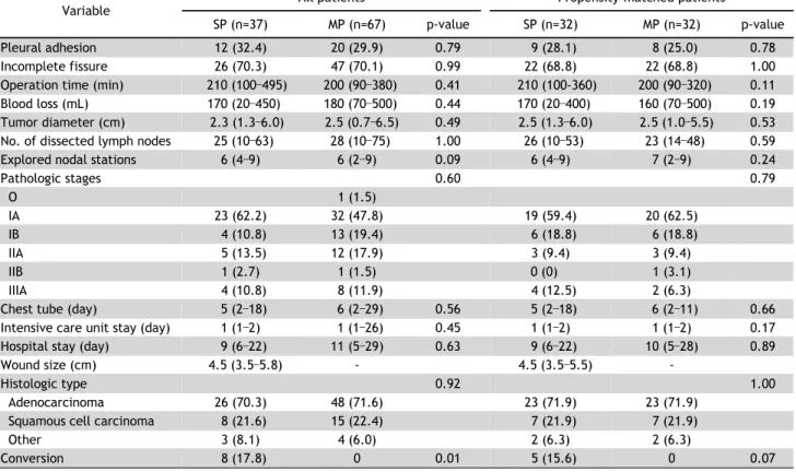
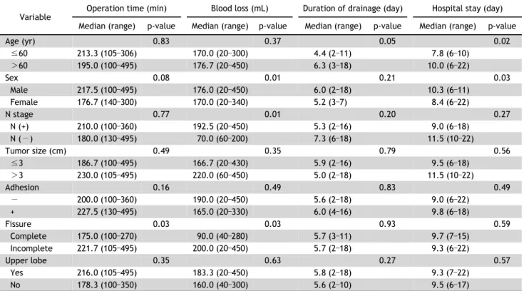
Both groups were compared in terms of the operation time, number of wedge resections, amount of chest tube drainage during the first 24 hours after surgery,

CC This is an open access article distributed under the terms of the Creative Commons Attribution Non-Commercial License

In order to evaluate the feasibility of sin- gle-port VATS for SSP, the postoperative results of single-port VATS (n=15) in patients with emphysema were compared with those

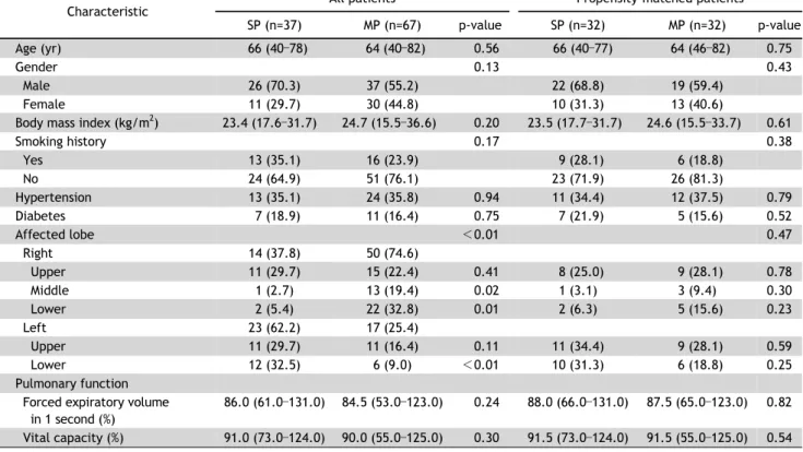
Background: We evaluated the feasibility and outcomes of pulmonary resection and mediastinal node dis- section (MND) by video-assisted thoracoscopic surgery (VATS)