Copyrights © 2016 The Korean Society of Radiology
331
Case Report
pISSN 1738-2637 / eISSN 2288-2928 J Korean Soc Radiol 2016;74(5):331-334 http://dx.doi.org/10.3348/jksr.2016.74.5.331
INTRODUCTION
Video-assisted thoracic surgery (VATS) is a minimally invasive technique commonly used worldwide for biopsy or resection of pulmonary lesions. Although VATS procedures are considered safe and effective, they have potential complications such as pro- longed air leak, bleeding, infection, and port site metastasis. Port site metastasis is a rare complication after VATS for a malignancy, with a few reports of mesothelioma cases and others (1-4). How- ever, to our knowledge, it has not been reported in a breast can- cer patient in the English literature. We reported the first case of iatrogenic port site metastasis in a breast cancer patient who re- ceived VATS wedge resection for pulmonary metastasis.
CASE REPORT
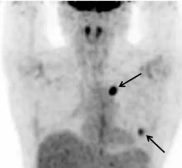
We experienced a case of a 57-year-old woman who had un- dergone modified radical mastectomy for clinical stage T3N1M0 triple negative subtype invasive ductal carcinoma (IDC) of the right breast. After 2 years, 2 pulmonary metastatic nodules were suspected in her left lung on a surveillance positron emission to- mography-computed tomography (PET-CT) scan (Fig. 1). She underwent VATS wedge resection and the lung nodules were pathologically confirmed as metastatic IDC.
Six months after VATS, she presented at our hospital with a small palpable mass on the left breast periphery. Ultrasonogra- phy (US) revealed an approximately 2.0 cm sized, circumscribed, round, and complex echoic mass with internal vascularity (on color Doppler US) in the lower outer quadrant periphery of the left breast just beneath the VATS port site scar (Fig. 2). The dif-
Port Site Metastasis of Breast Cancer after Video-Assisted Thoracic Surgery for Pulmonary Metastasis of Breast Cancer: A Case Report
수술 후의 혈종으로 오인된 유방암의 폐 전이에 대한 비디오 흉강경 절제술 이후 발생한 포트 자리 전이: 증례 보고Meehyun Park, MD, Ji-Young Hwang, MD*, Su Jeong Hyun, MD, Yul Lee, MD, Ji Young Woo, MD, Ik Yang, MD, Hye Sook Hong, MD, Han Myun Kim, MD
Department of Radiology, Kangnam Sacred Heart Hospital, Hallym University College of Medicine, Seoul, Korea
We reported a case of port site metastasis in a 57-year-old patient who underwent video-assisted thoracic surgery (VATS) resection of pulmonary metastasis from breast cancer. Port site metastasis after VATS is very rare in patients with breast cancer. However, when suspicious lesions are detected near the port site in patients who have undergone VATS for pulmonary metastasis, port site metastasis should be considered in the differential diagnosis.
Index terms Neoplasm Metastasis Neoplasm Seeding Breast Neoplasm
Video-Assisted Thoracoscopic Surgery
Received September 3, 2015 Revised September 18, 2015 Accepted November 3, 2015
*Corresponding author: Ji-Young Hwang, MD Department of Radiology, Kangnam Sacred Heart Hospital, Hallym University College of Medicine, 1 Singil-ro, Yeongdeungpo-gu, Seoul 07441, Korea.
Tel. 82-2-829-5241 Fax. 82-2-832-1845 E-mail: [email protected]
This is an Open Access article distributed under the terms of the Creative Commons Attribution Non-Commercial License (http://creativecommons.org/licenses/by-nc/3.0) which permits unrestricted non-commercial use, distri- bution, and reproduction in any medium, provided the original work is properly cited.
332
Port Site Metastasis of Breast Cancer after Video-Assisted Thoracic Surgery for Pulmonary Metastasis
jksronline.org
J Korean Soc Radiol 2016;74(5):331-334 ferential diagnosis included tumor spread, metachronous or
contralateral secondary primary cancer; however, since the mass was detected at the site of the postoperative scar on the first fol- low-up US after VATS resection with no abnormal uptake on the previous post-op PET-CT taken 4 months prior, the possi- bility of postoperative organizing hematoma was also consid-
ered. Consequently, the lesion was initially rated as category 4a (low suspicion for malignancy) according to the Breast Imaging- Reporting and Data System; and as a precaution, follow up US was decided upon.
After 6 months, the follow-up US revealed an increase in the mass size (3.8 × 3 × 3.4 cm), as compared to the previous US (Fig.
3A). In addition, a consecutively performed contrast enhanced CT revealed a necrotic rim enhancing lobulated mass and PET- CT scan showed hot uptake with maximum standardized uptake value of 5.0 at the previous VATS port site (Fig. 3B, C). Subsequent excisional biopsy was confirmed histopathologically as metastatic IDC consistent with primary triple negative breast cancer (Fig.
3D). Despite complete resection of the port site metastatic mass and chemotherapy, recurrent pulmonary metastasis and pleural seeding developed and the patient expired 2 years later.
DISCUSSION
VATS is currently a routinely performed procedure for the management of metastatic lung tumor, especially when the le- sion is located peripherally and is small in size and number.
Compared with open thoracotomy, VATS provides many advan- tages: smaller incisions, better visualization of the pleural cavity, less postoperative pain, less surgical morbidity, shorter length of hospitalization, fewer adhesions, shorter interval between sur- gery, and adjuvant therapy with better treatment compliance. But Fig. 1. Positron emission tomography-computed tomography of a
59-year-old woman 2 years after mastectomy. Two pulmonary nod- ules are seen in the left lung with maximum standardized uptake value of 5.0, suggestive of metastasis (arrows). These nodules were con- firmed as metastatic invasive ductal carcinoma from the breast after video-assisted thoracic surgery resection.
Fig. 2. Ultrasonography (US) performed 6 months after video-assisted thoracic surgery (VATS) resection for evaluation of a palpable mass in the left breast.
A. Transverse US scan reveals a 2.0 × 1.8 × 1.9 cm size, circumscribed round mass in lower outer quadrant of left breast around the port insertion area. The mass shows complex cystic and solid echogenicity. Port site scar after VATS (arrow) is demonstrated as subtle linear hypoechoic tract between the mass and the skin.
B. On color Doppler US, the mass shows internal vascularity.
A B
333
Meehyun Park, et al
jksronline.org J Korean Soc Radiol 2016;74(5):331-334 there are also complications such as prolonged air leak, bleeding, postoperative wound infections, and metastasis at the port site (5). Port site metastasis is a rare complication of VATS for malig- nancy with an overall incidence of 0.12–0.57% (5, 6). The port site metastasis is an indicator of multifocal disease with poor prognosis and increasing mortality (6). While the mechanism of port site metastasis has not been clarified, tumor implantation in the port wound is considered as the most likely cause (1).
There are only a few reported cases of port site metastasis after VATS procedures for pulmonary malignancies due to pulmo- nary adenocarcinoma, mesothelioma, metastatic colorectal car- cinoma, metastatic endometrial carcinoma, metastatic medul-
lary thyroid carcinoma, metastatic melanoma (1-4).
However, to our best knowledge, port site metastasis of breast cancer has not yet been reported in the English literature. In gen- eral, for newly developed masses in the contralateral breast after mastectomy for breast cancer, the first differential diagnosis is synchronous or metachronous breast cancer, or second primary breast cancer. This is because women with unilateral breast can- cer are at an increased risk for the development of contralateral breast cancers, with a 1–5% incidence of synchronous cancer and a 3–13% incidence of metachronous cancer (7, 8). Also, the risk of developing a second primary breast cancer for women with unilateral breast cancer is 2–6 times higher than the general pop-
Fig. 3. Follow-up ultrasonography (US), computed tomography (CT), and positron emission tomography-computed tomography (PET-CT) after video-assisted thoracic surgery resection of pulmonary metastasis of breast cancer, and histopathologic image of surgically resected specimen.
A. The mass in the lower outer quadrant periphery of the left breast shows an increase in size, as compared with previous US performed 6 months earlier, measuring 3.8 × 3 × 3.4 cm.
B. Contrast-enhanced CT demonstrates rim enhancing lobulated mass with central necrosis at the port site.
C. PET-CT shows fluorodeoxyglucose (FDG) uptake in the periphery of the mass with maximum standardized uptake value of 5.0. An enlarged right internal mammary lymph node is also noted with increased FDG uptake.
D. Photomicrograph shows prominent nuclear pleomorphism, no identifiable tubule formation, and rampant mitotic activity, indicating invasive ductal carcinoma with nuclear grade 3 and modified Bloom-Richardson histologic grade III/III (hematoxylin and eosin stain, × 200).
A B
C D
334
Port Site Metastasis of Breast Cancer after Video-Assisted Thoracic Surgery for Pulmonary Metastasis
jksronline.org
J Korean Soc Radiol 2016;74(5):331-334 ulation (9). In our case, a palpable mass developed at the port
site, 6 months after VATS, which was detected at the first post- operative follow-up US, therefore, differential diagnosis includ- ed not only contralateral metachronous or second primary breast cancer, but also postoperative organizing hematoma. All of the above lesions can be seen as growing masses, tend to have well- demarcated contours, and can demonstrate similar sonographic findings as complex echoic lesions. However, organizing hema- toma usually shows no flow on color Doppler US.
In conclusion, this was a rare case of port site metastasis after resection of pulmonary metastasis from breast cancer by VATS.
Thus, radiologists should consider port site metastasis in the differential diagnosis in cases of newly developed masses at the port site following VATS for pulmonary metastasis. Furthermore, clinicians should not hesitate to perform a biopsy for early de- tection and diagnosis of the potentially fatal complication of port site metastasis.
REFERENCES
1. Downey RJ, McCormack P, LoCicero J 3rd. Dissemination of malignant tumors after video-assisted thoracic surgery: a report of twenty-one cases. The Video-Assisted Thoracic Surgery Study Group. J Thorac Cardiovasc Surg 1996;111:
954-960
2. Thurer RL. Video-assisted thoracic surgery. Ann Thorac Surg
1993;56:199-200
3. Walsh GL, Nesbitt JC. Tumor implants after thoracoscopic resection of a metastatic sarcoma. Ann Thorac Surg 1995;
59:215-216
4. Fry WA, Siddiqui A, Pensler JM, Mostafavi H. Thoracoscopic implantation of cancer with a fatal outcome. Ann Thorac Surg 1995;59:42-45
5. Imperatori A, Rotolo N, Gatti M, Nardecchia E, De Monte L, Conti V, et al. Peri-operative complications of video-assist- ed thoracoscopic surgery (VATS). Int J Surg 2008;6 Suppl 1:S78-S81
6. Johnstone PA, Rohde DC, Swartz SE, Fetter JE, Wexner SD.
Port site recurrences after laparoscopic and thoracoscopic procedures in malignancy. J Clin Oncol 1996;14:1950-1956 7. Jobsen JJ, van der Palen J, Ong F, Meerwaldt JH. Synchro-
nous, bilateral breast cancer: prognostic value and inci- dence. Breast 2003;12:83-88
8. Gao X, Fisher SG, Emami B. Risk of second primary cancer in the contralateral breast in women treated for early-stage breast cancer: a population-based study. Int J Radiat Oncol Biol Phys 2003;56:1038-1045
9. Hankey BF, Curtis RE, Naughton MD, Boice JD Jr, Flannery JT. A retrospective cohort analysis of second breast cancer risk for primary breast cancer patients with an assessment of the ef- fect of radiation therapy. J Natl Cancer Inst 1983;70:797-804
수술 후의 혈종으로 오인된 유방암의 폐 전이에 대한 비디오 흉강경 절제술 이후 발생한 포트 자리 전이: 증례 보고
박미현 · 황지영* · 현수정 · 이 열 · 우지영 · 양 익 · 홍혜숙 · 김한면
저자들은 폐 전이로 인해 비디오 흉강경 수술을 받은 57세 유방암 환자에서의 포트 삽입 부위 전이의 증례를 보고하고자 한 다. 유방암 환자에서 폐 전이로 인한 비디오 흉강경 수술 후 절개 자리 전이는 매우 드물지만, 폐 전이에 대해 비디오 흉강경 수술을 받은 환자에서 포트 자리 주위에서 의심스러운 병변이 발견된다면, 포트 자리 전이가 감별진단으로 고려되어야 한다.
한림대학교 의과대학 강남성심병원 영상의학과