ISSN: 2233-601X (Print) ISSN: 2093-6516 (Online)
Received: May 4, 2017, Revised: October 31, 2017, Accepted: October 31, 2017, Published online: February 5, 2018
Corresponding author: Yong Soo Choi, Department of Thoracic and Cardiovascular Surgery, Samsung Medical Center, Sungkyunkwan University School of Medicine, 81 Irwon-ro, Gangnam-gu, Seoul 06351, Korea
(Tel) 82-2-3410-6542 (Fax) 82-2-3410-6986 (E-mail) [email protected]
© The Korean Society for Thoracic and Cardiovascular Surgery. 2018. All right reserved.
This is an open access article distributed under the terms of the Creative Commons Attribution Non-Commercial License (http://creativecommons.org/
licenses/by-nc/4.0) which permits unrestricted non-commercial use, distribution, and reproduction in any medium, provided the original work is properly cited.
Outcomes of Pulmonary Resection and Mediastinal Node Dissection by Video-Assisted Thoracoscopic Surgery
Following Neoadjuvant Chemoradiation Therapy for Stage IIIA N2 Non-Small Cell Lung Cancer
Yeong Jeong Jeon, M.D. 1 , Yong Soo Choi, M.D. 1 , Kyung Jong Lee, M.D. 2 , Se Hoon Lee, M.D. 3 , Hongryull Pyo, M.D. 4 , Joon Young Choi, M.D. 5
1
Department of Thoracic and Cardiovascular Surgery, Divisions of
2Pulmonary and Critical Care Medicine and
3
Hematology-Oncology, Department of Medicine, Departments of
4Radiation Oncology and
5Nuclear Medicine, Samsung Medical Center, Sungkyunkwan University School of Medicine
Background: We evaluated the feasibility and outcomes of pulmonary resection and mediastinal node dis- section (MND) by video-assisted thoracoscopic surgery (VATS) following neoadjuvant therapy for stage IIIA N2 non-small cell lung cancer (NSCLC). Methods: From November 2009 to December 2013, a total of 35 consecutive patients with pathologically or radiologically confirmed stage IIIA N2 lung cancer underwent pul- monary resection and MND, performed by a single surgeon, following neoadjuvant chemoradiation.
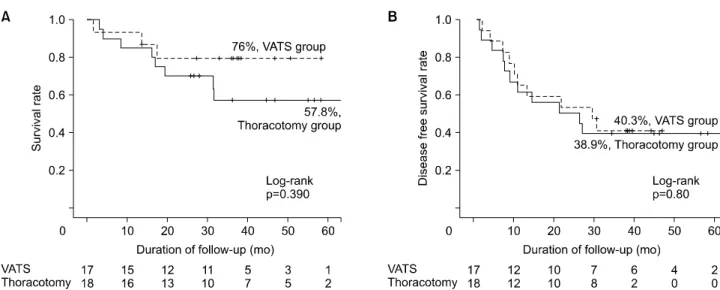
Preoperative patient characteristics, surgical outcomes, postoperative drainage, postoperative complications, and mortality were retrospectively analyzed. Results: VATS was completed in 17 patients. Thoracotomy was performed in 18 patients, with 13 planned thoracotomies and 5 conversions from the VATS approach. The median age was 62.7±7.9 years in the VATS group and 60±8.7 years in the thoracotomy group. The patients in the VATS group tended to have a lower diffusing capacity for carbon monoxide (p=0.077). There were no differences between the 2 groups in the method of diagnosing the N stage, tumor response and size after induction, tumor location, or histologic type. Complete resection was achieved in all patients. More total and mediastinal nodes were dissected in the VAT S group than in the thoracotomy group (p<0.05). The median chest tube duration was 5.3 days (range, 1 to 33 days) for the VATS group and 7.2 days (range, 2 to 28 days) for the thoracotomy group. The median follow-up duration was 36.3 months. The 5-year survival rates were 76% in the VATS group and 57.8% in the thoracotomy group (p=0.39). The 5-year disease-free surviv- al rates were 40.3% and 38.9% in the VATS and thoracotomy groups, respectively (p=0.8). Conclusion: T he VATS approach following neoadjuvant treatment was safe and feasible in selected patients for the treatment of stage IIIA N2 NSCLC, with no compromise of oncologic efficacy.
Key words: 1. Non-small-cell lung carcinoma 2. Neoadjuvant therapy
3. Video-assisted thoracoscopic surgery 4. Lobectomy
https://doi.org/10.5090/kjtcs.2018.51.1.29
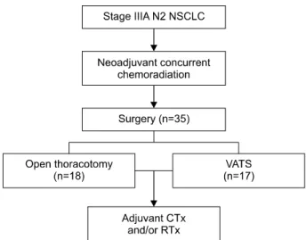
Fig. 1. Study design. NSCLC, non-small cell lung cancer; VATS, video-assisted thoracoscopic surgery; CTx, chemotherapy;
RTx, radiotherapy.
Introduction
Lung resection by video-assisted thoracoscopic sur- gery (VATS) was first reported in 1992 [1]. Subse- quently, VATS has been increasingly performed worldwide for early-stage non-small cell lung cancer (NSCLC). Multiple studies have demonstrated that VATS lobectomy may reduce postoperative pain, the length of hospitalization, and the incidence of compli- cations [2,3]. However, the application of VATS in pa- tients with stage IIIA N2 NSCLC who have undergone neoadjuvant therapy remains controversial. Concerns have been articulated regarding the technical diffi- culty of mediastinal node dissection (MND), which could compromise the oncologic outcomes [4].
However, as experience with VATS has increased, surgeons have successfully performed VATS lung re- section and MND with comparable outcomes [5-7].
We evaluated the feasibility of lobectomy and MND by VATS following neoadjuvant therapy for stage IIIA N2 NSCLC.
Methods
1) Patient selection
A retrospective analysis of 35 consecutive patients with stage IIIA N2 NSCLC was performed. Patients who underwent pulmonary resection and MND, which were performed by a single surgeon, following neoadjuvant chemoradiation at Samsung Medical Center between November 2009 and December 2013 were eligible. All cases had pathologically or radio- logically confirmed T and N staging. The histologic evaluation of mediastinal nodes was conducted by mediastinoscopy or endobronchial ultrasound. The imaging studies included computed tomography and positron emission tomography. The TNM (tumor- node-metastasis) staging was based on the seventh edition of the American Joint Committee on Cancer lung cancer staging guidelines. All patients completed preoperative concurrent chemoradiotherapy (CCRT) and underwent anatomic resection and MND. The op- erative technique was chosen by the surgeon.
2) Statistical analysis
Preoperative patient characteristics, surgical out- come, postoperative drainage, postoperative complica- tions, recurrence rate, and mortality were compared
between the VATS and thoracotomy groups. Measure- ments are presented as mean±standard deviation for continuous variables or number of patients and per- centages for categorical variables. Intergroup compar- isons were made with the Mann-Whitney test for continuous variables or the Pearson chi-square and Fisher exact tests for categorical variables. The 5-year survival rate was analyzed with the Kaplan-Meier method, and statistical significance was calculated us- ing the log-rank test. All p-values <0.05 were con- sidered to indicate statistical significance. All analyses were performed on an intent-to-treat basis using IBM SPSS ver. 21.0 (IBM Corp., Armonk, NY, USA).
Results
Between November 2009 and December 2013, a total of 35 patients with stage III N2 NSCLC under- went pulmonary resection after induction therapy.
Pulmonary resection and MND by VATS were at- tempted in 22 patients, while 13 patients underwent the same procedure through an open thoracotomy.
Five patients were converted to a thoracotomy due to anthracofibrotic nodes (4 patients) or tight adhe- sions (1 patient); these patients were included in the thoracotomy group. As a result, 17 patients were in- cluded in the VATS group and 18 in the thoracotomy group. All patients underwent neoadjuvant CCRT (Fig.
1). The patients received cisplatin/paclitaxel (TP) or
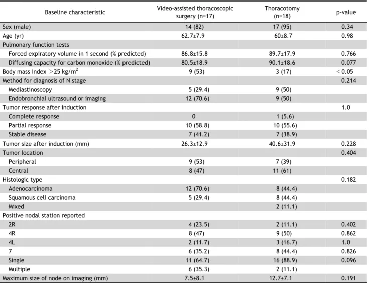
Table 1. Baseline characteristics
Baseline characteristic Video-assisted thoracoscopic surgery (n=17)
Thoracotomy
(n=18) p-value
Sex (male) 14 (82) 17 (95) 0.34
Age (yr) 62.7±7.9 60±8.7 0.98
Pulmonary function tests
Forced expiratory volume in 1 second (% predicted) 86.8±15.8 89.7±17.9 0.766
Diffusing capacity for carbon monoxide (% predicted) 80.5±18.9 90.1±18.6 0.077
Body mass index >25 kg/m
29 (53) 3 (17) <0.05
Method for diagnosis of N stage 0.214
Mediastinoscopy 5 (29.4) 9 (50)
Endobronchial ultrasound or imaging 12 (70.6) 9 (50)
Tumor response after induction 1.0
Complete response 0 1 (5.6)
Partial response 10 (58.8) 10 (55.6)
Stable disease 7 (41.2) 7 (38.9)
Tumor size after induction (mm) 26.3±12.9 40.6±31.9 0.228
Tumor location 0.404
Peripheral 9 (53) 7 (39)
Central 8 (47) 11 (61)
Histologic type 0.182
Adenocarcinoma 12 (70.6) 8 (44.4)
Squamous cell carcinoma 5 (29.4) 8 (44.4)
Mixed 2 (11.1)
Positive nodal station reported
2R 4 (23.5) 2 (11.1) 0.402
4R 8 (47) 9 (50) 0.862
4L 2 (11.7) 3 (16.7) 1.0
7 6 (35.2) 8 (44.4) 0.826
Single 11 (64.7) 16 (88.9) 0.096
Multiple 6 (35.3) 2 (11.1)
Maximum size of node on imaging (mm) 7.5±8.1 12.7±7.1 0.191
Values are presented as number (%) or mean±standard deviation.
cisplatin/docetaxel (DP) for 5 weeks and a total dose of 44 Gy with 2.0 Gy/fraction of radiation, concur- rently. The median age w as 62.7±7.9 years in the VATS group and 60±8.7 years in the thoracotomy group. The patients in the VATS group tended to have a lower diffusing capacity for carbon monoxide (DLCO) (p=0.077). More patients in the VATS group had a body mass index (BMI) >25 kg/m
2(53% ver- sus 17%, p<0.05). The method of the preoperative diagnosis of the N stage, tumor response and size af- ter induction, tumor location, histologic type, max- imum size of the node, and number of perioperative positive nodal stations reported were not sig- nificantly different between the 2 groups (Table 1).
Most patients underwent lobectomy (88.2% in the VATS group and 77.8% in the thoracotomy group)
(Table 2). Two patients in the VATS group and 2 pa- tients in the thoracotomy group underwent bilobec- tomy, and 2 patients in the thoracotomy group un- derwent sleeve lobectomy. There were no differences in the anatomic distribution of the resected lobes, and all patients underwent complete resection. The patients in the VATS group had significantly more to- tal (24±8.1 versus 13.5±9.4, p<0.05) and mediastinal nodes dissected (13±6.5 versus 6±6.2, p<0.05). There was no difference in mediastinal clearance (nodal downstaging to ypN0 and ypN1) between the 2 groups.
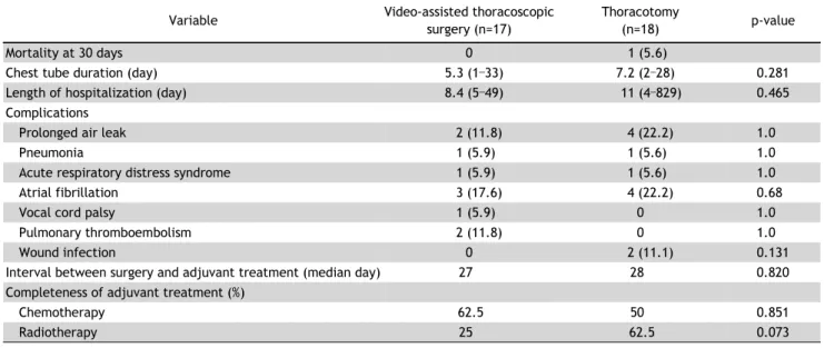
In terms of postoperative complications, the me-
dian chest tube duration was 5.3 days (range, 1 to
33 days) in the VATS group and 7.2 days (range, 2
to 28 days) in the thoracotomy group (Table 3). The
duration of hospitalization was 8.4 days (range, 5 to
Table 2. Surgical outcomes
Surgical outcome Video-assisted thoracoscopic surgery (n=17)
Thoracotomy
(n=18) p-value
Surgical procedure
Lobectomy 15 (88.2) 14 (77.8)
Bilobectomy 2 (11.8) 2 (11.1)
Sleeve lobectomy 2 (11.1)
Resected lobe
Left upper lobe 3 (17.6) 3 (16.7) 1.0
Left lower lobe 2 (11.8) 2 (11.1) 1.0
Right upper lobe 7 (41.2) 8 (44.4) 0.845
Right lower lobe 3 (17.6) 3 (16.7) 1.0
Right middle/lower lobe 2 (11.8) 2 (11.1) 1.0
Resection margin (R0) 17 (100) 18 (100)
No. of nodes dissected
Total 24±8.1 13.5±9.4 0.004
Mediastinal 13±6.5 6±6.2 0.04
Mediastinal clearance (yes) 8 (47.1) 12 (66.7) 0.241
Extracapsular extension of node (present) 6 (35.3) 8 (44.4) 0.581
Values are presented as number (%) or mean±standard deviation.
Table 3. Perioperative outcomes and complications