Downloadedfromhttp://journals.lww.com/md-journalbyBhDMf5ePHKav1zEoum1tQfN4a+kJLhEZgbsIHo4XMi0hCywCX1AWnYQp/IlQrHD3i3D0OdRyi7TvSFl4Cf3VC1y0abggQZXdtwnfKZBYtws=on04/27/2021
Downloadedfrom http://journals.lww.com/md-journalby BhDMf5ePHKav1zEoum1tQfN4a+kJLhEZgbsIHo4XMi0hCywCX1AWnYQp/IlQrHD3i3D0OdRyi7TvSFl4Cf3VC1y0abggQZXdtwnfKZBYtws=on
04/27/2021
Comparison of the intraoperative analgesic ef ficacy between ultrasound-guided deep and super ficial serratus anterior plane block during video-assisted thoracoscopic lobectomy
A prospective randomized clinical trial
Suyoung Moon, MD
a, Jungwon Lee, MD
b, Hyuckgoo Kim, MD
b, Jeongeun Kim, MD
b, Jiseob Kim, MD
c, Saeyoung Kim, MD, PhD
a,∗Abstract
Background:
The serratus anterior plane block (SAPB) is a novel method that provides lateral chest wall analgesia. There are 2 methods of SAPB; deep and super ficial SAPB. Each of these methods has been demonstrated to provide effective perioperative analgesia in thoracic surgery. The aim of this study was to compare the intraoperative hemodynamic and analgesic bene fits of deep versus super ficial SAPB during video-assisted thoracic surgery (VATS) lobectomy.
Methods:
We performed a prospective, randomized, patient/assessor-blinded trial. We included patients who were 20 to 75 years of age and scheduled to undergo VATS lobectomy with American Society of Anesthesiologists physical status 1 or 2. Patients were randomly allocated to receive either ultrasound-guided deep SAPB (Group D) or super ficial SAPB (Group S). The primary outcome was intraoperative remifentanil consumption. We also recorded intraoperative systolic blood pressure (SBP), heart rate (HR), emergence time, and doses of rescue drugs used to manage hemodynamic instability.
Results:
Data for 50 patients undergoing 3-port VATS lobectomy were analyzed. Intraoperative remifentanil consumption did not differ signi ficantly between Group D (n=25, 715.62±320.36 mg) and group S (n=25, 721.08±294.48mg) (P=.97). Additionally, there were no signi ficant differences between the 2 groups in SBP and HR at any time point, emergence time, or amount of rescue drugs used.
Conclusion:
Our study suggests that the intraoperative analgesic efficacy is similar for deep and superficial SAPB during VATS lobectomy.
Abbreviations:
HR = heart rate, SBP = systolic blood pressure, SPB = serratus plane block, VATS = video-assisted thoracic surgery.
Keywords:
analgesia, intraoperative care, nerve block, thoracic surgery, ultrasonography, video-assisted
1. Introduction
Sufficient intraoperative pain control is critical to stabilizing a patient ’s hemodynamics during surgery and to reduce the incidence of complications and length of hospitalization.
[1–3]For effective intraoperative pain relief, intravenous opioid administration is usually used. However, reducing opioid
consumption has become important because of its side effects such as delayed recovery from general anesthesia, opioid-induced hyperalgesia, sedation, nausea, and respiratory depression. To reduce intraoperative opioid use during thoracic surgery, local anesthetic in filtration at the incision site, thoracic epidural block, paravertebral block, and intercostal nerve block can be used alone or in combination.
[4–7]Editor: Joho Tokumine.
The authors have no conflicts of interest to disclose.
The datasets generated during and/or analyzed during the current study are available from the corresponding author on reasonable request.
aDepartment of Anesthesiology and Pain Medicine, School of Medicine, Kyungpook National University,bDepartment of Anesthesiology and Pain Medicine, Yeungnam University College of Medicine,cDepartment of Anesthesiology and Pain Medicine, Keimyung University School of Medicine, Daegu, Republic of Korea.
∗Correspondence: Saeyoung Kim, Department of Anaesthesiology and Pain Medicine, School of Medicine, Kyungpook National University, 130, Dongdeok-ro, Jung- gu, Daegu 41944, Republic of Korea (e-mail: [email protected]).
Copyright© 2020 the Author(s). Published by Wolters Kluwer Health, Inc.
This is an open access article distributed under the terms of the Creative Commons Attribution-Non Commercial License 4.0 (CCBY-NC), where it is permissible to download, share, remix, transform, and buildup the work provided it is properly cited. The work cannot be used commercially without permission from the journal.
How to cite this article: Moon S, Lee J, Kim H, Kim J, Kim J, Kim S. Comparison of the intraoperative analgesic efficacy between ultrasound-guided deep and superficial serratus anterior plane block during video-assisted thoracoscopic lobectomy: a prospective randomized clinical trial. Medicine 2020;99:47(e23214).
Received: 25 May 2020 / Received infinal form: 12 October 2020 / Accepted: 16 October 2020 http://dx.doi.org/10.1097/MD.0000000000023214
Clinical Trial/Experimental Study Medicine
OPEN
Ultrasound-guided serratus anterior plane block (SAPB) is a new method characterized by a long blocking time, wide blocking range, and low risk of serious complications associated with the procedure. The efficacy of SAPB in perioperative pain manage- ment during thoracic surgery has already been well docu- mented.
[8–13]There are 2 variations of SAPB: Superficial SAPB has its target point in the super ficial serratus plane between the latissimus dorsi muscle and the serratus anterior muscle while deep SAPB has its target point in the deep serratus plane between the serratus anterior muscle and rib/external intercostal muscle.
Intercostal nerves run between the internal intercostal muscles and the innermost intercostal muscles. On the lateral aspect of the chest wall, the lateral cutaneous branches derived from the intercostal nerves pierce the external intercostal muscles and serratus anterior muscle, then divide into anterior and posterior branches. SAPB provide analgesic effect on lateral chest wall by spreading local anesthetics along the super ficial or deep serratus plane through which the lateral branches of the intercostal nerves pass. Super ficial SAPB has been known to have a longer duration and a wider area of blockage than deep SAPB.
[6]Thus, we speculated that super ficial SAPB could provide a more effective intraoperative pain control than deep SAPB during video-assisted thoracic surgery (VATS) lobectomy; therefore, super ficial SAPB might reduce intraoperative opioid usage more effectively than deep SAPB. In a previous study, during VATS lobectomy, super ficial SAPB was found to reduce intraoperative remifentanil usage as compared with a control group and provide hemodynamic stability.
[10]However, no study on the intraop- erative analgesic efficacy of deep SAPB has been published to date.
Therefore, the aim of this study was to compare intraoperative hemodynamic changes and opioid consumption to determine which of the 2 variants of SAPB results in more effective intraoperative pain relief in VATS lobectomy.
2. Methods
The present prospective randomized trial was approved by the local hospital ethics committee (KNUH2019-05-003-01) and registered in ClinicalTrials.gov (NCT04252378). All patients in this study provided written informed consent.
Patients had to meet the following inclusion criteria: Physical status 1 or 2 according to the American Society of Anesthesi- ologists; age, 20 to 75 years; and elective 3-port VATS lobectomy.
Patients with the following conditions or issues were excluded from the study: a history of opioid or local anesthetic allergy;
local infection at the injection site or systemic infection;
coagulopathy; dif ficulty in understanding the study protocol;
and refusal to participate.
We randomly divided the patients by a 1:1 ratio into a “deep SAPB group ” (Group D) and a “superficial SAPB group” (Group S), using computer-generated block randomization. The alloca- tion sequence was concealed in sequentially numbered, sealed, opaque envelops by an assistant who was not associated with this study. Before the induction of general anesthesia, a sequential envelope was opened by a nurse and SAPB was performed according to the allocation in the preanesthesia room.
All SAPBs were performed by 1 practitioner. After placing the patient in the lateral decubitus position, the fifth rib was identified using a high-frequency linear ultrasound probe (Edge instrument 8–16MHz linear transducer; SonoSite Inc, Bothell, WA) at the mid-axillar line. The latissimus dorsi muscle, serratus anterior muscle, and external intercostal muscle between the ribs were easily distinguished. In Group D, a 22-gauge Tuohy needle was advanced to the plane between the serratus anterior muscle and the fifth rib using the in-plane technique. After hydrodissection with normal saline to con firm that the needle tip was placed in the interfascial space, 20 mL of 0.375% ropivacaine solution was injected (Fig. 1A). In Group S, after the needle was placed in the interfascial space between the latissimus dorsi muscle and the serratus anterior muscle, 20 mL of 0.375% ropivacaine solution was injected (Fig. 1B). Because the device, needle insertion site, injectate, and blocking process were identical in the 2 methods (except for the position of the needle tip), the patients remained blinded with regard to their group allocation. We defined successful block as the loss of pinprick sensation from T5 to T8 dermatomes on the lateral chest wall.
In the operating room, standard monitoring was performed, and we started induction with propofol (2 mg/kg), remifentanil (0.3–1.0 mg/kg/min), and rocuronium (0.8mg/kg). We performed intubation with a double-lumen tube and inserted a catheter into the radial artery for continuous blood pressure (BP) monitoring.
Figure 1. Ultrasound views for the deep and superficial serratus anterior plane blocks. The needle (arrows) tips were placed in the deep serratus plane (A) and superficial serratus plane (B). Dotted line shows the spread of local anesthetics. LA=local anesthetics, LD=latissimus dorsi muscle, SA=serratus anterior muscle.
After intubation, anesthesia was maintained by titrating the propofol concentration to a bispectral index of 40 to 60 and by remifentanil infusion to achieve BP and heart rate (HR) values within 70% to 130% of the baseline values. When severe hemodynamic changes occurred beyond 70% to 130% of the baseline values and were difficult to control by remifentanil adjustment, appropriate medications, including ephedrine, phenylephrine, nicardipine, esmolol, and glycopyrrolate, were administered. During surgery, rocuronium (0.2 mg/kg) was administered every 30 minutes.
The VATS lobectomy was performed by a single surgeon using the conventional 3-port technique. Two incisions (1 –1.5cm in length) were made at the 7th intercostal space on the anterior axillary line and the 8th intercostal space on the posterior axillary line. Another incision (4–5cm in length) was made at the 5th intercostal space on the mid-axillary line. After completed surgery, all participants received sugammadex (4 mg/kg), and propofol administration and remifentanil infusion were discon- tinued. After con firmation of a bispectral index above 80, opening of the eyes, sufficient spontaneous respiration, and full recovery of muscle strength, extubation was carried out. Patients were transferred to the recovery room with monitoring of vital signs.
The primary outcome was intraoperative remifentanil use. The secondary outcomes were systolic blood pressure (SBP), HR, emergence time, and doses of rescue drugs used to manage hemodynamic instability. Emergence time was defined as the time period between surgical incision closure and extubation. We recorded the SBP and HR before induction, immediately after incision, and at 5, 15, 30, 60, and 120 min during surgery. Data were collected by the attending anesthesiologists who were blinded to the allocation.
In the pilot study, the intraoperative remifentanil consumption of 8 patients in Group D was 762.2±345.1mg. A reduction of the intraoperative remifentanil consumption by 50% in Group S was considered clinically relevant. The sample size was estimated based on the requirement of Type I and II errors <0.05 and<0.2, respectively. Considering a 10% dropout rate, we included 28 patients in each group.
Statistical analyses were performed using IBM SPSS software (version 25.0; IBM Corp, Armonk, NY). For continuous variables, after assessment of normality using Shapiro –Wilk test, normally distributed data were analyzed using Student t test and non-normally distributed data were analyzed using the Mann–Whitney U test. We compared categorical variables using Fisher exact test. A probability P value<.05 was considered to indicate statistical significance.
3. Results
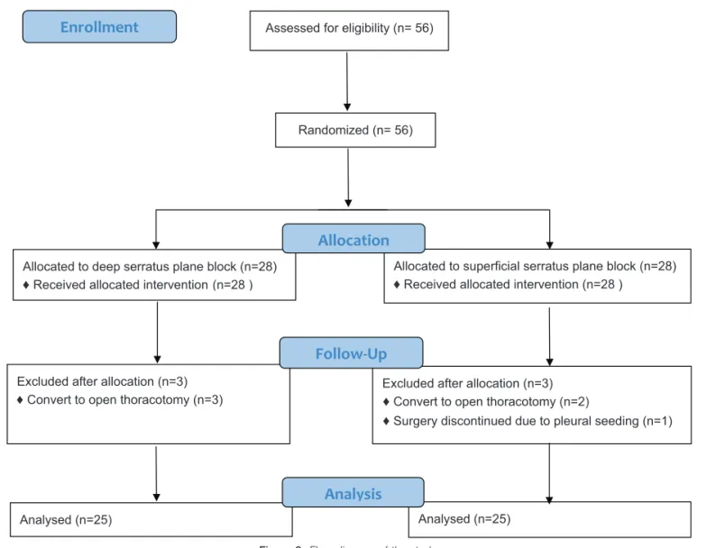
Fifty-six patients were enrolled in this study. Twenty-eight patients received superficial SAPB and the remainder received deep SAPB. All blocks were successful. We analyzed data for 50 out of 56 participants. In Group D, 3 patients were excluded from the analysis owing to conversion to open thoracotomy. In Group S, 2 patients were excluded owing to conversion to open thoracotomy and 1 patient was excluded because of operation termination due to malignant pleural seeding (Fig. 2). Demo- graphic characteristics, duration of anesthesia, and preanesthetic SBP and HR did not differ signi ficantly between the 2 groups (Table 1).
There was no signi ficant difference in the intraoperative remifentanil consumption between Group D (715.62±320.36mg) and Group S (721.08 ±294.48mg) (P=.97).
During surgery, there were no significant differences in SBP and HR between the 2 groups (Figs. 3 and 4). Emergence time did not differ significantly between Group D (10.00±4.06min) and Group S (11.11 ±5.55min) (P=.32). Additionally, there were no differences in the amounts of rescue drugs used to control BP and HR.
All patients showed no complications related to SAPB, such as systemic local anesthetic toxicity, pneumothorax, bleeding, and focal infection.
4. Discussion
In the present study, the 2 SAPB methods were not seen to differ significantly with regard to intraoperative remifentanil consump- tion and hemodynamic parameters, including SBP and HR, during VATS lobectomy.
Thoracic surgery is known as one of the most painful of surgeries. The pain is caused by traction of the incision site, resection of the ribs, damage to the intercostal nerve caused by dislocation of the costovertebral joint, irritation of the pleura by the chest tube, and excessive stretching of the ipsilateral brachial plexus and associated shoulder muscles.
[14,15]VATS has been developed with a view to reducing surgical stress by using a less invasive technique. Although VATS has several advantages over thoracotomy, including reduced postoperative pain, reduced perioperative bleeding, and shorter hospital stay, the surgery is still painful.
[5,16]Inadequate management of pain associated with surgical procedures induces peripheral and central sensitizations, which cause an intensification of postoperative pain. Preventive analgesia is considered critical to reducing short- and long-term postoperative pain. This practice refers to the administration of analgesic treatment throughout the perioperative period. The rationale of preventive analgesia is that adequate analgesia throughout the perioperative period prevents sensitization of pain, thereby reducing postoperative pain. Therefore, intraop- erative pain management is an important component of preventive analgesia, which is performed using analgesic medication and regional block.
[17,18]Epidural block and paravertebral block have been widely
performed in patients undergoing thoracic surgery. These blocks
are effective for pain relief but are technically difficult and are
associated with some serious complications. An epidural block is
linked to the risk of dural puncture, compromised hemodynam-
ics, epidural hematoma and abscess, and respiratory depression,
and a paravertebral block can lead to pneumothorax and
hemodynamic instability.
[19]Therefore, a regional block, which
is less dif ficult and more safe, may be appropriate for
thoracoscopic surgery, considering the fact that there is relatively
less pain than in thoracotomy, and it would facilitate early
recovery. Recently, Blanco et al
[6]introduced SAPB. It provides
analgesic effects at approximate levels T2 –T9 by blocking the
lateral cutaneous branches of the intercostal nerves that traverse
through the serratus plane. Because the target point of the
procedure is super ficial and does not involve any major vessels,
SAPB is easy to perform and safe. In the past few years, many
reports on SAPB have documented its ef ficacy for post-VATS
pain management.
[9,20–22]In recent studies, SAPB was reported
to be superior to local anesthetic infiltration and noninferior to paravertebral block for reducing postoperative pain.
[13,20]Consequently, SAPB has now been acknowledged as an alternative approach of nerve blocking in patients undergoing VATS.
SAPB is performed by spreading local anesthetics within the plane either deep of or super ficial to the serratus anterior muscle.
Blanco et al
[6]described the mean duration of analgesia as 752 min in super ficial SAPB and 386min in deep SAPB. Recent studies reported that both deep and superficial SAPB performed at induction of anesthesia lasted 8 hours postoperatively.
[20,23]Several studies have reported successful reduction of postopera- tive pain following either superficial or deep SAPB.
[9,13,20–22,24]A previous study demonstrated that super ficial SAPB reduced intraoperative opioid consumption and maintained hemodynam- ic stability during VATS lobectomy.
[10]However, only few reports comparing the clinical effectiveness between superficial and deep SAPB are available. Abdallah et al
[25]demonstrated that deep SAPB was as effective as super ficial SAPB with regard to opioid consumption and pain severity after breast surgery. One case series reported that deep SAPB was ef ficacious in patients undergoing ineffective superficial SAPB for postmastectomy pain syndrome.
[26]Our trial is the first to compare the intraoperative analgesic ef ficacy of superficial versus deep SAPB. The results did not indicate statistically significant differences in opioid consumption and hemodynamics between the 2 methods. Because these 2 variants of SAPB both target the lateral branches of the intercostal nerves, the extent to which the injectate spreads within the plane determines the effects of the 2 methods. A recent study using cadavers demonstrated that the extent of dye spread was independent on the targeted plane of the serratus anterior
Table 1Demographics of the study patients.
Demographics Group D (n =25) Group S (n =25)
ASA class (I/II) 1/24 0/25
Sex (M/F) 10/15 8/17
Age (y) 62.61±10.69 65.61±8.35
Height (cm) 162.23±7.65 162.05±6.65
Weight (kg) 63.61±10.26 64.71±10.01
Anesthesia time (min) 208.79±68.89 203.00±65.94
Surgery time (min) 158.86±64.07 154.46±57.22
Preanesthetic SBP (mm Hg) 162.39±21.03 157.07±21.95 Preanesthetic HR (beats/min) 69.14±11.56 73.36±16.23 Group D, patients who received deep serratus anterior plane block; Group S, patients who received superficial serratus plane block.
ASA=American Society of Anesthesiologists, F=female, HR=heart rate, M=male, SBP=systolic blood pressure.
Excluded after allocation (n=3)
♦ Convert to open thoracotomy (n=3)
Excluded after allocation (n=3)
♦ Convert to open thoracotomy (n=2)
♦ Surgery discontinued due to pleural seeding (n=1) Assessed for eligibility (n= 56)
Analysed (n=25)
Allocated to deep serratus plane block (n=28)
♦ Received allocated intervention (n=28 )
Allocated to superficial serratus plane block (n=28)
♦ Received allocated intervention (n=28 )
Analysed (n=25)
Allocation
Analysis Follow-Up
Randomized (n= 56)
Enrollment
Figure 2. Flow diagram of the study.
muscle (super ficial vs. deep plane).
[27]This result supports our observations, which indicate similar effectiveness between the 2 methods. The result of the study by Abdallah et al
[25]is consistent with the finding of our study, although the period and type of surgery differ.
There are several limitations in our study. First, the sample size was relatively small, although we conducted this study after calculating the minimum sample with adequate statistical power according to a pilot study. Second, this study was performed at a single center. To validate the findings, further studies on various
Figure 3. The box and whisker plot depicts systolic blood pressure during video-assisted thoracic surgery lobectomy. The middle line in the box represents the median; the upper and lower margins of the box represent the 75th and 25th percentiles, respectively, and the whiskers represent the maximum and minimum observations. There were no significant differences between Group D and Group S. Group D, patients who received deep serratus anterior plane block; Group S, patients who received superficial serratus plane block.Figure 4. The box and whisker plot depicts the heart rate during video-assisted thoracic surgery lobectomy. The middle line in the box represents the median; the upper and lower margins of the box represent the 75th and 25th percentiles, respectively, and the whiskers represent the maximum and minimum observations.
There were no significant differences between Group D and Group S. Group D, patients who received deep serratus anterior plane block; Group S, patients who received superficial serratus plane block.
demographic populations across multiple centers are needed.
Finally, postoperative analgesic consumption and pain scores were not evaluated; therefore, we did not compare the effectiveness for postoperative pain relief of the 2 SAPB methods.
Although no data were collected on pain score immediately after recovery from anesthesia, the success of block was confirmed preoperatively and the known duration of SAPB was suf ficiently longer than the surgery time in this study. Thus, the SAPB effect was considered to last throughout the surgery.
In conclusion, the present study suggests that the intraoper- ative analgesic efficacy of superficial versus deep SAPB during VATS lobectomy is similar.
Author contributions
Conceptualization: Saeyoung Kim.
Data curation: Suyoung Moon, Jeongeun Kim.
Formal analysis: Suyoung Moon.
Investigation: Jeongeun Kim, Saeyoung Kim.
Methodology: Jungwon Lee, Saeyoung Kim.
Project administration: Jungwon Lee.
Supervision: Saeyoung Kim.
Validation: Hyuckgoo Kim, Jiseob Kim, Saeyoung Kim.
Writing – original draft: Suyoung Moon.
Writing – review & editing: Jungwon Lee, Hyuckgoo Kim, Jiseob Kim, Saeyoung Kim.
References
[1] Ochroch EA, Gottschalk A. Impact of acute pain and its management for thoracic surgical patients. Thorac Surg Clin 2005;15:105–21.
[2] Ochroch EA, Gottschalk A, Augostides J, et al. Long-term pain and activity during recovery from major thoracotomy using thoracic epidural analgesia. Anesthesiology 2002;97:1234–44.
[3] Rogers ML, Henderson L, Mahajan RP, et al. Preliminaryfindings in the neurophysiological assessment of intercostal nerve injury during thoracotomy. Eur J Cardiothorac Surg 2002;21:298–301.
[4] Pintaric TS, Potocnik I, Hadzic A, et al. Comparison of continuous thoracic epidural with paravertebral block on perioperative analgesia and hemodynamic stability in patients having open lung surgery. Reg Anesth Pain Med 2011;36:256–60.
[5] Kaplowitz J, Papadakos PJ. Acute pain management for video-assisted thoracoscopic surgery: an update. J Cardiothorac Vasc Anesth 2012;26:
312–21.
[6] Blanco R, Parras T, McDonnell JG, et al. Serratus plane block: a novel ultrasound-guided thoracic wall nerve block. Anaesthesia 2013;68:
1107–13.
[7] Gottschalk A, Cohen SP, Yang S, et al. Preventing and treating pain after thoracic surgery. Anesthesiology 2006;104:594–600.
[8] Okmen K, Okmen BM, Uysal S. Serratus anterior plane (SAP) block used for thoracotomy analgesia: a case report. Korean J Pain 2016;29:189–92.
[9] Park MH, Kim JA, Ahn HJ, et al. A randomised trial of serratus anterior plane block for analgesia after thoracoscopic surgery. Anaesthesia 2018;73:1260–4.
[10] Lee J, Kim S. The effects of ultrasound-guided serratus plane block, in combination with general anesthesia, on intraoperative opioid consump- tion, emergence time, and hemodynamic stability during video-assisted thoracoscopic lobectomy: a randomized prospective study. Medicine (Baltimore) 2019;98:e15385.
[11] Kim DH, Oh YJ, Lee JG, et al. Efficacy of ultrasound-guided serratus plane block on postoperative quality of recovery and analgesia after video-assisted thoracic surgery: a randomized, triple-blind, placebo- controlled study. Anesth Analg 2018;126:1353–61.
[12] Khalil AE, Abdallah NM, Bashandy GM, et al. Ultrasound-guided serratus anterior plane block versus thoracic epidural analgesia for thoracotomy pain. J Cardiothorac Vasc Anesth 2017;31:152–8.
[13] Wang L, Wang Y, Zhang X, et al. Serratus anterior plane block or thoracic paravertebral block for postoperative pain treatment after uniportal video-assisted thoracoscopic surgery: a retrospective propen- sity-matched study. J Pain Res 2019;12:2231–8.
[14] Hughes R, Gao F. Pain control for thoracotomy. Contin Educ Anaesth Crit Care Pain 2005;5:56–60.
[15] Elmore B, Nguyen V, Blank R, et al. Pain management following thoracic surgery. Thorac Surg Clin 2015;25:393–409.
[16] Bendixen M, Jørgensen OD, Kronborg C, et al. Postoperative pain and quality of life after lobectomy via video-assisted thoracoscopic surgery or anterolateral thoracotomy for early stage lung cancer: a randomised controlled trial. Lancet Oncol 2016;17:836–44.
[17] Rosero EB, Joshi GP. Preemptive, preventive, multimodal analgesia:
what do they really mean? Plast Reconstr Surg 2014;134:85S–93S.
[18] Vadivelu N, Mitra S, Schermer E, et al. Preventive analgesia for postoperative pain control: a broader concept. Local Reg Anesth 2014;7:17–22.
[19] Baidya DK, Khanna P, Maitra S. Analgesic efficacy and safety of thoracic paravertebral and epidural analgesia for thoracic surgery: a systematic review and meta-analysis. Interact Cardiovasc Thorac Surg 2014;18:
626–35.
[20] Chen G, Li Y, Zhang Y, et al. Effects of serratus anterior plane block for postoperative analgesia after thoracoscopic surgery compared with local anesthetic infiltration: a randomized clinical trial. J Pain Res 2019;12:
2411–7.
[21] Ökmen K, Metin Ökmen B. Evaluation of the effect of serratus anterior plane block for pain treatment after video-assisted thoracoscopic surgery. Anaesth Crit Care Pain Med 2018;37:349–53.
[22] Viti A, Bertoglio P, Zamperini M, et al. Serratus plane block for video-assisted thoracoscopic surgery major lung resection: a ran- domized controlled trial. Interact Cardiovasc Thorac Surg 2020;30:
366–72.
[23] Razek AA, AbouAllo MM, Abd El Hamid SA, et al. Ultrasound-guided pectoral nerve blocks versus serratus intercostal plane block in breast surgeries. Res Opin Anesth Intensive Care 2018;5:162–9.
[24] Yao Y, Li J, Hu H, et al. Ultrasound-guided serratus plane block enhances pain relief and quality of recovery after breast cancer surgery: a randomised controlled trial. Eur J Anaesthesiol 2019;36:436–41.
[25] Abdallah FW, Cil T, MacLean D, et al. Too deep or not too deep?: A propensity-matched comparison of the analgesic effects of a superficial versus deep serratus fascial plane block for ambulatory breast cancer surgery. Reg Anesth Pain Med 2018;43:480–7.
[26] Piracha MM, Thorp SL, Puttanniah V, et al. A tale of two planes”: deep versus superficial serratus plane block for postmastectomy pain syndrome. Reg Anesth Pain Med 2017;42:259–62.
[27] Biswas A, Castanov V, Li Z, et al. Serratus plane block: a cadaveric study to evaluate optimal injectate spread. Reg Anesth Pain Med 2018;43:
854–8.