KJTCVS
The Korean Journal of Thoracic and Cardiovascular SurgeryClinical Research Nodal Outcomes of Uniportal versus Multiportal Video-Assisted Thoracoscopic Surgery for Clinical Stage I Lung Cancer
Jung Suk Choi, M.D.
1, Jiyun Lee, M.D.
1, Young Kyu Moon, M.D., Ph.D.
2, Seok Whan Moon, M.D., Ph.D.
1, Jae Kil Park, M.D., Ph.D.
1, Mi Hyoung Moon, M.D., Ph.D.
11
Department of Thoracic and Cardiovascular Surgery, Seoul St. Mary’s Hospital, College of Medicine, The Catholic University of Korea;
2Department of Thoracic and Cardiovascular Surgery, Eunpyeong St. Mary’s Hospital, College of Medicine, The Catholic University of Korea, Seoul, Korea
ARTICLE INFO
Received August 26, 2019 Revised October 18, 2019 Accepted October 30, 2019 Corresponding author Mi Hyoung Moon Tel 82-2-2258-6768 Fax 82-2-594-8644
E-mail [email protected] ORCID
https://orcid.org/0000-0003-2799-4570
Background: Accurate intraoperative assessment of mediastinal lymph nodes is a critical aspect of lung cancer surgery. The efficacy and potential for upstaging implicit in these dissections must therefore be revisited in the current era of uniportal video-assisted tho- racoscopic surgery (VATS).
Methods: A retrospective study was conducted in which 544 patients with stage I (T1abc–T2a, N0, M0) primary lung cancer were analyzed. To assess risk factors for nodal upstaging and to limit any imbalance imposed by surgical choices, we constructed an inverse probability of treatment-weighted (IPTW) logistic regression model (in addition to non-weighted logistic models). We also evaluated risk factors for early locoregional recur- rence using IPTW logistic regression analysis.
Results: In the comparison of uniportal and multiportal VATS, the resected lymph node count (14.03±8.02 vs. 14.41±7.41, respectively; p=0.48) and rate of nodal upstaging (6.5%
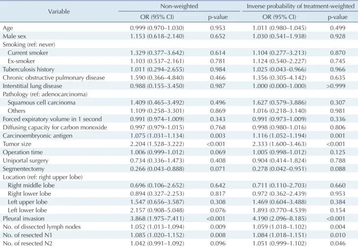
vs. 8.7%, respectively; p=0.51) appeared similar. Predictors of nodal upstaging included tu- mor size (odds ratio [OR], 1.74; 95% confidence interval [CI], 1.12–2.70), carcinoembryonic antigen level (OR, 1.11; 95% CI, 1.04–1.18), and histologically confirmed pleural invasion (OR, 3.97; 95% CI, 1.89–8.34). The risk factors for locoregional recurrence within 1 year were found to be number of resected N2 nodes, age, and nodal upstaging.
Conclusion: Uniportal and multiportal VATS appear similar with regard to accuracy and thoroughness, showing no significant difference in the extent of nodal dissection.
Keywords: Lung neoplasms, Nodal upstaging, Single port video-assisted thoracic sur- gery, Uniportal, Video-assisted thoracic surgery
Copyright
© The Korean Society for Thoracic and Cardiovascular Surgery. 2020. All right reserved.
This is an Open Access article distributed under the terms of the Creative Commons Attribution Non-Commercial License (http://creativecommons.org/licenses/
Introduction
Mediastinal lymph node staging is an essential part of the assessment and management of patients with early- stage lung cancer. Since the extent of lymph node involve- ment is the most important prognostic factor in these pa- tients, it heavily influences therapeutic strategies [1].
According to the current consensus and guidelines, preop- erative invasive mediastinal lymph node assessment is rec- ommended via endobronchial ultrasound-transbronchial needle aspiration, endoscopic ultrasound, mediastinosco- py, or video-assisted thoracoscopic surgery (VATS). Pa- tients with peripherally-situated clinical stage IA tumors, normal-sized lymph nodes, and negative position-emission
tomography and computed tomography studies (PET/CT) are exceptions [2]. However, clinical practice patterns seem to vary substantially and often depart from these guide- lines. Inaccurate preoperative staging heightens the role of mediastinal node removal at the time of resection in stag- ing [3,4].
Controversy lingers regarding the survival benefit of dis- secting versus sampling mediastinal nodes, but systematic nodal dissection is recommended in all cases to ensure compliance with evidence-based guidelines adopted in Eu- rope and the United States [1,5]. Although the extent of nodal dissection has yet to be stipulated, most current guidelines and studies recommend that at least 3 N2 nodes be examined, and to ensure pathologic N0 status, at least 6
https://doi.org/10.5090/kjtcs.2020.53.3.104 pISSN: 2233-601X eISSN: 2093-6516
Korean J Thorac Cardiovasc Surg. 2020;53(3):104-113
Jung Suk Choi, et al. Uniportal Thoracoscopic LN Dissection KJTCVS
nodes from the hilar and mediastinal stations should be removed [5,6].
Since the first VATS lobectomy for lung cancer (in the early 1990s), the feasibility, safety, benefits and oncologic outcomes of VATS have compared favorably with thoracot- omy [7]. Advances in instrument technology and video-as- sisted surgery now allow for fewer ports than before (now 1 or 2). The first report of uniportal VATS was published in 2011. Subsequent data on clinical outcomes, feasibility, and safety have since dispelled concerns of inferiority [8-10].
However, single-port mediastinal lymph node dissection (MLND) presents a technical challenge that few studies have attempted to address [11,12].
The purpose of this study was to investigate uniportal and multiportal VATS in patients with clinical T1abc–T2a N0 lung cancer by analyzing differences in the extent of MLND and the incidence of nodal upstaging. We also as- sessed the likelihood and clinical implications of nodal up- staging.
Methods
Patient cohort
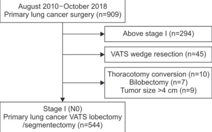
This retrospective review drew upon prospectively-col- lected institutional data. We identified patients who under- went surgical treatment of primary lung cancer with cura- tive intent between August 2010 and October 2018. The grounds for exclusion were as follows: (1) T stage T2b or higher, (2) clinical N1 or N2 disease, (3) wedge resection or surgery with non-curative intent, (4) bilobectomy, and (5) surgical thoracotomy or conversion of VATS to open tho- racotomy (Fig. 1). To emphasize the standard practice of lymph node dissection, we intentionally excluded data from patients who underwent wedge resection (n=45), bi- lobectomy (n=7), or VATS-to-thoracotomy conversion (n=10). We also excluded patients with sizeable (>40-mm) tissue-confirmed tumors that were preoperatively assessed as clinical stage I but proved instead to be stage II (n=9).
One-sleeve lobectomy was categorized simply as lobecto- my.
Both chest CT and PET/CT findings were used to define clinical N0 lung cancer. A lymph node with a short-axis diameter of less than 1 cm on an axial CT scan and no sig- nificant focal fluorodeoxyglucose uptake on PET/CT was defined as clinical N0 [7].
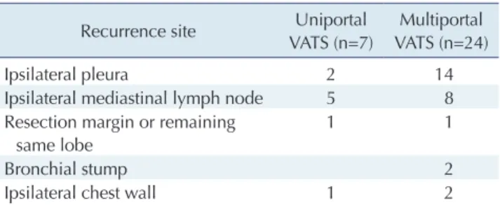
Locoregional recurrence was defined as recurrent disease at the bronchial stump, stapled margin, ipsilateral hilum or mediastinum, ipsilateral pleura, or ipsilateral chest wall
[13,14]. Early locoregional recurrence was defined in this study as recurrence within 1 year after surgery. Distant metastasis was equated with a recurrent tumor of the ipsi- lateral or contralateral lung, contralateral mediastinum, supraclavicular lymph node, or extrathoracic location. The first date upon which imaging studies raised suspicion of recurrence was considered the date of recurrence for our purposes.
This study was approved by the institutional review board of Seoul St. Mary’s Hospital (approval no., KC19RE- SI0521). The requirement for the informed consent of indi- vidual patients was waived on the basis of the retrospective design of this study.
Surgical techniques used in video-assisted thoracoscopic surgery
Our uniportal VATS program began in 2017. Each pa- tient was placed in the lateral decubitus position with a semi-flexed arm, as in multiportal VATS procedures. In our uniportal approach, a working incision of 3–5 cm was made at the fourth or fifth intercostal space at the anterior axillary line as described previously [15]. A 10-mm 30°
thoracoscope was used. The procedure was otherwise sim- ilar to multiportal VATS, in which 2 (n=22), 3 (n=2), or 4 (n=367) ports were created. For 2-port and 4-port VATS, the camera ports were placed over the seventh and eighth intercostal spaces, respectively, on the mid-axillary line.
The utility incision for 4-port VATS was made at the fifth intercostal space, with 2 instrumental ports at the seventh intercostal space anteriorly and the remaining 2 at the sixth intercostal space posteriorly.
Thoracotomy conversion (n=10) Bilobectomy (n=7) Tumor size >4 cm (n=9) August 2010 October 2018
Primary lung cancer surgery (n=909)
Stage I (N0)
Primary lung cancer VATS lobectomy /segmentectomy (n=544)
Above stage I (n=294) VATS wedge resection (n=45)
Fig. 1. Study flowchart, with counts and reasons for exclusion.
VATS, video-assisted thoracoscopic surgery.
https://doi.org/10.5090/kjtcs.2020.53.3.104
KJTCVS
Statistical analysis
Baseline characteristics were expressed conventionally, using mean±standard deviation for continuous variables and frequencies with percentages for categorical variables.
The distributions of continuous variables were compared via the Student t-test or the Mann-Whitney U-test, de- pending on the results of the normality test. The chi- square test or the Fisher exact test were utilized to compare categorical variables. Risk factors for nodal upstaging were assessed via binary logistic regression analysis. Receiver operating curve analyses were performed to calculate the area under the curve for the tumor size and serum carci- noembryonic antigen (CEA) level in the prediction of nod- al upstaging.
Given the observational nature of this study, surgical al- location to either the uniportal or the multiportal group was not random. To address this, we first used propensity scores (PS) to balance baseline variables and maximize the balance between the 2 groups. Variables included in the PS model were those that (in our institutional experience) in- fluenced the decision to perform uniportal surgery: age, sex, presence of chronic obstructive pulmonary disease or interstitial lung disease, clinical tumor-node-metastasis stage, and type of surgery planned (lobectomy or segmen- tectomy). Because the distribution of PS within the 2 groups differed considerably, and the study cohort was of limited size, inverse probability of treatment weighting (IPTW) was applied [16,17]. The IPTW method has been shown to outperform simple logistic regression in the con- text of case-mix adjustment. Weights were extracted from the PS model to calculate the IPTW, after which the IPTW was utilized in each case to produce an average treatment among the treated (ATT) estimate. The ATT estimate ac- counted for the unavoidable non-random selection of sur- gical patients and facilitated coherence in PS matching [18].
In the assessment of the clinical significance of nodal up- staging, considering the differences in follow-up time, we used early locoregional recurrence within a 1-year period after surgery as another endpoint. The factors associated with early locoregional recurrence were assessed with IPTW logistic regression analysis. Two-sided p-values less than 0.05 were used to indicate statistical significance, and variables that demonstrated statistical significance in uni- variate analysis were incorporated into a multivariate mod- el. All computations were facilitated by R freeware ver. 3.6.1 (R Project for Statistical Computing, Vienna, Austria;
https://www.R-project.org/).
Results
General characteristics of the patients and clinical outcomes
Ultimately, 544 patients (men, 242; women, 302) quali- fied for this study, all diagnosed with clinical stage I (T1abc or T2a N0M0) lung cancer. The mean age was 63.91 years (range, 30–85 years). Uniportal VATS was preferentially performed over multiportal VATS in women (65.0% versus 52.2%, p=0.016) and in patients with lower forced expirato- ry volume in 1 second (93.0±15.66 L versus 96.6±17.83 L, p=0.037), lower diffusing capacity for carbon monoxide as a percentage of predicted value (92.1%±15.07% versus 88.1%±
18.02%, p=0.015), and smaller tumor size (1.86±0.83 cm versus 2.11±0.83 cm, p=0.002). There were no group-wise differences in age; smoking history; history of tuberculosis, chronic obstructive pulmonary disease, or interstitial lung disease; or history of contralateral lung surgery or extent of surgery (segmentectomy versus lobectomy). The median follow-up time for the overall cohort was 34 months. The total number of dissected lymph node was 14.03±8.02 for the uniportal group and 14.41±7.41 for the multiportal group (p=0.48).
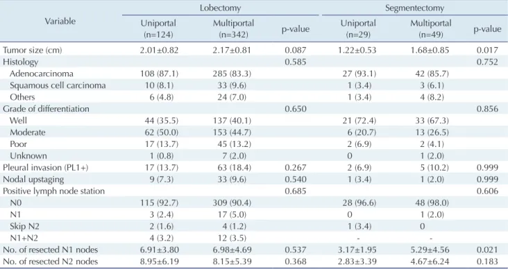
Detailed data on patient enrollees are shown in Table 1 according to the type of surgery and the number of ports.
Among members of the lobectomy group, no differences were observed between the uniportal and multiportal groups with regard to the distribution of major demo- graphic characteristics, underlying lung disease, tumor histotype, preoperative forced expiratory volume in 1 sec- ond, CEA level, or the location or size of the tumor. The same was true of the segmentectomy group, with the ex- ception that T1a/b/c tumors were more prevalent in the uniportal lobectomy group than in the multiportal lobec- tomy group (p=0.003).
Surgical outcomes are presented in Table 2. Regardless of the type of surgery performed, there were no significant differences in terms of operative time, postoperative mor- bidity, adjuvant treatment, or crude locoregional recur- rence. For each type of surgery, the uniportal group exhib- ited lower blood loss volumes and shorter hospital stays than the multiportal group. Operative mortality was 0.55%
(3 of 544), including 2 patients in the uniportal lobectomy
group (1.6%) and 1 patient in the multiportal lobectomy
group (0.3%). The causes of death were pneumonia and
acute respiratory distress syndrome. No deaths occurred in
the segmentectomy group.
Jung Suk Choi, et al. Uniportal Thoracoscopic LN Dissection KJTCVS
Nodal upstaging
Pathologic outcomes are shown in Table 3. The overall incidence of nodal upstaging in this cohort was 8.09% (44 of 544). From the perspective of uniportal versus multipor- tal surgery, the number of resected lymph nodes differed among the resected lobes, as follows: the right upper lobe (RUL), 17.2±9.2 versus 14.4±7.7, respectively); right middle lobe (RML), 9.7±4.8 versus 14.3±7.5; right lower lobe (RLL), 15.9±4.8 versus 16.4±8.2; left upper lobe (LUL), 10.7±7.2 versus 12.8±6.5; and left lower lobe (LLL), 11.5±6.0 versus 14.0±6.1. After Bonferroni adjustment, statistical signifi- cance remained present only between the uniportal LUL versus multiportal RLL and uniportal RUL versus multi- portal LUL pairs. Regardless of the type of surgery per- formed, uniportal and multiportal VATS showed no signif- icant differences in either nodal upstaging (10 [6.5%] versus 34 [8.7%], respectively; p=0.5) or resected node counts
(14.03 versus 14.41, p=0.48). Likewise, the nature of surgery had no significant impact on nodal upstaging (Table 3).
More lymph nodes were dissected in the lobectomy group than in the segmentectomy group, but the lymph node to- tals at each station did not differ significantly by number of ports within the lobectomy or segmentectomy groups.
No significant differences were observed in the frequency of single or multi-station nodal metastasis according to the number of ports (p=0.999), type of surgery (p=0.999), his- tological findings (p=0.758), or resected lobe (p=0.384).
Factors associated with nodal upstaging
The factors associated with nodal upstaging were ana- lyzed using univariate and multivariate logistic regression methods. Additionally, to reduce the imbalances associated with choice of treatment, we calculated PS and constructed the IPTW model before logistic regression was performed Table 1. Patient characteristics by type of surgery and number of ports
Characteristic
Lobectomy Segmentectomy
Uniportal (n=124)
Multiportal
(n=342) p-value Uniportal (n=29)
Multiportal
(n=49) p-value
Male sex 46 (37.1) 162 (47.4) 0.062 9 (31.0) 25 (51.0) 0.138
Age (yr) 64.19±11.20 63.68±10.09 0.637 66.79±9.64 63.06±9.16 0.092
Smoking (pack-years)
a)26.49±18.76 33.86±20.14 0.025 33.07±15.28 30.35±21.32 0.459
Smoking status 0.236 0.726
Current smoker 5 (4.0) 29 (8.5) 2 (6.9) 4 (8.2)
Ex-smoker 33 (26.6) 94 (27.5) 12 (41.4) 15 (30.6)
Never smoked 86 (69.4) 219 (64.0) 15 (51.7) 30 (61.2)
Tuberculosis history 11 (8.9) 30 (8.8) 0.999 5 (17.2) 3 (6.1) 0.239
Chronic obstructive pulmonary disease 3 (2.4) 19 (5.6) 0.245 2 (6.9) 1 (2.0) 0.552
Interstitial lung disease 3 (2.4) 17 (5.0) 0.346 1 (3.4) 4 (8.2) 0.646
Previous lung operation 2 (1.6) 0 0.070 3 (10.3) 6 (12.2) 0.999
Forced expiratory volume in 1 second (% of predicted)
94.13±14.66 97.18±18.03 0.088 88±18.85 92.76±15.98 0.239
Diffusing capacity for carbon monoxide (%) 92.63±15.23 88.18±17.75 0.014 89.69±14.35 87.24±19.94 0.566
Carcinoembryonic antigen (ng/mL) 2.63±4.80 2.74±5.51 0.139 3.77±11.17 1.77±1.56 0.587
Clinical T stage 0.003 0.194
Tis 4 (3.2) 12 (3.5) 4 (13.8) 7 (14.3)
T1a 12 (9.7) 24 (7.0) 12 (41.4) 11 (22.4)
T1b 61 (49.2) 121 (35.4) 11 (37.9) 20 (40.8)
T1c 32 (25.8) 92 (26.9) 2 (6.9) 5 (10.2)
T2a 15 (12.1) 93 (27.2) 0 6 (12.2)
Tumor location 0.627 0.532
Right upper lobe 44 (35.5) 136 (39.8) 5 (17.2) 9 (18.4)
Right middle lobe 11 (8.9) 31 (9.1) 0 0
Right lower lobe 30 (24.2) 68 (19.9) 5 (17.2) 13 (26.5)
Left upper lobe 18 (14.5) 61 (17.8) 11 (37.9) 20 (40.8)
Left lower lobe 21 (16.9) 46 (13.5) 8 (27.6) 7 (14.3)
Values are presented as number (%) or mean±standard deviation.
a)
Data were derived from 161 available patients.
https://doi.org/10.5090/kjtcs.2020.53.3.104
KJTCVS
Table 2. Clinical outcomes by type of surgery and number of ports Variable
Lobectomy Segmentectomy
Uniportal (n=124)
Multiportal
(n=342) p-value Uniportal (n=29)
Multiportal
(n=49) p-value
Operation time (min) 146.40±42.02 141.40±41.56 0.194 150.60±40.78 171.10±44.54 0.056
Blood loss (mL) 78.14±109.59 180.9±275.52 <0.001 86.55±120.04 161±157.90 0.001
Numerical Rating Scale on postoperative day 1
3.06±1.71 2.02±1.77 <0.001 3.28±1.91 1.55±1.44 <0.001
Postoperative complications 19 (15.3) 61 (17.8) 0.619 6 (20.7) 11 (22.4) 0.999
Prolonged air leak 9 (7.3) 47 (13.7) 0.082 2 (6.9) 9 (18.4) 0.196
Pneumonia 2 (1.6) 6 (1.8) 0.999 2 (6.9) 0 0.135
Bleeding
a)2 (1.6) 2 (0.6) 0.289 0 0 -
Chylothorax 1 (0.8) 6 (1.8) 0.681 2 (6.9) 0 0.135
Hoarseness 1 (0.8) 4 (1.2) 0.999 1 (3.4) 1 (2.0) 0.999
Others 2 (1.6) 4 (1.2) 0.659 0 1 (2.0) 0.999
Adjuvant treatments 0.523 0.999
None 116 (93.5) 313 (91.5) 28 (96.6) 46 (93.9)
Chemotherapy 8 (6.5) 24 (7.0) 1 (3.4) 3 (6.1)
Chemoradiation 0 5 (1.5) -
Hospital stay duration (day) 5.2±4.44 6.5±5.84 0.003 4.9±5.50 7.5±9.63 0.029
Locoregional recurrence 6 (4.8) 20 (5.8) 0.848 1 (3.4) 4 (8.2) 0.646
Distant metastasis 5 (4.0) 30 (8.8) 0.129 0 5 (10.2) 0.151
In-hospital death 2 (1.6) 1 (0.3) 0.345 0 0 -
Follow-up duration (mo) 14.2±6.36 52.0±25.94 <0.001 14.9±6.77 53.4±21.88 <0.001
Values are presented as mean±standard deviation or number (%).
a)