ISSN: 2233-601X (Print) ISSN: 2093-6516 (Online)
1
Department of Thoracic and Cardiovascular Surgery, Seoul National University Hospital,
2Department of Thoracic and Cardiovascular Surgery, Center for Lung Cancer, Research Institute and Hospital, National Cancer Center,
3Department of Thoracic and Cardiovascular Surgery, Seoul National University Bundang Hospital
†This paper was presented at the 46th Annual Meeting of the Korean Society for Thoracic and Cardiovascular Surgery, Yeosu, Oct 23rd–25th, 2015.
Received: December 26, 2014, Revised: July 22, 2015, Accepted: July 23, 2015, Published online: December 5, 2015
Corresponding author: Sukki Cho, Department of Thoracic and Cardiovascular Surgery, Seoul National University Bundang Hospital, 82 Gumi-ro 173beon-gil, Bundang-gu, Seongnam 13620, Korea
(Tel) 82-31-787-7132 (Fax) 82-31-787-4050 (E-mail) [email protected]
C
The Korean Society for Thoracic and Cardiovascular Surgery. 2015. All right reserved.
CC
This is an open access article distributed under the terms of the Creative Commons Attribution Non-Commercial License (http://creative- commons.org/licenses/by-nc/4.0) which permits unrestricted non-commercial use, distribution, and reproduction in any medium, provided the original work is properly cited.
Single-Port Video-Assisted Thoracic Surgery for Secondary Spontaneous Pneumothorax: Preliminary Results
Min-Seok Kim, M.D.
1, Hee Chul Yang, M.D.
2, Mi-Kyung Bae, M.D.
3, Sukki Cho, M.D., Ph.D.
3, Kwhanmien Kim, M.D., Ph.D.
3, Sanghoon Jheon, M.D., Ph.D.
3Background: The aim of this study was to evaluate the feasibility of single-port video-assisted thoracic surgery (VATS) in the treatment of secondary spontaneous pneumothorax (SSP). Methods: Twenty-four patients who were scheduled to undergo single-port VATS for SSP were studied. The medical records of the patients were retro- spectively reviewed. The mean follow-up duration was 26.1±19.8 months. In order to evaluate the feasibility of sin- gle-port VATS for SSP, the postoperative results of single-port VATS (n=15) in patients with emphysema were compared with those of emphysematous patients who underwent three-port VATS (n=15) during the study period.
Results: Single-port VATS was feasible in 19 of 24 patients (79.2%), while an additional port was needed in five patients. In the single-port VATS patients, the median operation time, duration of chest tube drainage, and hospital stay were 84.0 minutes, one day, and two days, respectively. Postoperative complications included prolonged chest tube drainage for more than five days (n=1), wound infection (n=1), and vocal fold palsy (n=1). No recurrence of pneumothorax was observed during the follow-up period. The median operation time, duration of chest tube drain- age, and hospital stay of the emphysematous patients who underwent single-port VATS were shorter than those who underwent three-port VATS group (p<0.05 for all parameters). Conclusion: Single-port VATS proved to be a feasible procedure in the treatment of patients with secondary spontaneous pneumothorax.
Key words: 1. Video-assisted thoracic surgery (VATS) 2. Secondary spontaneous pneumothorax 3. Lung wedge resections
INTRODUCTION
Secondary spontaneous pneumothorax (SSP) is associated with underlying pulmonary pathologies, such as chronic obstructive pulmonary disease, in contrast to primary spontaneous pneu- mothorax (PSP), which occurs in the absence of underlying lung disease [1]. Patients with SSP tend to exhibit more severe
symptoms due to their underlying lung disease and frequently require active intervention, because the air leakage is less likely to stop spontaneously than in patients with PSP [2,3].
Since its introduction, video-assisted thoracic surgery (VATS)
has proven to be as effective as thoracotomy in the treatment
of pneumothorax, with an acceptable recurrence rate and few
complications [4]. Recently, the single-port approach has been
http://dx.doi.org/10.5090/kjtcs.2015.48.6.387
Table 1. Characteristics of patients scheduled for single-port surgery (N=24)
Variable Value
Age (yr) 50.8±16.8
Sex (male:female) 19:5
Smoking history (pack-year) 20.0±20.4 Etiology
Emphysema 18
Catamenial 4
Others 2
Site
Right 12
Left 11
Bilateral 1
Previous episodes of pneumothorax
None 9
One 13
Two or more 2
Follow-up duration (mo) 16.2±6.8
Values are presented as mean±standard deviation or number.
introduced as an alternative to conventional multi-port VATS for the treatment of PSP [5]. Single-port VATS has shown to have advantages over three-port VATS, including lower com- plication rates, shorter hospital stays, and less postoperative pain and paresthesia, and does not lead to increased recur- rence rates [6-8]. Although single-port VATS is currently ac- cepted as a treatment modality for PSP, its application to SSP is still limited due to the nature and clinical characteristics of the underlying disease processes. The aim of this study was to evaluate the feasibility of single-port VATS in the treatment of SSP.
METHODS
The study protocol was reviewed by our institutional re- view board and approved as a minimal-risk retrospective study (approval number, H-1212-002-445) that did not require individual consent based on the institutional guidelines for waiving consent.
Twenty-four SSP patients who were initially scheduled to undergo single-port VATS from July 2011 to December 2013 were enrolled. The etiologies of SSP were emphysema (n=18), catamenial pneumothoraces (n=4), and lymphangioleiomyoma- tosis (n=2) (Table 1). A high-resolution computed tomography
scan of the chest was performed preoperatively in each case to examine the location and extent of bullae and blebs. The indications for surgery were recurrent pneumothorax, massive air leakage with a collapsed lung or increasing subcutaneous emphysema, and persistent air leakage for more than 48 hours despite chest tube drainage. The postoperative management protocol included the application of thoracic suction (−15 cm H
2O) until the morning of postoperative day 1, a change to water-sealed drainage of the chest tube if no air leak existed, removal of the chest tube in the afternoon of postoperative day 1, and discharge of the patient. The perioperative pain management protocol included a preemptive intercostal nerve block using bupivacaine (20 mL of 0.5% bupivacaine), and oral medications every six hours postoperatively. If the regu- lar oral medication was not sufficient for postoperative an- algesic purposes, intravenous analgesics were administered.
Epidural or intravenous patient-controlled analgesia was not used during the study period. Pain scores were evaluated us- ing the visual analog scale, ranging from 0 (no pain) to 10 (worst pain ever experienced). Pain scores were obtained ev- ery eight hours by the assigned nurse. Patients underwent regular postoperative follow-up at our outpatient clinic at three- or four-month intervals. A retrospective review of their medical records was performed, including sex, age, under- lying etiology, site of the pneumothorax, number of episodes, operation time, the duration of chest tube drainage, the length of their hospital stay, postoperative complications, post- operative pain, and whether recurrences took place. Operation time was defined as the time from the skin incision to the end of skin closure.
In addition, the perioperative parameters of emphysematous SSP patients who underwent single-port VATS (n=15) were compared with those who underwent three-port VATS (n=15) during the same study period, in order to demonstrate the feasibility of single-port VATS for the treatment of SSP. The operations were all performed by a single surgeon, and the surgical method was determined according to the surgeon’s preference. The indications for surgery, postoperative manage- ment protocols, and perioperative pain management protocols were similar between the single-port VATS and the three-port VATS groups.
The patients underwent postoperative follow-up examina-
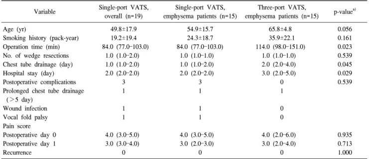
Table 2. Operative outcomes of the single-port VATS and three-port VATS groups Variable Single-port VATS,
overall (n=19)
Single-port VATS, emphysema patients (n=15)
Three-port VATS,
emphysema patients (n=15) p-value
a)Age (yr) 49.8±17.9 54.9±15.7 65.8±4.8 0.056
Smoking history (pack-year) 19.2±19.4 24.3±18.7 35.9±22.1 0.161
Operation time (min) 84.0 (77.0–103.0) 84.0 (77.0–103.0) 114.0 (98.0–151.0) 0.023
No. of wedge resections 1.0 (1.0–2.0) 1.0 (1.0–1.0) 1.0 (1.0–1.0) 0.539
Chest tube drainage (day) 1.0 (1.0–2.0) 1.0 (1.0–2.0) 2.0 (2.0–4.0) 0.045
Hospital stay (day) 2.0 (2.0–2.0) 2.0 (2.0–2.0) 3.0 (2.0–5.0) 0.029
Postoperative complications 3 3 0 0.539
Prolonged chest tube drainage (>5 day)
1 1 1
Wound infection 1 1 0
Vocal fold palsy 1 1 0
Pain score
Postoperative day 0 4.0 (3.0–5.0) 4.0 (3.0–5.0) 4.0 (2.0–6.0) 0.935
Postoperative day 1 3.0 (3.0–4.0) 3.0 (2.0–3.0) 3.0 (2.0–4.0) 0.713
Recurrence 0 0 0 1.000
Values are reported as mean±standard deviation or as median (interquartile range).
VATS, video-assisted thoracic surgery.
a)