pISSN 2005-9159 eISSN 2093-0569
INTRODUCTION
Video-assisted thoracoscopic surgery (VATS) has many
po-tential advantages over thoracotomy, such as early mobi-lization, a more cosmetic incision type, less postoperative pain, and a shorter length of hospital stay. Although VATS
Clinical Research Article
This is an open-access article distributed under the terms of the Creative Commons Attribution Non-Commercial License (http://cre-ativecommons.org/licenses/by-nc/4.0/), which permits unrestricted non-commercial use, distribution, and reproduction in any medium, provided the original work is properly cited.
© The Korean Pain Society, 2021
Author contributions: Merve Sena Baytar: Methodology; Canan Yılmaz: Study conception; Derya Karasu: Writing/manuscript preparation; Çağdaş Baytar: Writing/manuscript preparation.
Comparison of ultrasonography guided serratus anterior
plane block and thoracic paravertebral block in video-assisted
thoracoscopic surgery: a prospective randomized double-blind
study
Merve Sena Baytar1, Canan Yılmaz2, Derya Karasu2, and Çağdaş Baytar3
1Department of Anesthesiology and Reanimation, Zonguldak Atatürk State Hospital, Zonguldak, Turkey
2Department of Anesthesiology and Reanimation, Bursa Yüksek Ihtisas Training and Education Hospital, Health Sciences University, Bursa, Turkey 3Department of Anaesthesiology and Reanimation, Zonguldak Bülent Ecevit University Medicine Faculty, Zonguldak, Turkey
Received November 25, 2020 Revised January 26, 2021 Accepted January 28, 2021
Handling Editor: Jeong-Gill Leem
Correspondence
Çağdaş Baytar
Department of Anaesthesiology and Reanimation, Zonguldak Bülent Ecevit University Medicine Faculty, İbni Sina Kampüsü, Esenköy-Kozlu, Zonguldak 67600, Turkey
Tel: +90-554-225-59-60 Fax: +90-372-261-02-64
E-mail: [email protected]
Background: Various truncal block techniques with ultrasonography (USG) are becoming widespread to reduce postoperative pain and opioid requirements in video-assisted thoracoscopic surgery (VATS). The primary aim of our study was to determine whether the USG-guided serratus anterior plane block (SAPB) is as ef-fective as the thoracic paravertebral block (TPVB) in VATS. Our secondary aim was to evaluate patient and surgeon satisfaction, block application time, first analgesic time, and length of hospital stay.
Methods: Patients in Group SAPB received 0.4 mL/kg bupivacaine with a guid-ed SAPB, and patients in Group TPVB receivguid-ed 0.4 mL/kg bupivacaine with a USG-guided TPVB. We recorded the pain scores, the timing of the first analgesic require-ment, the amount of tramadol consumption, and postoperative complications for 24 hours. We also recorded the block application time and length of hospital stay. Results: A total of 62 patients, with 31 in each group (Group SAPB and Group TPVB) completed the study. Between the two groups, there were no significant differences in rest and dynamic pain visual analog scale scores at 0, 1, 6, 12, and 24 hours after surgery. The total consumption of tramadol was significantly lower in the TPVB group (P = 0.026). The block application time was significantly shorter in Group SAPB (P < 0.001).
Conclusions: An SAPB that is applied safely and rapidly as a part of multimodal analgesia in patients who undergo VATS is not inferior to the TPVB and can be an alternative to it.
Key Words: Analgesia, Patient-Controlled; Analgesics, Opioid; Bupivacaine; Interme-diate Back Muscles; Nerve Block; Pain, Postoperative; Paraspinal Muscles; Thoracic Surgery, Video-Assisted; Tramadol; Ultrasonography.
is a minimally invasive surgery and causes less postopera-tive pain than thoracotomy, it should be treated carefully in terms of both chronicity and disruption of the patient’s healing process [1]. Various blocks are performed with the widespread use of ultrasonography (USG) to relieve post-operative pain and reduce the need for opioids in VATS.
The serratus anterior plane block (SAPB) provides an-algesia in the chest wall by blocking the lateral branches of the intercostal nerves, usually between the T2-T9 levels [2]. The paravertebral block (PVB) has been used for many years in the treatment of breast, thorax, and abdominal surgeries; rib fractures; and cancer pain [3]. The PVB was found to be as effective as a thoracic epidural in postop-erative pain control in thoracic surgery [4]. Both blocks are applied more safely with the increasing use of USG.
The primary aim of our study was to determine whether the USG-guided SAPB is as effective as the thoracic para-vertebral block (TPVB) in VATS. Our secondary aim was to evaluate patient and surgeon satisfaction, block applica-tion time, first analgesic time, postoperative complica-tions, and length of hospital stay.
MATERIALS AND METHODS
1. Study design and patient selection
For our study, we obtained approval from the Uludag University Faculty of Medicine Research Ethics Commit-tee (Local Ethics CommitCommit-tee Ethical number: 2018-7/27, Clinical Trials.gov identifier: NCT04235530), and informed patient consent was obtained from all participants. Our study was a single-center, prospective, randomized, double-blind study conducted on patients who underwent VATS for wedge resection. Patients who were aged 18-65 years, were American Society of Anesthesiologists physi-cal status I-II, and were undergoing elective VATS were included in the study. Patients who had a bleeding diathe-sis, mental or psychiatric disorders, an allergy to the drugs used, any contraindications for SAPB and TPVB applica-tion, an inability to speak Turkish, or whose body mass index above 35.0 kg/m2 were not included in the study. Patients who converted to an open thoracotomy, were dis-charged before 24 hours, had problems with the patient-controlled analgesia (PCA) device, or who experienced block failure (inappropriate local anesthetic distribution and USG image) were also excluded from the study. All patients were informed about the PCA device and visual analog scale (VAS) that were be used in the postoperative period. Patients were randomized into two groups: SAPB (n = 40) and TPVB (n = 40). The randomization list, as well as sealed and opaque envelopes were prepared using a
com-puter program before starting the study by a researcher who was not included in the study.
2. Anesthesia management
We administered 0.01-0.02 mg/kg midazolam to the pa-tients for premedication. Routine monitoring was applied. After monitoring, the patients were intubated after induc-tion with 1-2 mcg/kg of fentanyl, 2-3 mg/kg of propofol, and 0.6 mg/kg of rocuronium. Sevoflurane was used for maintenance in a mixture of 50% air and 50% O2 with a minimum alveolar concentration of 1. At the end of the surgery, the patients were transferred to the postoperative recovery room following extubation. Patients with a Modi-fied Aldrete Score ≥ 9 were transported to the thoracic sur-gery clinic.
3. Block procedure
The blocks were administered by a single experienced an-esthesiologist by USG (MyLab30Gold Cardiovascular; Es-aote, Florence, Italy) guidance before the beginning of the surgical procedure, after intubation. After the area where the block was applied was sterilized with an antiseptic so-lution, the linear probe was wrapped with sterile gloves.
1) Group SAPB (n = 34)
While the patient was in the supine position, a high-fre-quency linear ultrasound probe was placed horizontally on the mid-axillary line at the level of 4th or 5th ribs on the side of the block. The serratus anterior, latissimus dorsi, and intercostal muscles were identified. The block needle (22-gauge 80 mm, Stimuplex Ultra; B. Braun Melsungen AG, Melsungen, Germany) was advanced below the ser-ratus anterior muscle (SAM) towards the fifth rib (using in-plane technique). The prepared 0.25% bupivacaine was administered at 0.4 mL/kg (max. 20 mL) between the SAM and the rib. It was observed that the solution of local anes-thesia was spread between the SAM and the rib (Fig. 1).
2) Group TPVB (n = 36)
A high-frequency linear ultrasound probe was placed between transverse processes from the T4 level in the paramedian plane while patients were in the lateral de-cubitus position. The transverse processes, superior cos-totransverse ligaments, and pleura were visualized. The block needle (22 gauge 80 mm, Stimuplex Ultra; B. Braun Melsungen AG) was advanced until it crossed the superior costotransverse ligament. The prepared 0.25% bupiva-caine was administered at 0.4 mL/kg (max. 20 mL) in the
thoracic paravertebral space. Depression of the pleura was observed as a result of the spread of local anesthetic (Fig. 2).
4. Analgesia management
After induction, intravenous (IV) 1 g of paracetamol and IV 20 mg of tenoxicam was administered to all patients 10 minutes before the end of the surgery. An IV PCA device (CADD-Legacy® PCA; Smiths Medical, Saint Paul, MN) was used for postoperative pain control. A 54 mL saline + 6 mL tramadol (50 mg/mL) IV solution was prepared. The device was set to a 5 mL bolus dose and a 30 minutes lock time, without basal infusion and loading dose. All patients were administered IV 20 mg tenoxicam every 12 hours in the postoperative period and 1 g paracetamol every 8 hours.
5. Outcomes
The primary outcomes of our study were the total amount of opioid consumption and postoperative VAS and dy-namic (during coughing) VAS (DVAS) scores of patients in the recovery room (0 hr) and at postoperative 1, 6, 12, and 24 hours. Secondary outcomes included patient and sur-geon satisfaction, block application time, first analgesic time, postoperative complications, and length of hospital stay. The description of the block application time is from needle puncture to the end of local anesthetic injection. The satisfaction of the patients and surgeons was recorded according to the postoperative pain status with a 4-point scale.
6. Statistical analysis
In order to determine the sample size of the study, the minimum sample size was 78 individuals according to the results of the pilot study using a reference power = 0.80 and a confidence interval = 0.95. The SPSS ver. 22.0 (IBM Co., Armonk, NY) was used for statistical analysis. In the descriptive statistics of the data, mean, standard deviation for quantitative data, and percentage values for qualita-tive data were used. In the distribution of variables, the Kolmogorov–Smirnov normal distribution test was used. Mann–Whitney U, Kruskal–Wallis, and chi-square tests were used in the analysis of data that did not match the normal distribution. P < 0.05 was considered statistically significant as the level of significance in the assessment.
RESULTS
Seventy of the 80 patients who underwent VATS were in-cluded in the study, and 62 patients were evaluated statis-tically (Fig. 3). The demographic data of the patients were
statistically similar between the groups (Table 1). There
were no significant differences in hemodynamic param-eters and vital signs between the groups, perioperatively.
1. Primary outcomes
There were no significant differences in terms of resting VAS and DVAS scores at 0-1-6-12-24 hours between the TPVB and SAPB groups (Table 2). When the total amount
of tramadol consumed was compared, there was a
statis-LDM SAM LA 5th rib Needl e
Fig. 1. Serratus anterior plane block application. LDM: latissimus dorsi muscle, SAM: serratus anterior muscle, LA: local anesthetic.
TM
ESM
PVS
4th TP Needle
Fig. 2. Thoracic paravertebral block application. TM: trapezius muscle, ESM: erector spinae muscle, TP: transverse process, PVS: paravertebral space.
tically significant difference between the SAPB and the TPVB groups (P = 0.026, Table 3).
2. Secondary outcomes
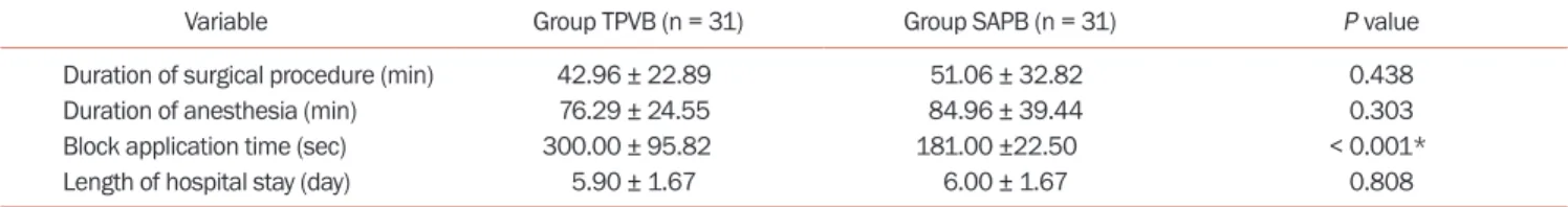
No significant difference was found between the groups with regard to the first analgesic time (P = 0.651). No sig-nificant differences were found between the groups in operative and anesthesia time, or length of hospital stay
(Table 4). The block application time was significantly
shorter in Group SAPB (P < 0.001, Table 4). There were no significant differences between the groups in terms of patient and surgeon satisfaction (Table 5). There were no
significant differences between the groups regarding post-operative complications (Table 6).
DISCUSSION
This prospective, randomized, and double-blinded study was performed on patients who underwent VATS with the intention of wedge resection under general anesthesia. As
part of multimodal analgesia, the SAPB or TPVB were ap-plied to patients before the surgery. Although the postop-erative opioid consumption of the TPVB group was signifi-cantly lower than that of the SAPB group, the consumption of both groups was substantially low at 24 hours (Group
Excluded (n = 10) Language problem (n = 7) Declined to participate (n = 3) Assessed for eligibility (n = 80)
Group TPVB (n = 33) Discontinued intervention (n = 2)
PCA-related problems (n = 2) Group TPVB (n = 36) Did not receive intervention (n = 3) USG-related problem (n = 1) Converted to open thoracotomy (n = 2)
Group TPVB (n = 31) Enrollment Intervention Follow-up Analysis Randomized (n = 70) Group SAPB (n = 34) Did not receive intervention (n = 1)
USG-related problem (n = 1)
Group SAPB (n = 33) Discontinued intervention (n = 2)
PCA-related problems (n = 2)
Group SAPB (n = 31)
Fig. 3. Trial flow diagram. TPVB: thoracic paravertebral block, SAPB: serratus an-terior plane block, USG: ultrasonography, PCA: patient-controlled analgesia.
Table 1. Demographic and clinical data Variable Group TPVB (n = 31) Group SAPB (n = 31) P value Age (yr) 51.2 ± 19.3 47.6 ± 16.9 0.375 Sex (Male/Female) 13/18 11/20 0.764 BMI (kg/m2) 27.2 ± 5.3 26.3 ± 6.0 0.237 ASA (I/II) 3/28 1/30
Values are presented as mean ± standard deviation or number only. TPVB: thoracic paravertebral block, SAPB: serratus anterior plane block, BMI: body mass index, ASA: American Society of Anaesthesiologists.
Table 2. VAS-DVAS scores
Time (hr) VAS/DVAS Group TPVB (n = 31) Group SAPB (n = 31) P value 0 VAS DVAS 2 (0-6) 3 (0-8) 2 (0-6) 3 (0-8) 0.307 0.334 1 VAS DVAS 1 (0-5) 2 (0-5) 1 (0-6) 2 (0-8) 0.0530.080 6 VAS DVAS 1 (0-5) 2 (0-7) 1 (0-5) 2 (0-8) 0.205 0.336 12 VAS DVAS 0 (0-2) 1 (0-3) 0 (0-6) 1 (0-8) 0.2170.166 24 VAS DVAS 0 (0-4) 0 (0-5) 0 (0-4) 1 (0-6) 0.225 0.104 Values are presented as median (minimum-maximum).
VAS: visual analog scale, DVAS: dynamic visual analog scale, TPVB: tho-racic paravertebral block, SAPB: serratus anterior plane block.
Table 3. Comparison of patient controlled analgesia in groups
PCA Group TPVB
(n = 31)
Group SAPB (n = 31) P value First analgesic time (hr) 2.45 ± 1.57 2.12 ± 1.46 0.651 Tramadol consumption
during 24 hr (mg)
18.54 ± 16.08 31.12 ± 23.08 0.026* Values are presented as mean ± standard deviation.
PCA: patient-controlled analgesia, TPVB: thoracic paravertebral block, SAPB: serratus anterior plane block.
SAPB: 31.12 mg, Group TPVB: 18.54 mg). Furthermore, the postoperative median VAS scores of both groups were under 3 and there was no statistically significant differ-ence between the two groups. While the SAPB application time was 182 seconds, the TPVB application time was 300 seconds. The duration of the SAPB application was signifi-cantly shorter than that of the TPVB group (P < 0.001).
Piraccini et al. [5] reported that a PVB was superior to intravenous analgesia in pain control and preservation of postoperative pulmonary function, and it was also equal to thoracic epidural analgesia. We planned our study to investigate whether the SAPB is as effective as the TPVB in pain management after VATS.
SAPB can be applied with two different techniques, deep and superficial. In the superficial technique there is an injection of local anesthetic between the latissimus dorsi muscle and the SAM, while in the deep technique, the injection of local anesthetic is made between the SAM and the external intercostal muscles [6]. By applying the deep SAPB, the anterior and lateral cutaneous branches of the thoracic intercostal nerves are blocked [6-9]. It is known that performing the superficial SAPB also blocks the thoracicus longus nerve and consequently a winged scapula can occur [10]. Piracha et al. [11] applied deep SAPB to four patients who had previously undergone the superficial SAPB for post-mastectomy pain syndrome, to compare deep with superficial SAPBs. The patients stated that they benefited more from the second application and were more satisfied. In addition, they concluded that the
deep SAPB is more effective. The outcomes of the SAPB may differ depending on the type, volume, concentra-tion, and target point of the local anesthetic. In superficial or deep SAPB applications, 10-30 mL of 0.125%-0.375% ropivacaine or bupivacaine were administered and it was reported to provide adequate analgesia for rib fractures, VATS, thoracotomy, breast surgery [2,12,13]. In order to avoid the development of a winged scapula and local an-esthetic systemic toxicity, we administered 20 mL of 0.25% bupivacaine by USG guidance to apply a deep SAPB.
Wang et al. [14] conducted a retrospective study similar to our study by dividing 123 patients into three groups: SAPB, TPVB, and a control group for postoperative pain treatment in patients who underwent single incision (uniportal) VATS. At postoperative hours 1, 2, 4, and 6, the VAS scores of the SAPB and TPVB groups were found to be significantly lower than those of the control group, but no difference was found at the 24 hr and 48 hr timepoints. There was no significant difference between the SAPB and TPVB groups in terms of VAS scores. In the present study, there were no significant differences in rest and DVAS scores at postoperative hours 0, 1, 6, 12, and 24. The VAS scores were below 3 for both groups.
Wang et al. [14] stated in their study that the total opioid consumption of the SAPB group was similar to that of the TPVB group, and that the SAPB was as effective as the TPVB. They discussed that the reason for this result was that the operation was performed with a single incision. The TPVB is known to induce deep analgesia depend-ing on the spread of local anesthetics. Local anesthetic injections to the thoracic paravertebral space can block Table 4. Duration of surgical procedure and anesthesia, block application time, length of hospital stay
Variable Group TPVB (n = 31) Group SAPB (n = 31) P value
Duration of surgical procedure (min) 42.96 ± 22.89 51.06 ± 32.82 0.438
Duration of anesthesia (min) 76.29 ± 24.55 84.96 ± 39.44 0.303
Block application time (sec) 300.00 ± 95.82 181.00 ±22.50 < 0.001*
Length of hospital stay (day) 5.90 ± 1.67 6.00 ± 1.67 0.808
Values are presented as mean ± standard deviation.
TPVB: thoracic paravertebral block, SAPB: serratus anterior plane block. *P < 0.05.
Table 5. Patient-surgeon satisfaction Parameter
Patient satisfaction Surgeon satisfaction TPVB (n = 31) (n = 31)SAPB (n = 31)TPVB (n = 31)SAPB Very satisfied 24 28 26 29 Satisfied 6 1 5 2 Undecided 1 1 0 0 Unsatisfied 0 1 0 0 P value 0.135 0.212
TPVB: thoracic paravertebral block, SAPB: serratus anterior plane block.
Table 6. Postoperative complications Complication Group TPVB
(n = 31)
Group SAPB
(n = 31) P value
Nausea and vomiting 2 1 0.500
Hypertension 3 6 0.236
Hypotension 1 0 0.500
Tachycardia 0 1 0.500
the sympathetic chain by spreading directly to the spinal nerves, laterally to the intercostal nerves, and by spread-ing through the intervertebral foramina into the epidural space located in the medial region [15-17]. In a randomized controlled trial conducted by Aly et al. [18] for post-thora-cotomy analgesia, the SAPB and TPVB were compared in terms of both resting and DVAS scores. In the SAPB group, the DVAS score at the 12th and 18th hours and total mor-phine consumption were found to be significantly higher. In this study, it was emphasized that the SAPB might not affect the posterior cutaneous branches of the intercostal nerves and does not involve autonomic blockade. In the present study, all operations were performed with a 3-port incision and one of the port incisions was located poste-riorly. Therefore, we consider that there is more tramadol consumption in the SAPB group than in the TPVB group. However, the total doses were acceptable for thoracic sur-gery.
In two meta-analyses investigating the analgesic effi-cacy of adding the SAPB to general anesthesia, the authors stated that combining the USG-guided SAPB with general anesthesia provides more effective postoperative analge-sia in VATS [2,19]. They stated that combining the USG-guided SAPB with general anesthesia provides more effec-tive postoperaeffec-tive analgesia. We also believe that the SAPB can be applied effectively as a part of multimodal analge-sia in VATS, along with to non-steroidal anti-inflammatory drugs and paracetamol.
Gupta et al. [20] performed the SAPB and TPVB under USG guidance on patients undergoing modified radi-cal mastectomy under general anesthesia. Morphine consumption, first analgesia time, and VAS scores were recorded in the postoperative period. At the end of the study, while VAS scores were found to be similar, it was determined that the SAPB group consumed more mor-phine. There was no significant difference between the two groups in terms of the first analgesia time. In the pres-ent study, there was similarly no significant difference between the groups in terms of the first analgesia time. The opioid preference in our study was tramadol, a weak opioid with a lower side effect profile. Owing to this prefer-ence, we achieved minimal opioid-related adverse effects in our patients.
Aly et al. [18] compared the SAPB and TPVB for pain af-ter thoracotomy, and no significant difference was found in terms of intraoperative hemodynamic changes, pa-tient satisfaction, and complication incidence. In another study, preoperative the USG-guided TPVB was applied to patients who underwent open cholecystectomy and it was reported that a total spinal block developed after a sudden clouding of consciousness [21]. In our study, there was no significant difference in terms of intraoperative
hemody-namic changes, and the changes were within the clinically acceptable range. There were no complications in either group. In addition, our patient and surgeon satisfaction was high, and there was no significant difference between the two groups.
Aly et al. [18] compared USG-guided SAPB and TPVB application times and found that the application time was significantly shorter in the SAPB. They mentioned that the SAM is very superficial and easily distinguishable from other structures in the image obtained when the USG probe is placed longitudinally in the mid-axillary line, causing this difference. Similar to this study, the duration of the SAPB application was significantly shorter in the present study. In addition to anatomical placement and distinction, we believe that targeting the rib while advanc-ing the needle makes the performer feel safe and is effec-tive in applying the block without wasting time.
Multimodal analgesia is defined as the administration of two or more analgesic agents, with different modes of action, using one or more routes of administration, and providing superior analgesia with fewer side effects by acting synergistically. Multimodal analgesia strategies are very important for effective postoperative analgesia and rehabilitation [22]. In our study, we planned to apply a multimodal analgesia regimen with a preoperative trun-cal block, intraoperative IV paracetamol, as well as non-steroidal anti-inflammatory drugs (NSAIDs), opioids, and postoperative opioids using IV PCA, IV paracetamol, and NSAID administration. We believe that our successful use of multimodal analgesia methods is the reason why opioid consumption in the 24 hours postoperative period was at very low doses.
The limitations of our study include the absence of a control group, the failure to evaluate the effects of the blocks on the onset time, and the affected dermatome lev-els. Another limitation is that opioid consumption was not measured over time.
In our study, we concluded that the SAPB, applied safely and rapidly as a part of multimodal analgesia in patients who will undergo VATS, is not inferior to the TPVB and can be an alternative to it.
CONFLICT OF INTEREST
No potential conflict of interest relevant to this article was reported.
FUNDING
ORCID
Merve Sena Baytar, https://orcid.org/0000-0002-4829-4779 Canan Yılmaz, https://orcid.org/0000-0002-6626-3626 Derya Karasu, https://orcid.org/0000-0003-1867-9018 Çağdaş Baytar, https://orcid.org/0000-0001-7872-9676
REFERENCES
1. Kaya FN, Turker G, Mogol EB, Bayraktar S. Thoracic paraver-tebral block for video-assisted thoracoscopic surgery: single injection versus multiple injections. J Cardiothorac Vasc Anesth 2012; 26: 90-4.
2. De Cassai A, Boscolo A, Zarantonello F, Piasentini E, Di Gregorio G, Munari M, et al. Serratus anterior plane block for video-assisted thoracoscopic surgery: a meta-analysis of randomised controlled trials. Eur J Anaesthesiol 2021; 38: 106-14.
3. Ng SC, Chazapis M, West S. Ultrasound-guided paraverte-bral block [Internet]. London: World Federation of Societies of Anesthesiologists; 2018. Available at: https://wfsa.traver-stodd.com/wp-content/uploads/376_english.pdf.
4. Yeung JH, Gates S, Naidu BV, Wilson MJ, Gao Smith F. Para-vertebral block versus thoracic epidural for patients under-going thoracotomy. Cochrane Database Syst Rev 2016; 2: CD009121.
5. Piraccini E, Pretto EA Jr, Corso RM, Gambale G. Analgesia for thoracic surgery: the role of paravertebral block. HSR Proc Intensive Care Cardiovasc Anesth 2011; 3: 157-60.
6. Blanco R, Parras T, McDonnell JG, Prats-Galino A. Serratus plane block: a novel ultrasound-guided thoracic wall nerve block. Anaesthesia 2013; 68: 1107-13.
7. Diéguez P, Fajardo M, López S, Alfaro P. BRILMA methylene blue in cadavers. Anatomical dissection. Rev Esp Anestesiol Reanim 2016; 63: 307-8.
8. Diéguez P, Casas P, López S, Fajardo M. [Ultrasound guided nerve block for breast surgery]. Rev Esp Anestesiol Reanim 2016; 63: 159-67. Spanish.
9. Torre PA, Jones JW Jr, Álvarez SL, Garcia PD, Miguel FJG, Rubio EMM, et al. [Axillary local anesthetic spread after the thoracic interfacial ultrasound block - a cadaveric and ra-diological evaluation]. Rev Bras Anestesiol 2017; 67: 555-64. Portuguese.
10. Hanley C, Wall T, Bukowska I, Redmond K, Eaton D, Ní Mhuircheartaigh R, et al. Ultrasound-guided continu-ous deep serratus anterior plane block versus continucontinu-ous thoracic paravertebral block for perioperative analgesia in videoscopic-assisted thoracic surgery. Eur J Pain 2020; 24:
828-38.
11. Piracha MM, Thorp SL, Puttanniah V, Gulati A. “A tale of two planes”: deep versus superficial serratus plane block for postmastectomy pain syndrome. Reg Anesth Pain Med 2017; 42: 259-62.
12. Okmen K, Okmen BM, Uysal S. Serratus anterior plane (SAP) block used for thoracotomy analgesia: a case report. Korean J Pain 2016; 29: 189-92.
13. Paul S, Bhoi SK, Sinha TP, Kumar G. Ultrasound-guided ser-ratus anterior plane block for rib fracture-associated pain management in emergency department. J Emerg Trauma Shock 2020; 13: 208-12.
14. Wang L, Wang Y, Zhang X, Zhu X, Wang G. Serratus anterior plane block or thoracic paravertebral block for postoperative pain treatment after uniportal video-assisted thoracoscopic surgery: a retrospective propensity-matched study. J Pain Res 2019; 12: 2231-8.
15. Cheema SP, Ilsley D, Richardson J, Sabanathan S. A thermo-graphic study of paravertebral analgesia. Anaesthesia 1995; 50: 118-21.
16. Conacher ID. Resin injection of thoracic paravertebral spac-es. Br J Anaesth 1988; 61: 657-61.
17. Cowie B, McGlade D, Ivanusic J, Barrington MJ. Ultrasound-guided thoracic paravertebral blockade: a cadaveric study. Anesth Analg 2010; 110: 1735-9.
18. Aly AA, Abd Ellatif SE. Comparison of ultrasound-guided serratus plane block and thoracic paravertebral block for postoperative analgesia after thoracotomy: a randomized controlled trial. Res Opin Anesth Intensive Care 2018; 5: 314-22.
19. Zhang X, Zhang C, Zhou X, Chen W, Li J, Wang H, et al. Anal-gesic effectiveness of perioperative ultrasound-guided ser-ratus anterior plane block combined with general anesthesia in patients undergoing video-assisted thoracoscopic surgery: a systematic review and meta-analysis. Pain Med 2020; 21: 2412-22.
20. Gupta K, Srikanth K, Girdhar KK, Chan V. Analgesic efficacy of ultrasound-guided paravertebral block versus serratus plane block for modified radical mastectomy: a randomised, controlled trial. Indian J Anaesth 2017; 61: 381-6.
21. Beyaz SG, Özocak H, Ergönenç T, Erdem AF, Palabıyık O. Total spinal block after thoracic paravertebral block. Turk J Anaesthesiol Reanim 2014; 42: 43-5.
22. Hanna MN, Ouanes JPP, Tomas VG. Postoperative pain and other acute pain syndromes. In: Practical management of pain. 5th ed. Edited by Benzon HT, Rathmell JP, Wu CL, Turk DC, Argoff CE, Hurley RW. Philadelphia, Elsevier/Mosby. 2014, pp 271-97.e11.