www.jkfas.org pISSN 1738-3757 eISSN 2288-8551 J Korean Foot Ankle Soc 2014;18(3):129-132 http://dx.doi.org/10.14193/jkfas.2014.18.3.129
복재신경 분지와 유착되어 발생한 족부의 혈관내 유두내피 증식증: 증례 보고
이상형, 김창희, 정승효
동국대학교 의과대학 일산병원 정형외과학교실
Intravascular Papillary Endothelial Hyperplasia in Foot Adherent to a Saphenous Nerve Branch: A Case Report
Sang Hyeong Lee, Chang Hee Kim, Seung Hyo Jung
Department of Orthopaedic Surgery, Dongguk University Ilsan Hospital, Dongguk University College of Medicine, Goyang, Korea
Intravascular papillary endothelial hyperplasia (IPEH) has appeared in the literature under a variety of names, including Masson’s tu- mor, Masson’s hemangioma, and Masson’s pseudoangiosarcoma. It is a benign lesion of the skin and subcutaneous tissue characterized by reactive proliferation of vascular endothelial cells with papillary formations. The clinical picture is not specific and the lesion resem- bles malignant angiosarcoma clinically and histopathologically. Therefore, it is often mistaken for angiosarcoma and a group of other benign and malignant vascular lesions. We report on a case of IPEH adherent to peripheral nerve treated with operative excision.
Key Words: Foot, Vascular endothelial cells, Saphenous nerve
증례 보고
34세 여자 환자가 우측 발의 종괴를 주소로 내원하였다. 종괴는 특별한 외상력 없이 약 3년 전부터 발생하였으며 환자는 해당 부 위의 동통을 호소하였고 보행 시 증상은 악화되었다. 간헐적인 저 린감이 동반되었으나 감각저하 등 다른 신경학적 증세는 호소하 지 않았다. 이학적 검사상 우측 엄지 발가락 중족지 배내측 부위 에 약 2×2 cm 크기의 유동성이 있는 원형의 종괴가 촉지되었으며 병변 위 피부의 색깔 변화는 관찰되지 않았다(Fig. 1). 종괴는 압박 시 사라지고 압박 제거 시 다시 발생하는 양상을 보였다. 티넬 징 후(Tinel sign)는 불분명하였다. 결절종(ganglion), 혈관종(heman- gioma) 등을 포함한 연부조직의 양성 종양 의심하에 추가적인 검 사를 진행하였다.
단순 방사선 검사에서 증가된 연부조직 음영이 관찰되었으며, 종괴 주변으로 골성 병변은 관찰되지 않았고 종괴 내부에도 특별 한 음영 변화는 관찰되지 않았다(Fig. 2). 초음파 검사상에서 내부 에 액체-액체층(fluid-fluid level)이 관찰되는 2.2×0.7×1.4 cm 크 기의 낭성 종괴가 관찰되었으며 혈류순환은 관찰되지 않았다(Fig.
3). 이학적 검진 및 영상학적 검사상 결절낭종 진단하에 자기공명 혈관내 유두내피 증식증(intravascular papillary endothelial hy-
perplasia)은 인체의 몸통, 두경부, 상지에 주로 발생하는 드문 양 성 종양 중의 하나이다. 1) Masson 2) 은 혈관 내강(vascular lumen) 세 포의 증식에 의해서 발생하는 종양으로 설명하였다. 드물지만 몇 몇 저자들에 의해 족부에서 발생한 유두내피 증식증이 보고되었으 나 3) 대부분 족저 피부(plantar skin)에서 발생하였으며, 신경조직과 유착되어 발생한 유두내피 증식증에 대해서는 보고된 바가 많지 않다. 저자들은 말초신경집 종양(peripheral nerve sheath tumor)과 오인하기 쉬운, 복재신경(saphenous nerve)의 정상신경 분지와 유 착되어 발생한 드문 증례의 유두내피 증식증에 대해서 보고하고자 한다. 본 증례 보고와 관련된 자료의 사용에 대해서는 환자에게 알 리고 동의를 받았다.
Received March 30, 2014 Revised April 21, 2014 Accepted May 29, 2014 Corresponding Author: Sang Hyeong Lee
Department of Orthopaedic Surgery, Dongguk University Ilsan Hospital, 27 Dongguk-ro, Ilsandong-gu, Goyang 410-773, Korea
Tel: 82-31-961-7296, Fax: 82-31-961-7290, E-mail: [email protected] Financial support: None.
Conflict of interest: None.
Case Report
This is an Open Access article distributed under the terms of the Creative Commons Attribution Non-Commercial License (http://creativecommons.org/licenses/
CCby-nc/3.0) which permits unrestricted non-commercial use, distribution, and reproduction in any medium, provided the original work is properly cited.
Copyright 2014 Korean Foot and Ankle Society. All rights reserved. ⓒ
130 Vol. 18 No. 3, September 2014
영상 등 추가적인 검사의 진행 없이 수술적 치료를 진행하였다.
수술은 척추마취하에 지혈대를 시행 후 진행하였다. 종괴 위 피 부에 절개를 가한 후 조심스럽게 연부조직을 박리하여 얇은 막에 둘러싸여 있는 2.5×2×1 cm 크기의 검붉은 종괴 소견을 관찰할 수 있었다. 종괴의 원위부 및 근위부로 연결된 족부의 내측 및 배 측 신경을 지배하는 복재신경의 정상 분지를 확인할 수 있었으며, 이는 종괴의 연결 부위로 늘어나 있었다(Fig. 4). 종괴를 둘러싸고 있는 신경외막(epineural)에 절개를 가하자 혈전(thrombus)이 노 출되었다. 현미경하 미세 수술 기구를 이용하여 정상신경 분지와 종괴를 분리하기 위하여 노력하였으나 종괴는 신경 분지와 완전히 유착되어 있었으며 종괴 내부 및 외부에 정상신경 분지가 관찰되 지 않아 신경다발을 분리할 수 없었다. 원위부 및 근위부의 정상신 Figure 1.
Figure 1. Photograph shows a protruding mass lesion on dorsomedial aspect of right foot.
Figure 2.
Figure 2. Plain radiograph shows a round soft tissue lesion on medial aspect of medial cuneiform. There is no bony erosion or calcification.
Figure 3.
Figure 3. Ultrasonography shows an ovoid-shape, 2.2×0.7×1.4 cm size cystic mass on dorsomedial aspect of right foot. Sagittal (A) and Axial (B).
Figure 4.
Figure 4. The mass is connected to the branch of saphenous nerve proximally and distally.
Figure 5.
Figure 5. Photograph shows a dark-reddish cystic mass of 2×1×1 cm
size.
www.jkfas.org 131 Sang Hyeong Lee, et al. Intravascular Papillary Endothelial Hyperplasia
감별하기는 힘들며, 따라서 연부조직에서 발생할 수 있는 지방종 (lipoma), 신경초종(schwannoma), 결정낭종, 과오종(hamartoma), 건초 거대세포종(giant cell tumor of tendon sheath) 등 다른 종양 질환을 고려해야 한다. 5) 특히 육안적으로 혈종이나 다른 정맥의 기형과 비슷하게 관찰되며, 조직학적으로 악성 혈관육종(malig- nant angiosarcoma)과 유사한 양상을 보이므로 반드시 조직 병리 학적으로 확진 검사를 시행해야만 한다. 6) 조직학적으로 유두내피 증식증은 혈관육종과 구분되는 점이 있는데, 유두내피 증식증은 혈관육종에서 보이는 괴사(necrosis), 궤양(ulceration) 및 유사분열 (mitosis)이 드물며 혈관 내강으로 증식된 돌기는 다른 주변 조직으 로 침윤하는 모습을 보이지 않으며 혈관 내강에 국한되어 있다. 7)
혈관내 유두내피 증식증의 진단 방법으로는 조직 병리학적 검사 가 필수적이나 초음파 및 자기공명영상 촬영 등을 통하여 감별진 단을 할 수 있다. Lee 등 8) 이 기술한 사지에서 발생한 유두내피 증 식증에 대한 영상학적 분석 결과에서, 혼합형 병변의 경우 초음파 상에서 혈관 분포 및 고에코성(hyperechoic)의 격막(septum)을 가 지는 저에코성(hypoechoic) 종괴 병변을 보인다. 또 자기공명영상 에서는 T1에서 고강도 신호(hyperintense signal)의 결절(nodule) 양상의 병소를 포함하는 동등 또는 조금 증가된 강도의 신호를 보 이는 종괴를 나타내고 T2에서는 저강도(hyperintense signal)의 결 절 병소를 포함하는 고강도 신호의 종괴를 보인다. 조영증강영상 에서는 주변과 격막 또는 중심부에 조영증강을 나타낸다.
가장 효과적인 치료는 통증을 유발하는 종괴에 대해 완전 절제 를 시행하는 것이다. 5) 병변에 지속적인 압력을 가하는 경우 일부 의 경우에서 호전될 수 있으나 재발의 확률이 높으며, 펀치 생검 (punch biopsy)은 불충분한 종괴의 제거 가능성 및 종괴가 동맥에 서부터 기원한 경우 다량의 출혈이 발생할 수 있어 권유되지 않는 다. 본 증례의 경우에서 유두내피 증식증은 신경혈관 근처부터 기 원한 것으로, 치료에 있어서 처음부터 통상적으로 완전 절제를 시 행하기 힘든 면이 있었다. 경우는 다르지만 Mestdagh 등 9) 은 후경 골 신경에서 기원한 혈관종의 치료에 있어서 현미경적 신경다발 경 분지에 표시 봉합실(tagging suture)로 묶은 후 두 봉합사 사이
에서 절제하여 종괴를 완전히 제거하였다(Fig. 5). 근위부 신경 분 지는 신경종(neuroma)의 발생을 예방하기 위하여 심부조직 안쪽 으로 묻혀있도록 하였다. 이후 지혈대를 풀어 출혈 여부를 확인하 였으며, 수술 부위에 심한 출혈은 관찰되지 않았다. 연부조직을 흡 수성 봉합사로 봉합하고 배액관(drain)을 삽입 후 다시 피부를 일 차 봉합하였다. 조직 병리학적 검사를 통하여 혈관 내피세포로 구 성된 유두 모양의 세포들이 혈관 내강으로 증식된 전형적인 혈관 내 유두내피 증식증을 확진할 수 있었다. 면역염색 결과에서 슈반 세포(Schwann cell)에 염색되는 S-100 protein에 일부 염색된 부분 이 있었으며, 내피세포 표지 중의 하나인 CD31 염색에 양성 소견 이 관찰되었다(Fig. 6).
고 찰
Masson 2) 에 의해 보고된 혈관내 유두내피 증식증은 혈전 내에서 발생한 내피세포의 증식에 의한 신생물 병변이다. Hashimoto 등 1) 은 이를 세 가지 유형으로 분류하였는데, 첫 번째는 확장된 혈관에 서 발생한 내피세포의 증식이며, 두 번째는 이전부터 존재하였던 혈관종이나 동정맥기형(ateriovenous malformation) 등의 혈전에 서부터 내피세포가 증식한 혼합형 병변, 세 번째는 드물지만 혈관 외에서 기원한 병변이다.
말초신경에는 신경외막에 존재하는 혈관에 의한 외인성(extrin- sic) 체계와 신경내막(endoneural)에 분포하는 혈관에 의한 내인성 (intrinsic) 체계가 서로 문합(anastomosis)하며 혈관망을 구성하고 있으며, 이러한 말초신경 내에 분포하는 혈관에서부터 혈관종 등 혈관 기원 질환이 발생할 수 있다고 보고된 바 있다. 4)
임상적으로 피하조직에 발생한 경계가 잘 지어진 종괴 형태를 보이며, 그 크기가 크지 않다면 증상이 없을 수 있으나 종괴의 크 기가 커지게 되면 종괴 자체에 의한 불편감 및 압통이 발생할 수 있다. 대부분의 유두내피 증식증은 임상 증상만으로 다른 종양과
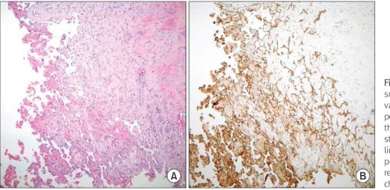
Figure 6.
Figure 6. Pathologic features. (A) Myriad
small delicate papillae project into the
vascular lumen. The papillae are com-
posed of a single layer of vascular endo-
thelium surrounding a stromal core (H&E
stain, ×100). (B) The vascular endothe-
lium of papillae is well demonstrated by
positive (brown) immunohistochemical
reaction to CD31 antibody (immunohisto-
chemistry stain, ×100).
132 Vol. 18 No. 3, September 2014
J Dermatopathol. 1983;5:539-46.
2. Masson P. Hemangioendotheliome vegetant intravasculaire. Bull Soc Anat (Paris). 1923;93:517-23.
3. Fink B, Temple HT, Mizel MS. Intravascular papillary endothelial hyperplasia: a pseudotumor presenting on the plantar foot. Foot Ankle Int. 2003;24:871-4.
4. Papagelopoulos PJ, Mavrogenis AF, Skarpidi E, Nikolaou I, Sou- cacos PN. A 56-year-old woman with a right arm mass. Clin Orthop Relat Res. 2008;466:2892-8.
5. Miyamoto H, Nagatani T, Mohri S, Nakajima H. Intravas- cular papillary endothelial hyperplasia. Clin Exp Dermatol.
1988;13:411-5.
6. Clearkin KP, Enzinger FM. Intravascular papillary endothelial hyperplasia. Arch Pathol Lab Med. 1976;100:441-4.
7. Amérigo J, Berry CL. Intravascular papillary endothelial hyper- plasia in the skin and subcutaneous tissue. Virchows Arch A Pathol Anat Histol. 1980;387:81-90.
8. Lee SJ, Choo HJ, Park JS, Park YM, Eun CK, Hong SH, et al. Im- aging findings of intravascular papillary endothelial hyperplasia presenting in extremities: correlation with pathological findings.
Skeletal Radiol. 2010;39:783-9.
9. Mestdagh H, Lecomte-Houcke M, Reyford H. Intraneural hae- mangioma of the posterior tibial nerve. J Bone Joint Surg Br.
1990;72:323-4.
10. Lim R, Tay SC, Yam A. Intravascular papillary endothelial hy- perplasia (Masson's tumour) of the finger presenting as a digital nerve schwannoma. J Hand Surg Eur Vol. 2011;36:612-3.
내 박리술(intrafascicular microscopic dissection)을 시행하여 신경 의 전도성을 유지할 수 있다고 하였으며, 종괴의 완전 박리에 의하 여 운동신경에 장애가 발생 시 신경이식술이 필요하며, 단지 감각 신경에 장애가 있다면 신경절제술만으로 충분하다고 하였다. 수지 신경 근처에서 발생하여 신경종과 유사한 형상이 관찰된 유두내피 증식증이 보고된 바 있으나, 10) 본 증례와 같이 말초신경 분지와 유 착되어 정상신경 분지와 구분되지 않는 유두내피 증식증에 대해서 는 보고된 바가 없었다. 저자들은 복재신경의 정상신경 다발 박리 를 위해 노력하였으나 신경 분지가 종괴와 완전히 유착되어 분리 할 수 없었기에 종괴의 완전 절제를 시행하여 재발을 방지하도록 하였다.
신경학적 증상이 명확하지 않은 하지의 종괴에 있어서는 일반적 으로 흔한 질환들뿐만 아니라 신경 주변에서 발생할 수 있는 여러 종괴에 대한 감별이 필요하다. 하지의 종괴 치료 시 재발 방지를 고려하여 완전 절제를 시행할 경우 신경 분지에 따라 필요 시 신경 이식술 또는 가능하다면 신경다발 내 박리술 시행을 고려해야 할 것이다.
REFERENCES