Tuberc Respir Dis 2011;70:51-57
CopyrightⒸ2011. The Korean Academy of Tuberculosis and Respiratory Diseases. All rights reserved.
혈청 프로칼시토닌(serum procalcitonin) 측정을 통한 패혈증 진단 및 중등도 평가의 유용성
울산대학교 의과대학 서울아산병원
1중환자의학교실,
2호흡기내과학교실
박태진1, 임채만2, 고윤석2, 홍상범2
Utility of Serum Procalcitonin for Diagnosis of Sepsis and Evaluation of Severity
Taejin Park, M.D.
1, Chae-Man Lim, M.D.
2, Younsuck Koh, M.D.
2, Sang-Bum Hong, M.D.
2Departments of
1Critical Care Medicine,
2Pulmonary and Critical Care Medicine, Asan Medical Center, University of Ulsan College of Medicine, Seoul, Korea
Background: Early recognition and treatment of sepsis would improve patients' outcome. But it is difficult to distinguish between sepsis and non-infectious conditions in the acute phase of clinical deterioration. We studied serum level of procalcitonin (PCT) as a method to diagnose and to evaluate sepsis.
Methods: Between 1 March 2009 and 30 September 2009, 178 patients had their serum PCT tested during their clinical deterioration in the medical intensive care unit. These laboratories were evaluated, on a retrospective basis.
We classified their clinical status as non-infection, local infection, sepsis, severe sepsis, and septic shock. Then, we compared their clinical status with level of PCT.
Results: The number of clinical status is as follows: 18 non-infection, 33 local infection, 39 sepsis, 26 severe sepsis, and 62 septic shock patients. PCT level of non-septic group (non-infection and local infection) and septic group (sepsis, severe sepsis, septic shock) was 0.36±0.57 ng/mL and 18.09±36.53 ng/mL (p<0.001), respectively. Area under the curve for diagnosis of sepsis using cut-off value of PCT >0.5 ng/mL was 0.841 (p<0.001). Level of PCT as clinical status was statistically different between severe sepsis and septic shock (*severe sepsis; 4.53±6.15 ng/mL, *septic shock 34.26±47.10 ng/mL, *p<0.001).
Conclusion: Level of PCT at clinical deterioration showed diagnostic power for septic condition. The level of PCT was statistically different between severe sepsis and septic shock.
Key Words: Sepsis; Biomarkers; Procalcitonin; Diagnosis
Address for correspondence: San-Bum Hong, M.D.
Department of Pulmonary and Critical Care Medicine, Asan Medical Center, University of Ulsan College of Medicine, 388-1, Pungnap 2-dong, Songpa-gu, Seoul 138-736, Korea Phone: 82-2-3010-3893, Fax: 82-2-3010-4709
E-mail: [email protected] Received: Aug. 25, 2010 Accepted: Nov. 9, 2010
서 론
패혈증에 대한 다양한 노력에도 불구하고 아직 패혈증 으로 인한 사망률은 높다. Rangel-Frausto 등1의 연구에서 전신 염증반응 증후군(Systemic Inflammatory Response
Syndrome, SIRS), 패혈증(sepsis), 중증 패혈증(severe sepsis) 그리고 패혈증성 쇼크(septic shock)의 사망률은 각각 7%, 16%, 20% 그리고 46%로 나타났다. 또한 국내 중증 패혈증 환자의 사망률에 관한 연구에서도 64.6%의 높은 사망률을 보였다2. 이렇듯 높은 사망률의 원인 중 하 나로 패혈증 진단의 지연을 고려할 수 있다. 과거 연구에 서도 패혈증의 조기 진단 및 치료가 환자의 예후를 향상시 킨다고 보고되었다3. 그러나 급성 악화시기의 환자상태가 패혈증인지 다른 비 감염성 상태인지에 대한 감별이 어렵 다. 따라서 급성 악화시기에 적절한 치료가 시작되는데 어려움이 발생할 수 있다.
본 연구에서는 세균감염 시에 상승하는 것으로 알려진4
프로칼시토닌(procalcitonin, PCT)을 분석하여 패혈증 진 단의 유용성과 패혈증 중등도 감별의 유용성을 알아보고 자 하였다.
대상 및 방법
본 연구는 의무기록분석을 통한 후향성 연구(retrospec- tive study)로 28개 병상을 갖춘 한 곳의 3차 대학병원 내 과계 중환자실에서 이루어졌다. 서울아산병원 임상연구 심의위원회의 승인을 받았으며, 심의위원회와의 협의하 에 피험자의 서면동의 취득을 생략하였다.
1. 연구 대상
2009년 3월 1일부터 2009년 9월 30일까지 서울아산병 원 내과계 중환자실에서 입원치료를 받은 18세 이상의 성 인 환자 중에서 혈청 프로칼시토닌을 검사한 환자를 대상 으로 하였다. 이 환자들 중에서 악화시기 24시간 이후에 검사가 시행된 경우는 악화시기를 반영하기 어려운 것으 로 판단하여 제외하였다. 임상적 악화는 생체징후 및 검 사실 소견에 이상이 발생하거나 혹은 의식소실, 호흡곤란, 흉통, 발열 등과 같은 위중한 증상이 발생하여 임상의의 적극적인 진단과 처치가 필요한 상태로 본원에서 시행 중 인 응급의료지원팀(Medical Alert Team, MAT)에 지원요 청을 받은 경우로 정의하였다.
2. 대상 군 분류
환자군은 의무기록 분석을 통하여 임상적 악화의 수준 을 평가하였다. 평가 및 분류는 비패혈증 상태(non-sepsis state)와 패혈증 상태(sepsis state)로 크게 환자군을 구분 하였고, 비패혈증 상태는 비감염성 질환(non-infection) 및 국소 감염(local infection)으로 재분류하였다. 패혈증 상태는 패혈증(sepsis), 중증 패혈증(severe sepsis) 및 패 혈증성 쇼크(septic shock)로 재분류하였다. 이와 같은 분 류의 근거는 1992년 American College of Chest Physici- ans (ACCP)/Society of Critical Care Medicine (SCCM)에 서 정의한 패혈증 정의를 따랐다5. 해당 연구에서는 환자 상태를 전신 염증성 반응 증후군(SIRS; Systemic Inflam- matory Response Syndrome), 패혈증(sepsis), 중증 패혈 증(severe sepsis) 및 패혈증성 쇼크(septic shock)로 분류 하였다. SIRS는 다음의 기준에서 2개 이상을 보이는 경우 로 정의하였다. ① 체온 >38oC 혹은 <36oC; ② 심박동 수 >90회/분; ③ 호흡수 >20회/분 혹은 PaCO2 <32
mm Hg; ④ 백혈구수 12,000/μL 초과 또는 4,000/μL 미만 혹은 미성숙 호중구가 10% 초과되는 경우이다. 패혈 증의 경우 감염(infection)의 결과로 SIRS의 조건이 나타나 는 경우로 정의하였다. 중증 패혈증(severe sepsis)은 패 혈증으로 인해 장기 기능 부전, 저관류, 젖산 산혈증, 급성 의식변화 등의 증상이 동반되는 경우로 정의하였다. 패혈 증성 쇼크는 적절한 수액 소생술에도 불구하고 관류이상 을 동반한 패혈증-유발 저혈압이 지속되는 상태로 정의하 였다. 본 연구에서는 SIRS를 비패혈증 상태 중에서 감염증 이 없는 상태로 정의하였다.
3. 프로칼시토닌 측정
프로칼시토닌의 측정은 면역형광분석(immunolumino- metric assay)을 이용하였다(VIDAS BRAHMS PCT; bio- Mérieux SA, Marcy-l'Etoile, France). 본 검사는 프로칼시 토닌의 칼시토닌(calcitonin)과 카타칼신(katacalcin) 부위 에 붙는 두 개의 항원과 특수 단일클론 항체의 반응을 검 사하는 것으로 2∼3시간이 소요되며, 20 μL의 혈청이 필요하다.
4. 자료의 수집
의무기록 분석을 통해 입원 당시의 나이, 성별, 중환자 실 입실 시의 진단, 중환자실 및 병원 재원일수와 사망여 부, 28일 단기 사망여부를 조사하였으며, 입원 첫 24시간 Acute Physiology And Chronic Health Evaluation (APACHE) II 점수6와 Sequential Organ Failure Assess- ment (SOFA) 점수7를 계산하였다. 중환자실 입실 시의 진 단은 크게 호흡기, 심장, 위장, 내분비, 혈액, 신장, 기타 질환으로 구분하였다. 환자의 감염여부는 혈액, 소변, 대 변 및 체액 배양 검사 결과 및 특정 항체 검사 등을 이용하 였으며, 이를 통한 판단이 불가능한 경우에는 감염내과와 의 협진 및 영상의학적인 추정 진단을 통해 판단하였다.
5. 결과의 측정
본 연구의 일차 목적(primary endpoint)은 프로칼시토 닌 측정을 통한 패혈증 진단 및 패혈증 중등도 감별이고, 이차 목적(secondary endpoints)은 프로칼시토닌 농도에 따른 28일 단기사망률의 예측으로 하였다.
6. 통계분석
통계분석은 Window용 SPSS프로그램 version 14.0 (SPSS Inc., Chicago, IL, USA)을 이용하여 시행하였다.
Table 1. Baseline characteristics between non-sepsis group and sepsis group
Non-sepsis group (n=51)
Sepsis group (n=127) p-value
Age, yr 64.7±13.4 62.5±14.5 0.245
Sex (male/female) 30/21 89/38 0.150 APACHE, First 24 hr 24.2±9.0 26.0±8.3 0.204 SOFA, First 24 hr 8.2±3.5 11.2±4.0 <0.001
ICU LOS 17.8±32.3 13.5±16.3 0.904
Hospital LOS 35.0±32.9 46.5±43.7 0.157 ICU mortality, No. (%) 15 (29.4) 54 (42.5) 0.111 Hospital mortality, 17 (33.3) 59 (46.5) 0.106 No. (%)
Values are presented as mean±SD.
APACHE: acute physiology and chronic health evaluation;
SOFA: sequential organ failure assessment; ICU: intensive care unit; LOS: length of stay; SD: standard deviation.
Table 2. Infection focus of patients
Infection focus No. (%)
Lower respiratory tract 99 (55.6)
Abdominal 25 (14.0)
Others 21 (11.8)
Urogenital 5 (2.8)
Skin, soft tissue 5 (2.8)
Device-related 4 (2.2)
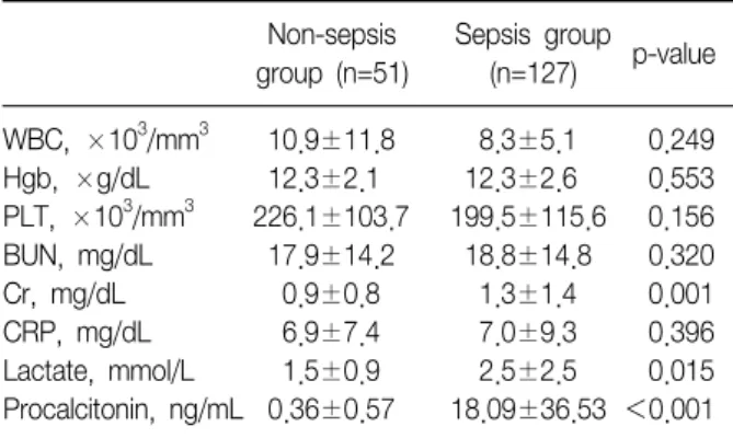
Table 3. Laboratory data of patients
Non-sepsis group (n=51)
Sepsis group (n=127) p-value WBC, ×10
3/mm
310.9±11.8 8.3±5.1 0.249 Hgb, ×g/dL 12.3±2.1 12.3±2.6 0.553 PLT, ×10
3/mm
3226.1±103.7 199.5±115.6 0.156 BUN, mg/dL 17.9±14.2 18.8±14.8 0.320 Cr, mg/dL 0.9±0.8 1.3±1.4 0.001 CRP, mg/dL 6.9±7.4 7.0±9.3 0.396 Lactate, mmol/L 1.5±0.9 2.5±2.5 0.015 Procalcitonin, ng/mL 0.36±0.57 18.09±36.53 <0.001 Values are presented as mean±SD.
Hgb: Hemogloblin; SD: standard deviation.
연속형 변수에 대한 모든 통계 값은 평균±표준편차로 표 기하였고, 범주형 변수의 경우에는 Chi-square test와 Fisher's exact test를 이용하였다. 프로칼시토닌 농도에 따 른 패혈증 진단의 유용성을 평가하기 위해 receiver oper- ating charateristic (ROC) curve를 그려 곡선 아래 면적 (area under curve, AUC)을 구하였다. 패혈증 상태에 따 른 프로칼시토닌 농도를 비교하고 이를 사후 검정을 통해 각 상태간에 농도의 차이가 있는지 비교하였다. 모든 통 계적인 분석은 p값이 0.05 미만인 경우를 통계적으로 유 의한 것으로 간주하였다.
결 과
1. 대상 환자 군의 분류
연구 대상기간 동안 내과계 중환자실에 입실하여 혈청 프로칼시토닌 검사를 시행한 환자는 총 307명이었다. 이 중 임상적 악화시기와 관련 없이 프로칼시토닌을 측정한 129명을 제외한 178명의 환자를 대상으로 의무기록을 분 석하였다. 제외된 환자는 수술 전후의 처치를 위해 입원 한 38명, 시술 전후의 처치를 위해 입원한 41명과 중환자 실 재원 중 추적 검사로 프로칼시토닌을 측정한 50명의 환자가 있었다.
대상 환자의 의무기록을 근거로 프로칼시토닌 검사가 시행된 상태의 임상상황을 구분하였다. 환자 군을 패혈증 여부로 구분하면 비패혈증 상태가 51명, 패혈증 상태가 127명으로 나타났다. 이들 환자 군을 중등도에 따라 분류
하면, 비감염성 질환(non-infection) 18명, 국소 감염(local infection) 33명, 패혈증(sepsis) 39명, 중증 패혈증(severe sepsis) 26명, 패혈증성 쇼크(septic shock) 62명으로 나타 났다.
2. 대상 환자 군의 기본적인 특성
비패혈증군과 패혈증군의 기본적인 특성에서 나이, 성 별, 중환자실 재원일, 병원 재원일, 중환자실 및 병원 사망 률에 있어 통계적인 차이를 보이지 않았다. SOFA 점수에 서는 두 군간에 통계적으로 유의하게 패혈증 군에서 높게 나타났다(Table 1). 대상 환자군의 추정 혹은 확진된 감염 병소로는 하부 호흡기계 감염이 99명(55.6%)으로 가장 높 게 나타났다(Table 2).
3. 검사실 검사
비패혈증군과 패혈증군으로 대상 군을 나누어 검사실 검사를 비교하였을 때, 두 군간에 유의한 차이를 보인 항 목은 혈청 크레아티닌, 젓산, 프로칼시토닌으로 나타났다.
반면 백혈구 수치와 C반응성 단백질은 두 군간에 차이를 보이지 않았다(Table 3). 대상 환자군의 임상적 중등도에
Figure 1. Mean values±SD of PCT in patients with non-infection (n=18), local infection (n=33), sepsis (n=39), severe sepsis (n=26), septic shock (n=62) at their clinical deterioration (1st ICU day). *p<0.001. SD: standard devi- ation; PCT: procalcitonin.
Table 4. Assessment of severity of sepsis by WBC, CRP and procalcitonin
Non infection
(n=18)
Local infection
(n=33) Sepsis (n=39) Severe sepsis (n=26)
Septic shock
(n=62) p-value
WBC, ×10
3/mm
38.8±4.0 12.1±14.3 8.1±4.9 9.2±5.8 8.0±4.9 0.650
CRP, mg/dL 7.8±8.4 6.4±6.8 8.2±10.8 6.8±7.4 6.4±8.9 0.813
Procalcitonin, ng/mL 0.18±0.20 0.45±0.68 1.44±1.33 4.53±6.15* 34.26±47.10* *<0.001 Values are presented as mean±SD.
WBC: white blood cell count; CRP: C-reactive protein; SD: standard deviation.
Figure 2. Diagnostic performance of procalcitonin for di- agnosis of sepsis. Using cut-off value of procalcitonin > 0.5 ng/mL for diagnosis of sepsis, area under curve (AUC) is 0.841 (95% confidence interval, 0.776∼0.907, p<0.001). ROC: receiver operating charateristic; PCT:
procalcitonin.
Table 5. Sensitivity and specificity as cut-off value of pro- calcitonin for diagnosis of septic state
Cut-off value, ng/mL
Sensitivity,
% 95% CI Specificity,
% 95% CI
>0.4 89.83 82.9∼94.6 63.93 50.6∼75.8
>0.5 85.59 77.9∼91.4 70.49 57.4∼81.5
>0.6 83.05 75.0∼89.3 72.13 59.2∼82.9
>0.9 77.97 69.4∼85.1 83.61 71.9∼91.8
CI: confidence interval.
따른 검사실 검사에서 프로칼시토닌은 중증 패혈증과 패 혈증성 쇼크상태간에 차이를 보였다(Table 4, Figure 1).
4. 패혈증 진단의 유용성
프로칼시토닌 농도와 패혈증 진단에 대한 ROC 곡선에 서 AUC는 0.841로 나타났다(p<0.001) (Figure 2). 패혈 증 진단에 대한 결정점(cut-off value)에 따른 민감도와 특 이도를 분석하였을 때, PCT >0.5 ng/mL인 경우 민감도 가 85.6%, 특이도가 70.5%로 나타났다(95% 신뢰구간 [confidence interval, CI], 0.776∼0.907) (Table 5).
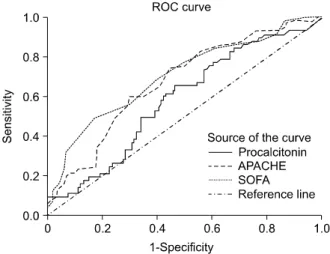
5. 패혈증 단기사망의 예측
연구 대상이 된 178명의 환자에서 추적 조사의 소실 없 이 급성 악화 후 28일 단기사망률을 조사하였다. 28일 단 기사망 여부에 따른 검사실 검사의 결과를 비교하였을 때, 프로칼시토닌은 통계적으로 유의한 차이를 보이지 않았
으나 유의수준 0.074로 사망한 경우에 있어서 높은 경향 을 나타냈다(Table 6). 또한 임상적 악화시기의 프로칼시 토닌 농도, APACHE II 점수 및 SOFA 점수에 따른 28일 단기사망률을 예측하였을 때, ROC 곡선의 곡선아래 면적
Table 6. Laboratory data between short term (28 days) sur- vivor and non-survivor group
Survivor (n=121)
Non-survivor
(n=57) p-value WBC, ×10
3/mm
39,268±8,255 8,500±6,354 0.291 CRP, mg/dL 7.2±9.1 6.7±7.9 0.869 Lactate, mmol/L 2.1±1.8 2.6±3.0 0.614 Procalcitonin, ng/mL 10.3±22.5 18.8±45.5 0.074 APACHE II score 23.9±8.2 29.0±8.1 <0.001 SOFA score 9.4±3.7 12.4±8.1 <0.001 The data are mean±SD.
APACH: acute physiology and chronic health evaluation; SOFA:
sequential organ failure assessment; ICU: intensive care unit;
SD: standard deviation.
Figure 3. Serum PCT measurment, APACHE Ⅱscore andSOFA score as a predictor of short-term (28 days) mortal- ity in critically ill patients: area under ROC-AUC with 95%
CI and p-value (PCT 0.583: 95% CI, 0.495∼0.671; p=
0.074; APACHE 0.673: 95% CI, 0.589∼0.756; p<
0.001; SOFA 0.703: 95% CI, 0.619∼0.787; p<0.001).
PCT: procalcitonin; APACHE: acute physiology and chronic health evaluation; SOFA: sequential organ failure assessment; ROC: receiver operating curve; AUC: area under curve; CI: confidence interval.
은 각각 0.583, 0.673 그리고 0.703으로 나타났다(p=
0.074, p<0.001, p<0.001) (Figure 3).
고 찰
본 연구에서 임상적 악화시기에 측정된 혈청 프로칼시 토닌의 농도가 비패혈증 상태와 패혈증 상태를 감별하는 데 유용하며, 패혈증 상태에서 중증 패혈증과 패혈증성 쇼크간의 감별에 유용한 생물학적 표지자(biomarker)임 을 알 수 있었다. 그러나 28일 단기사망률을 예측하는 표 지자로서 기존의 APACHE II 점수 및 SOFA 점수보다는 유용성이 떨어짐을 알 수 있었다.
프로칼시토닌은 12.6-kDa의 분자량을 갖는 칼시토닌의 전구체(precursor)이다. 정상적인 상태에서는 갑상선의 C-세포에서 활성화된 칼시토닌을 생성하고 분비하여 칼 슘의 대사에 관여한다8. 그러나 전신감염(systemic in- fection)이 발생하는 경우에는 모든 실질세포(parenchy- mal cell)에서 생성되는 것으로 알려져 있다9,10. 또한 프로 칼시토닌을 포함한 칼시토닌의 전구체들은 미생물 감염 이 있는 경우 그 농도가 수 천배까지 상승한다고 알려져 있다11.
기존의 백혈구 수 및 C 반응성 단백질 등은 항생제치료 가 필요한 중증 감염환자를 감별하는 역할을 하지 못한다 고 밝혀졌다12,13. 그러나 혈청 프로칼시토닌은 세균성 및 곰팡이 감염 그리고 호중구 감소성 발열(neutropenic fe- ver) 환자에서 감염과 비감염 상태를 감별할 수 있는 것으 로 알려져 있다4,14-16. 이 이외에도 프로칼시토닌은 패혈증 의 중등도 예측인자로서뿐만 아니라 사망률 예측에 도움
이 되는 생물학적 표지자로서 연구된 바 있다. Christ- Crain 등17은 82명을 대상으로 한 다기관 전향적 연구에서 SIRS, 패혈증, 중증 패혈증으로 대상을 구분하여 프로칼시 토닌의 농도를 측정하였고 결정점을 2.0 ng/mL로 하였을 경우 패혈증에 대한 진단 민감도가 94.7%, 특이도가 78.1%로 측정되었다. 또한 패혈증 및 중증 패혈증간의 감 별에도 도움이 된다고 하였다. 또한 Stocker 등18은 185명 을 대상으로 한 전향적 코호트 연구에서 패혈증의 중등도 에 따른 프로칼시토닌 농도를 측정하였고, 패혈증과 중증 패혈증 사이에 차이가 있다고 하였다. 사망률 예측에 있 어서 Meng 등19은 프로칼시토닌 키트(PCT-Q test)를 이용 하였고 농도가 10 ng/mL 이상인 경우 단기사망을 예측하 는 중요한 예측인자라고 발표하였다.
본 연구에서는 이러한 프로칼시토닌과 관련된 진단적 인 유용성 및 사망률 예측에 대해 연구하였다. 우선 본 연구에서는 진단적 가치로 첫 번째 패혈증 상태와 비패혈 증 상태간의 감별이 가능한지 확인하였다. 이전 연구들과 는 다르게 국소감염 진단에 대한 유용성을 배제하였고 임 상적으로 중요한 패혈증을 진단하고자 하였다. 이에 대한 결과 프로칼시토닌의 패혈증 진단에 대한 결정점 농도를
0.5 ng/mL 이상으로 설정한 경우 민감도가 85.6%, 특이 도가 70.5%로 다른 결정점에 비하여 높게 나타나서 패혈 증 가능성이 높다고 판단할 수 있었다. 즉 급성 악화상태 에서 측정한 초기의 프로칼시토닌 농도가 0.5 ng/mL 이 상인 경우 패혈증에 대한 조기치료를 수행하는데 활용할 수 있을 것으로 기대된다. 두 번째는 각 패혈증 상태간의 감별이 가능한지 확인하였다. 패혈증, 중증 패혈증, 패혈 증성 쇼크간에 농도 차이가 존재하였으나 이전 연구에서 처럼 임상적으로 중요한 패혈증과 중증 패혈증간의 감별 에 있어서는 통계적으로 유의점을 보이지는 않았다. 이는 이전의 연구가 전향적 연구였던 것에 비해 본 연구가 후향 적 연구로 의무기록 열람을 통한 분석의 한계점이 있을 것으로 생각된다. 즉 의무기록상 급성 악화시의 상태가 모두 적절하게 기록되지 않았을 수 있다. 마지막으로 28 일 단기사망률 예측인자로서의 가치를 확인하였다. 프로 칼시토닌 농도는 기존의 APACHE II 점수 및 SOFA 점수 에 비해 사망률 예측에 있어서 유용성을 보이지 않았다.
다만 사망 및 생존 군간에 프로칼시토닌 수치는 통계적인 유의한 차이를 보이지는 않았으나 높은 경향을 보였다.
이는 이전 연구와 차이를 보이는 부분으로 이에 대해서는 연구된 바가 적어 추가적인 대규모 연구가 필요할 것으로 생각된다.
참 고 문 헌
1. Rangel-Frausto MS, Pittet D, Costigan M, Hwang T, Davis CS, Wenzel RP. The natural history of the sys- temic inflammatory response syndrome (SIRS). A pro- spective study. JAMA 1995;273:117-23.
2. Hong SK, Hong SB, Lim CM, Koh Y. The characteristics and prognostic factors of severe sepsis in patients who were admitted to a medical intensive care unit of a ter- tiary hospital. Korean J Crit Care Med 2009;24:28-32.
3. Vincent JL, Bihari D. Sepsis, severe sepsis or sepsis syn- drome: need for clarification. Intensive Care Med 1992;
18:255-7.
4. Assicot M, Gendrel D, Carsin H, Raymond J, Guilbaud J, Bohuon C. High serum procalcitonin concentrations in patients with sepsis and infection. Lancet 1993;341:
515-8.
5. American College of Chest Physicians/Society of Critical Care Medicine Consensus Conference: definitions for sepsis and organ failure and guidelines for the use of innovative therapies in sepsis. Crit Care Med 1992;20:
864-74.
6. Knaus WA, Draper EA, Wagner DP, Zimmerman JE.
APACHE II: a severity of disease classification system.
Crit Care Med 1985;13:818-29.
7. Vincent JL, Moreno R, Takala J, Willatts S, De Mendonça A, Bruining H, et al. The SOFA (Sepsis-re- lated Organ Failure Assessment) score to describe or- gan dysfunction/failure. On behalf of the Working Group on Sepsis-Related Problems of the European Society of Intensive Care Medicine. Intensive Care Med 1996;22:707-10.
8. Becker KL, Nylén ES, White JC, Müller B, Snider RH Jr. Clinical review 167: Procalcitonin and the calcitonin gene family of peptides in inflammation, infection, and sepsis: a journey from calcitonin back to its precursors.
J Clin Endocrinol Metab 2004;89:1512-25.
9. Meisner M, Müller V, Khakpour Z, Toegel E, Redl H.
Induction of procalcitonin and proinflammatory cyto- kines in an anhepatic baboon endotoxin shock model.
Shock 2003;19:187-90.
10. Christ-Crain M, Müller B. Biomarkers in respiratory tract infections: diagnostic guides to antibiotic prescription, prognostic markers and mediators. Eur Respir J 2007;
30:556-73.
11. Christ-Crain M, Müller B. Procalcitonin in bacterial in- fections--hype, hope, more or less? Swiss Med Wkly 2005;135:451-60.
12. Herzum I, Renz H. Inflammatory markers in SIRS, sep- sis and septic shock. Curr Med Chem 2008;15:581-7.
13. Rau BM, Kemppainen EA, Gumbs AA, Büchler MW, Wegscheider K, Bassi C, et al. Early assessment of pan- creatic infections and overall prognosis in severe acute pancreatitis by procalcitonin (PCT): a prospective inter- national multicenter study. Ann Surg 2007;245:745-54.
14. Whang KT, Steinwald PM, White JC, Nylen ES, Snider RH, Simon GL, et al. Serum calcitonin precursors in sepsis and systemic inflammation. J Clin Endocrinol Metab 1998;83:3296-301.
15. Nakamura A, Wada H, Ikejiri M, Hatada T, Sakurai H, Matsushima Y, et al. Efficacy of procalcitonin in the early diagnosis of bacterial infections in a critical care unit. Shock 2009;31:586-91.
16. Sandri MT, Passerini R, Leon ME, Peccatori FA, Zorzino L, Salvatici M, et al. Procalcitonin as a useful marker of infection in hemato-oncological patients with fever.
Anticancer Res 2008;28:3061-5.
17. Christ-Crain M, Jaccard-Stolz D, Bingisser R, Gencay MM, Huber PR, Tamm M, et al. Effect of procalcito- nin-guided treatment on antibiotic use and outcome in
lower respiratory tract infections: cluster-randomised, single-blinded intervention trial. Lancet 2004;363:600-7.
18. Stocker M, Fontana M, El Helou S, Wegscheider K, Berger TM. Use of procalcitonin-guided decision-mak- ing to shorten antibiotic therapy in suspected neonatal early-onset sepsis: prospective randomized intervention
trial. Neonatology 2010;97:165-74.
19. Meng FS, Su L, Tang YQ, Wen Q, Liu YS, Liu ZF.
Serum procalcitonin at the time of admission to the ICU as a predictor of short-term mortality. Clin Bio- chem 2009;42:1025-31.