• 검색 결과가 없습니다.

Sepsis: Early Recognition and Optimized Treatment

N/A
N/A
Protected

Academic year: 2021

Share "Sepsis: Early Recognition and Optimized Treatment"

Copied!
9
0
0

로드 중.... (전체 텍스트 보기)

전체 글

(1)

of deaths was 215,000

2

. Recent large-scale epidemiological studies showed that the mortality rate of sepsis has decreased but its incidence continues to increase

3,4

. However, the true incidence of sepsis is likely to be underestimated.

On May 2017, the World Health Assembly (WHA) and World Health Organization (WHO) made sepsis a global health priority and adopted a resolution that urged the 194 United Nations Member States to improve the prevention, di- agnosis, and management of sepsis

5

. Accordingly, to improve patient outcomes, strategies that incorporate early recogni- tion and timely management of sepsis in hospitals are being implemented

5-8

.

In 2001, Rivers et al.

9

reported the groundbreaking study on early-goal directed therapy (EGDT). However, three sub- sequent multicenter randomized controlled trials (RCTs) did not show that EGDT reduced the sepsis mortality rate com- pared to usual care

10-12

. Recently, new sepsis definitions were issued by the Society of Critical Care Medicine (SCCM) and the European Society of Intensive Care Medicine (ESICM) for screening and early identification. However, their benefits have yet to be validated by prospective studies

3,4,13,14

, and ex- perts continue to place emphasis on the early administration of antibiotics and fluids for the initial resuscitation of patients

Introduction

Sepsis is a major cause of death from infection and repre- sents a substantial healthcare burden, accounting for 6.2% of total hospital costs in the United States 2011

1

. The estimated annual incidence of sepsis in the United States was 751,000 cases (3 cases/1,000 population) and the estimated number

Sepsis: Early Recognition and Optimized Treatment

Hwan Il Kim, M.D. and Sunghoon Park, M.D., Ph.D.

Division of Pulmonary, Allergy and Critical Care Medicine, Hallym University Sacred Heart Hospital, Anyang, Korea

Sepsis is a life-threatening condition caused by infection and represents a substantial global health burden. Recent epidemiological studies showed that sepsis mortality rates have decreased, but that the incidence has continued to increase. Although a mortality benefit from early-goal directed therapy (EGDT) in patients with severe sepsis or septic shock was reported in 2001, three subsequent multicenter randomized studies showed no benefits of EGDT versus usual care. Nonetheless, the early administration of antibiotics and intravenous fluids is considered crucial for the treatment of sepsis. In 2016, new sepsis definitions (Sepsis-3) were issued, in which organ failure was emphasized and use of the terms

“systemic inflammatory response syndrome” and “severe sepsis” was discouraged. However, early detection of sepsis with timely, appropriate interventions increases the likelihood of survival for patients with sepsis. Also, performance improvement programs have been associated with a significant increase in compliance with the sepsis bundles and a reduction in mortality. To improve sepsis management and reduce its burden, in 2017, the World Health Assembly and World Health Organization adopted a resolution that urged governments and healthcare workers to implement appropriate measures to address sepsis. Sepsis should be considered a medical emergency, and increasing the level of awareness of sepsis is essential.

Keywords: Compliance; Mortality; Sepsis; Treatment

Address for correspondence: Sunghoon Park, M.D., Ph.D.

Division of Pulmonary, Allergy and Critical Care Medicine, Hallym University Sacred Heart Hospital, 22 Gwanpyeong-ro 170beon-gil, Dongan-gu, Anyang 14068, Korea

Phone: 82-31-380-3715, Fax: 82-31-380-3973 E-mail: [email protected]

Received: Apr. 29, 2018 Revised: Jun. 29, 2018 Accepted: Jul. 20, 2018 Published online: Sep. 28, 2018

cc It is identical to the Creative Commons Attribution Non-Commercial License (http://creativecommons.org/licenses/by-nc/4.0/).

Copyright © 2019

The Korean Academy of Tuberculosis and Respiratory Diseases.

(2)

with sepsis.

Definitions and Early Identification of Sepsis

1. Change in sepsis definitions

The definition of sepsis has changed several times since 1992
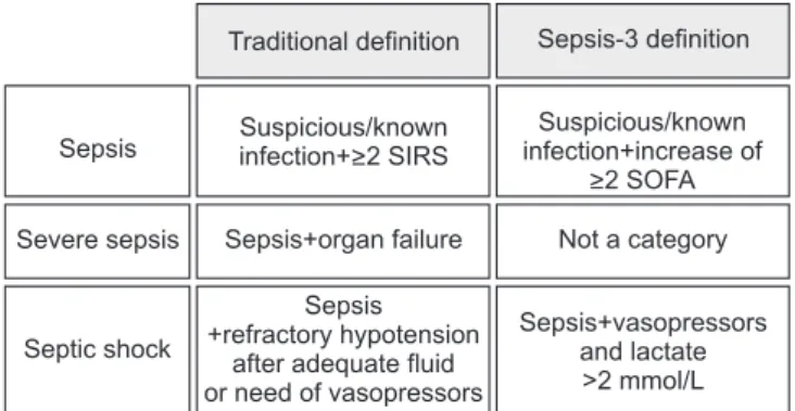
15,16

. The SCCM and ESICM revised the definition of sep- sis and septic shock in 2016. The new definitions (Sepsis-3) focused on a dysregulated host response to infection and or- gan dysfunction. Sepsis is defined as infected patients with an increase of ≥2 Sequential Organ Failure Score (SOFA) points

17

. Septic shock is defined as refractory hypotension requiring vasopressors with concurrent hyperlactemia (>2 mmol/L) despite adequate fluid resuscitation (Figure 1). Severe sepsis was excluded from the guidelines, and quick SOFA (qSOFA), instead of the systemic inflammatory response syndrome (SIRS), was adopted for screening purposes (Figure 2).

The Sepsis-3 definitions were based on a large database and were the first to be tested in derivation and validation datasets.

However, the definitions were not endorsed by some orga- nizations and there are several issues associated with them

6

. First, lactate was not retained in the sepsis definition. Hence, by the Sepsis-3 definitions, patients with an increased lactate level but no hypotension (or compensated septic shock) can be missed. In other words, we may miss patients in the early phase of sepsis. The prevalence of this phenotype (i.e., normo- tensive patients with hyperlactemia) was 26% in a previous multicenter trial

11

. In the Sepsis-3 datasets, the prevalence of normotensive hyperlactemia (>4 mmol/L) was 9.9% but their mortality rate was not low (29.9%). Therefore, the validity of

the Sepsis-3 definitions is suspect. Second, using the Sepsis-3 definitions, two components (the need for vasopressors and hyperlactemia) are needed concurrently to diagnose septic shock. That is, the lactate level is not a component of the definitions until the patient becomes hypotensive. Also, an infected patient with hypotension might not be considered to be in septic shock unless the lactate level was known. This implies that the utility of the Sepsis-3 definitions is limited in low-resource settings, where lactate levels are not frequently available. Therefore, further prospective studies are needed to demonstrate the validity of the Sepsis-3 definitions. Until then, it seems acceptable to use the pre-existing sepsis definitions.

2. Sepsis screening

Sepsis screening is reportedly associated with a decreased mortality rate

18,19

. The surviving sepsis campaign (SSC) guide-

Sepsis Suspicious/known infection+>2 SIRS

Suspicious/known infection+increase of

>2 SOFA Severe sepsis Sepsis+organ failure Not a category

Septic shock

Sepsis +refractory hypotension

after adequate fluid or need of vasopressors

Sepsis+vasopressors and lactate

>2 mmol/L Traditional definition Sepsis-3 definition

Figure 2. Comparison of traditional and revised (Sepsis-3) defini- tions for sepsis. SIRS: systemic inflammatory response syndrome;

SOFA: Sequential Organ Failure Assessment.

A patient with suspicious infection

SIRS (>2 criteria)

qSOFA (>2 criteria) Temperature

(>38 C or <36 C) Heart rate (>90 beats/min)

WBC

(<4x10 /L or >12x10 /L or >10%) Respiratory rate

(>20 breaths/min, PaCO <32 mm Hg)

9 9

2

Systolic blood pressure (<100 mm Hg) Respiration rate (>22 breaths/min) Altered mentation

(GCS<15)

"Sepsis"

"Being likely to be septic"

"Assess organ failure (SOFA score)"

Figure 1. Definitions for SIRS and qSOFA.

SIRS: systemic inflammatory response

syndrome; qSOFA: Sequential Organ

Failure Assessment; WBC: white blood

cell.

(3)

lines of 2016, as well as those of 2012, emphasize routine screening of potentially infected patients who are likely to be septic to improve the early identification and treatment of sepsis. They recommend that hospitals should have a perfor- mance improvement program that involves early recognition and management of sepsis

8

.

The SIRS criteria have, since 1992, been used to screen and identify sepsis patients

20

. To diagnose sepsis, at least two of the four SIRS criteria must be met. However, because SIRS can be triggered by a variety of infectious and noninfectious causes, it is insufficiently sensitive, and certainly not specific for sepsis.

Hence, some patients who satisfy the SIRS criteria may not have sepsis, and vice versa

21

. In this context, the Sepsis-3 Task Force discarded the concept of SIRS and introduced, instead, qSOFA for sepsis screening

17

. The qSOFA is a simplified ver- sion of the SOFA score that comprises only three variables, and patients with a qSOFA score of ≥2 should be considered for the possibility of sepsis (Figure 2). The qSOFA is a readily available bedside tool without laboratory tests, and has better performance in non‒intensive care unit (ICU) than ICU set- tings (area under the curve value, 0.81 vs. 0.66)

13

. The Sepsis-3 Task Force recommended that it be used to identify infected patients outside the ICU who are likely to be septic. However, a recent prospective study showed that a qSOFA score of ≥2 has high specificity (96.1% vs. 61.0% for SIRS ≥2) for organ dys- functions but its poor sensitivity (29.7% vs. 72.1% for SIRS ≥2) may limit its use as a bedside tool

22

. The authors insisted that the SIRS criteria can be still useful.

Clinical evidence indicates that patients with acute deterio- ration or sepsis manifest clinical signs or symptoms several hours before the condition worsens. Early warning scores, such as the Modified Early Warning Score (MEWS), Early Warning Scoring System (EWSS), or National Early Warning Score (NEWS), were developed to screen patients at high risk of deterioration

23-26

. Although strong evidence based on robust data is lacking, these scores showed a trend toward improved outcomes and, when coupled with an outreach service (i.e., rapid response teams or medical emergency teams), it facili- tates timely initiation of the optimal treatments upon recogni- tion of septic patients

25

. Although respiratory or cardiac prob- lems were the most common trigger for activations of such outreach teams

27,28

, one study showed that sepsis as a cause of activations accounted for 19.9%, and EGDT was undertaken in 22.7%

28

. Interestingly, Churpek et al.

29

compared several early warning scores, including qSOFA, among patients out- side the ICU. qSOFA had a higher specificity and lower sensi- tivity than SIRS, MEWS, and NEWS for predicting in-hospital death or ICU transfer. The SIRS criteria (≥2) were more rapid than qSOFA for identifying patients. Therefore, use of qSOFA may be premature, and the SIRS criteria are a sensitive and useful bedside tool for sepsis screening outside the ICU.

Early Treatments and Optimal Resuscitation

The EGDT was designed for the early detection of sepsis and timely optimization of hemodynamic parameters by continuous monitoring of central venous oxygen saturation (ScvO

2

, >70%), central venous pressure (8–12 mm Hg), mean arterial pressure (MAP, ≥65 mm Hg), and urine output (>0.5 mL/kg/h)

6,9

. This protocolized treatment, when administered to patients with severe sepsis or septic shock before admis- sion to the ICU, reduced the incidence of multi-organ dysfunc- tion and significantly decreased the in-hospital mortality rate compared with standard care

9

.

However, three international multicenter trials (Proto- colized Care for Early Septic Shock [ProCESS]

12

, Australasian Resuscitation in Sepsis Evaluation [ARISE]

11

, and Protocolized Management in Sepsis [ProMISe]

30

) did not show any signifi- cant survival benefit compared to usual care (Table 1). Also, in a meta-analysis of individual participants in the three RCTs, the EGDT did not result in better outcomes than usual care but was associated with increased hospitalization costs

31

. Therefore, the EGDT concept was weakened in the 2016 guidelines

8

. However, initial fluid resuscitation with crystal- loids is still being emphasized, and patients in the usual care groups received a considerable volume of fluids in these three RCTs (Table 1). Application of balanced crystalloids signifi- cantly decreased the rates of all-cause mortality, persistent renal insufficiency, and new dialysis treatments, compared to saline

32

. However, the 2016 guidelines emphasize, instead, the re-evaluation of volume status and tissue perfusion after the initial fluid resuscitation. This is because the persistence of a positive daily fluid balance over time was strongly associated with a higher mortality rate in patients with sepsis

33

. In this regard, the guidelines have recommended either repeated assessments of vital signs, cardiopulmonary status, capillary refill time, pulse, and skin findings, or measurement of the two of followings: CVP, ScvO

2

, bedside cardiovascular ultrasound, and dynamic assessment of fluid responsiveness with passive leg raise or fluid challenge.

A population-based study in the United States reported that

early central vein catheterization was associated with a lower

in-hospital mortality rate

34

. However, the three RCTs of EGDT

highlighted that there was no benefit of invasive hemody-

namic monitoring involving CVP and ScvO

2

if initial fluids and

adequate antibiotics were administered to septic patients in a

timely manner. Accordingly, the 2016 SSC guidelines do not

contain pre-specified treatment CVP and ScvO

2

targets. CVP

does not reflect intravascular volume status precisely and is

not predictive of the fluid response

35,36

. Instead, repeated mea-

surements of lactate (i.e., lactate clearance) enables evaluation

of the responsiveness to initial resuscitation

37

, and echocar-

diography is a noninvasive method of assessing the volume

status in patients on mechanical ventilator support

38

.

(4)

Ta ble 1. Com p ar is on of enr ollm en t, tr ea tm en t, an d o ut com e of E G DT s tu d ie s E G DT Pr oCE SS ARISE Pr oMIS e E G DT Con tr ol E G DT Pr ot oc ol- bas ed st an dar d T h era py

U sual c ar e E G DT U sual c ar e E G DT U sual c ar e Lo cation U nit ed S tat es U nit ed S tat es A us tr ali a, N ew Ze al and, Finl and, Ir el and, and H ong K ong

U nit ed K ing dom Enr olle d p atients 130 133 439 446 456 793 798 625 626 Ag e, yr 67.1±17.4 64.4±17.1 60±16.4 61±16.1 62±16.0 62.7±16.4 63.1±16.5 66.4±14.6 64.3±15.5 AP A CHE II scor e ( baseline) 21.4±6.9 20.4±7.4 20.8±8.1 20.6±7.4 20.7±7.5 15.4±6.5 15.8±6.5 18.7±7.1 18.0±7.1 Art eri al c athet er insertion, % R equir ed R equir ed R equir ed - - 91.4 76.3 74.2 62.2 C entr al vein c athet erization, % R equir ed R equir ed 93.6 56.5 57.9 90 61.9 92.1 50.9 Initi al l actat e >4 mmol/L , % 79 59 59.2 60.7 46 46.5 65.4 63.7 Total fluid within 6 hr , mL 4,981±2,984 3,499±2,438 5,059 5,511 4,362 4,479 4,304 4,100 4,074 Fluid prior t o r andomization (at ED)* - - 2,254±1,472 2,226±1,363 2,083±1,405 2,515±1,244 2,591±1,331 1,600 (1,000 – 2,500) 1,790 (1,000 – 2,500) U se of v asopr es sor s within 6 hr , % 27.4 30.3 54.9 52.2 44.1 66.6 57.8 53.3 46.6 RB C tr ansfus ion within 6 hr , % 64.1 18.5 14.4 8.3 7.5 13.6 7.0 8.8 3.8 Length of IC U s ta y, d ay* - - 5.1±6.3 5.1±7.1 4.7±5.8 2.8 (1.4 – 5.1) 2.8 (1.5 – 5.7) 2.6 (1.0 – 5.8) 2.2 (0.0 – 5.3) H os pital mortalit y, % 30.5 46.5 21.0 18.2 18.9 14.5 15.7 25.6 24.6 *M ean±s tand ar d de vi ation or me di an (int er quartile r ang e). EGD T : e ar ly g oal-dir ect ed ther apy; Pr oCE SS: Pr ot ocolize d C ar e for Ear ly S eptic S ho ck ; ARIS E: A us tr al as ian R es uscitation in S eps is Ev aluation; Pr oMIS e: Pr ot ocolize d M anag e- ment in S eps is ; AP A CHE II: A cut e P hy siology and C hr onic H ealth Ev aluation II; ED: emer genc y dep artment ; RB C : r ed blo od cell; IC U: int ens ive c ar e unit.

(5)

Delayed administration of empirical antibiotics after sepsis identification increases the in-hospital mortality rate

5,7

. Liu et al.

39

recently reported that a 1-hour delay in antibiotic initia- tion was associated with an increased odds of in-hospital mortality among patients who received antibiotics within 6 hours. Therefore, the SSC guidelines recommend the intra- venous administration of empiric antibiotics after obtaining blood culture results within 1 hour. Treatment with one or two broad-spectrum antibiotics and early de-escalation after clini- cal improvement or pathogen non-detection are recommended

8

. Early administration of vasopressors is associated with an increased survival rate in patients with septic shock

40

and is a component of the 6-hour sepsis bundle. Thus, norepi- nephrine, as the first choice, should be administered early to maintain a MAP ≥65 mm Hg, when hypotension does not respond to initial fluid resuscitation. In an open-label RCT, tar- geting a MAP of 80–85 mm Hg rather than 65–70 mm Hg did not increase the survival rate of patients with septic shock

41

. Thus, the target MAP should be determined according to the patients’ condition; a higher target may be needed for patients with chronic hypertension and a lower target for those with uncontrolled bleeding with trauma

42

.

The SSC guidelines do not include a target heart rate for pa- tients with septic shock

43

. However, due to the many adverse effects of tachycardia, such as diastolic dysfunction and myo- cardial ischemia, the heart rate should be maintained within the normal range in patients with septic shock

44

. Recently, Mo- relli et al.

45,46

demonstrated that esmolol can be safely used to reduce the heart rate (target rate, 80–94 beats/min), without increased adverse effects, and was associated with a reduced dose of norephinephrine and a lower mortality rate in patients with septic shock compared to the the controls. These results should be verified by further large-scale studies.

Further Efforts to Decrease the Sepsis- Related Mortality Mate

1. Increasing sepsis awareness

Although sepsis dates back to at least the time of Hip- pocrates, the term “sepsis” is not well known

47

. This has led to avoidable mortality and morbidity worldwide. On May 24, 2017, Sir Liam Donaldson, the WHO envoy for patient safety, reported that awareness of sepsis by the public and politicians must be increased during a WHA Side Event on sepsis

48

. He said further that sepsis is an important issue that has been ad- dressed effectively by clinicians and scientists, but is invisible to the public, political leaders, and leaders of healthcare systems.

Increased awareness, which leads to early presentation to hospital, can decrease the sepsis mortality rate by enabling timely and appropriate treatment. Experts recommend that the term “sepsis” be used frequently by healthcare profession-

als and patients to increase the level of awareness of the gen- eral public

49

. However, despite the significant impact, public awareness is currently very low; a survey in 2009 reported that 88% of respondents had never heard the term “sepsis”

50

. Since 2012, the Global Sepsis Alliance (GSA) has organized the an- nual World Sepsis Day, and many organizations or countries are undertaking sepsis awareness campaigns

51

. In healthcare facilities, continuous training and education are also needed to increase the level of awareness of sepsis on the part of healthcare providers, who must understand that the condition is a real medical emergency.

Most estimates of sepsis are based on studies in high- income countries, and data on low- and middle-income (or resource-limited) countries are scarce. So, increasing the awareness of sepsis is essential for controlling the sepsis bur- den in those countries

52,53

. In South Korea, on “World Sepsis Day”, annual symposia and field events have taken place since 2012

54

. However, further promotional or educational activities for the public, as well as support from the political leadership, are required to improve the situation.

2. Building a sepsis registry

Sepsis is frequently handled like a “garbage code” in the Global Burden of Disease Statistics. Most deaths due to sepsis are classified as their underlying infections, rather than sepsis itself

48

. Therefore, the burden of sepsis is likely to be underes- timated. Hence, improving the coding process for sepsis may facilitate estimation of the true burden.

Nationwide (or statewide) registry data collected prospec- tively can facilitate accurate estimation of the incidence of sep- sis, which can be used to improve performance and formulate future policies. A good example is the New York State sepsis registry. Beginning in 2014, all hospitals in New York State adopted sepsis protocols based on Rory’s regulations for the early diagnosis and treatment of sepsis

5,48

. They are required to report their performance (compliance), as well as clinical information, to the New York State Department of Health

5,48

. Surprisingly, after the onset of the initiative, the average rate of protocol compliance has increased progressively from 73.7%

in the second quarter of 2014 to 84.7% of adult sepsis patients in the third quarter of 2016

5

. This compliance is in contrast to Asian countries; a large multinational study of Asian ICUs reported rates of compliance with the resuscitation and man- agement bundles of 7.6% and 3.5%, respectively

55

.

The Core Outcome and Resource Evaluation (CORE) com-

mittee is a component of the Australia and New Zealand Inten-

sive Care Society (ANZICS)

56

. All ICUs in Australia and New

Zealand were invited to contribute to the ANZICS CORE regis-

tries in 1992. These registries have four registry domains: adult

patients, pediatric patients, critical care resources, and central

line-associated bloodstream infections. The CORE committee

collects comprehensive data on various aspects of ICUs and

(6)

reports back to the contributing ICUs. They also audit and ana- lyze the performance of ICUs for quality assurance purposes.

Therefore, Asian countries, including South Korea, need to benchmark the successful stories of Western countries. This would eventually lead to the performance improvement of healthcare workers and the reduction of sepsis mortality.

3. Implementation of performance improvement programs and sepsis care bundles

When several individual effective treatments are applied concurrently, we anticipate better outcomes than any of the individual treatments alone; e.g., the ventilator-associated pneumonia or central-line infection prevention bundles

57,58

. In 2004, the SSC group, in partnership with the Institute for Healthcare Improvement, developed the SSC bundle with the goal of reducing the mortality rate by 25%

59

. In 2012 and 2016, the 3-hour and 6-hour SSC bundles were introduced but, due to the negative results of three RCTs on EGDT, invasive monitoring, such as CVP and ScvO

2

, was excluded from the 6-hour bundle in 2016

8,60

. More recently, based on the 2016 SSC guidelines, a revised hour-1 bundle (2018 bundle) with five key elements was developed (Table 2)

61

.

The implementation of sepsis bundles is a cornerstone of sepsis performance improvement programs, which are as- sociated with a significant increase in compliance with the sepsis bundles and a reduction in the mortality rate. Levy et al.

62

reported low mortality rates in high-compliance hospi- tals during a 7.5-year observation. Analysis of the New York State registry also demonstrated that the compliance rate of the 3-hour sepsis bundle was associated with a lower risk- adjusted in-hospital mortality rate

5

. Among various factors, high-income countries, surgical ICUs, long duration of imple-

mentation, and presentation to an Emergency Department were associated with a high rate of compliance

55,62

, and lactate seemed frequently a non-compliant variable

62,63

. However, the rate of compliance should continue to increase during the first 2 years of implementation

62

, and the mortality rate may decrease even if bundle completion is delayed in sepsis pa- tients

64,65

.

A large Asian study reported a low rate of compliance with SSC bundles. In South Korea, lack of critical care personnel was significantly associated with low compliance rates (e.g., total compliance of 5.6%)

66

. Thus, sufficient critical care per- sonnel (e.g., intensivists and nurses) is an important factor for improving performance. Further studies should seek to iden- tify methods of improving bundle compliance, as well as ways to overcome other barriers.

Conclusion

Prevention and early recognition of sepsis are of paramount importance until novel emerging drugs (or interventions) are demonstrated to be effective. Early application of the optimal treatments and improved compliance with sepsis bundles are pre-requisites for improving patients’ outcomes. The validity of the Sepsis-3 definitions and that of qSOFA need to be dem- onstrated in large-scale prospective trials.

Authors’ Contributions

Conceptualization: Kim HI, Park S. Methodology: Kim HI, Park S. Formal analysis: Kim HI, Park S. Original draft prepara- tion: Kim HI, Park S. Review and editing: Kim HI, Park S. Ap- proval of final manuscript: all authors.

Conflicts of Interest

No potential conflict of interest relevant to this article was reported.

References

1. Torio CM, Moore BJ. National inpatient hospital costs: the most expensive conditions by payer, 2013. Statistical Brief

#204. Healthcare Cost and Utilization Project (HCUP) Statis- tical Briefs [Internet]. Rockville, MD: Agency for Healthcare Research and Quality; 2006 [cited 2018 Apr 29]. Available from: https://www.ncbi.nlm.nih.gov/books/NBK368492/.

2. Angus DC, Linde-Zwirble WT, Lidicker J, Clermont G, Carcillo J, Pinsky MR. Epidemiology of severe sepsis in the United States: analysis of incidence, outcome, and associated costs Table 2. Hour-1 surviving sepsis campaign bundle of care

The five key elements of hour-1 bundle

1. Measure lactate level. Remeasure if initial lactate is >2 mmol/L.

2. Obtain blood cultures prior to administration of antibiotics.

3. Administer broad-spectrum antibiotics.

4. Begin rapid administration of 30 mL/kg crystalloid for hypoten- sion or lactate ≥4 mmol/L.

5. Apply vasopressors if patient is hypotensive during or after fluid resuscitation to maintain MAP ≥65 mm Hg.

“Time zero” or “time of presentation” is defined as the time of tri- age in the Emergency Department, or if presenting from another care venue, from the earliest chart annotation consistent with all elements of sepsis (formerly severe sepsis) or septic shock ascer- tained through chart review.

Adapted from Levy et al. Crit Care Med. 2018;46:997-1000, with permission of Society of Critical Care Medicine

61

.

MAP: mean arterial pressure.

(7)

of care. Crit Care Med 2001;29:1303-10.

3. Kaukonen KM, Bailey M, Suzuki S, Pilcher D, Bellomo R. Mor- tality related to severe sepsis and septic shock among criti- cally ill patients in Australia and New Zealand, 2000-2012.

JAMA 2014;311:1308-16.

4. Martin GS, Mannino DM, Eaton S, Moss M. The epidemiology of sepsis in the United States from 1979 through 2000. N Engl J Med 2003;348:1546-54.

5. Seymour CW, Gesten F, Prescott HC, Friedrich ME, Iwashyna TJ, Phillips GS, et al. Time to treatment and mortality dur- ing mandated emergency care for sepsis. N Engl J Med 2017;376:2235-44.

6. Osborn TM. Severe sepsis and septic shock trials (ProCESS, ARISE, ProMISe): what is optimal resuscitation? Crit Care Clin 2017;33:323-44.

7. Pruinelli L, Westra BL, Yadav P, Hoff A, Steinbach M, Kumar V, et al. Delay within the 3-hour surviving sepsis campaign guideline on mortality for patients with severe sepsis and septic shock. Crit Care Med 2018;46:500-5.

8. Rhodes A, Evans LE, Alhazzani W, Levy MM, Antonelli M, Ferrer R, et al. Surviving sepsis campaign: international guidelines for management of sepsis and septic shock: 2016.

Intensive Care Med 2017;43:304-77.

9. Rivers E, Nguyen B, Havstad S, Ressler J, Muzzin A, Knoblich B, et al. Early goal-directed therapy in the treatment of severe sepsis and septic shock. N Engl J Med 2001;345:1368-77.

10. Lilly CM. The ProCESS trial: a new era of sepsis management.

N Engl J Med 2014;370:1750-1.

11. ARISE Investigators; ANZICS Clinical Trials Group, Peake SL, Delaney A, Bailey M, Bellomo R, et al. Goal-directed re- suscitation for patients with early septic shock. N Engl J Med 2014;371:1496-506.

12. ProCESS Investigators, Yealy DM, Kellum JA, Huang DT, Barnato AE, Weissfeld LA, et al. A randomized trial of protocol-based care for early septic shock. N Engl J Med 2014;370:1683-93.

13. Seymour CW, Liu VX, Iwashyna TJ, Brunkhorst FM, Rea TD, Scherag A, et al. Assessment of clinical criteria for sepsis: for the third international consensus definitions for sepsis and septic shock (Sepsis-3). JAMA 2016;315:762-74.

14. Shankar-Hari M, Phillips GS, Levy ML, Seymour CW, Liu VX, Deutschman CS, et al. Developing a new definition and as- sessing new clinical criteria for septic shock: for the third in- ternational consensus definitions for sepsis and septic shock (Sepsis-3). JAMA 2016;315:775-87.

15. American College of Chest Physicians/Society of Critical Care Medicine Consensus Conference: definitions for sepsis and organ failure and guidelines for the use of innovative therapies in sepsis. Crit Care Med 1992;20:864-74.

16. Levy MM, Fink MP, Marshall JC, Abraham E, Angus D, Cook D, et al. 2001 SCCM/ESICM/ACCP/ATS/SIS international sepsis definitions conference. Crit Care Med 2003;31:1250-6.

17. Singer M, Deutschman CS, Seymour CW, Shankar-Hari M,

Annane D, Bauer M, et al. The third international consen- sus definitions for sepsis and septic shock (Sepsis-3). JAMA 2016;315:801-10.

18. Gatewood MO, Wemple M, Greco S, Kritek PA, Durvasula R.

A quality improvement project to improve early sepsis care in the emergency department. BMJ Qual Saf 2015;24:787-95.

19. Hayden GE, Tuuri RE, Scott R, Losek JD, Blackshaw AM, Schoenling AJ, et al. Triage sepsis alert and sepsis protocol lower times to fluids and antibiotics in the ED. Am J Emerg Med 2016;34:1-9.

20. Bone RC, Balk RA, Cerra FB, Dellinger RP, Fein AM, Knaus WA, et al. Definitions for sepsis and organ failure and guide- lines for the use of innovative therapies in sepsis. The ACCP/

SCCM Consensus Conference Committee. American College of Chest Physicians/Society of Critical Care Medicine. Chest 1992;101:1644-55.

21. Beesley SJ, Lanspa MJ. Why we need a new definition of sep- sis. Ann Transl Med 2015;3:296.

22. Williams JM, Greenslade JH, McKenzie JV, Chu K, Brown AFT, Lipman J. Systemic inflammatory response syndrome, quick sequential organ function assessment, and organ dysfunc- tion: insights from a prospective database of ED patients with infection. Chest 2017;151:586-96.

23. Gardner-Thorpe J, Love N, Wrightson J, Walsh S, Keeling N.

The value of Modified Early Warning Score (MEWS) in surgi- cal in-patients: a prospective observational study. Ann R Coll Surg Engl 2006;88:571-5.

24. Smith GB, Prytherch DR, Meredith P, Schmidt PE, Feath- erstone PI. The ability of the National Early Warning Score (NEWS) to discriminate patients at risk of early cardiac ar- rest, unanticipated intensive care unit admission, and death.

Resuscitation 2013;84:465-70.

25. Roney JK, Whitley BE, Maples JC, Futrell LS, Stunkard KA, Long JD. Modified early warning scoring (MEWS): evaluat- ing the evidence for tool inclusion of sepsis screening criteria and impact on mortality and failure to rescue. J Clin Nurs 2015;24:3343-54.

26. Stark AP, Maciel RC, Sheppard W, Sacks G, Hines OJ. An early warning score predicts risk of death after in-hospital cardio- pulmonary arrest in surgical patients. Am Surg 2015;81:916- 21.

27. Al-Qahtani S, Al-Dorzi HM, Tamim HM, Hussain S, Fong L, Taher S, et al. Impact of an intensivist-led multidisciplinary extended rapid response team on hospital-wide cardiopul- monary arrests and mortality. Crit Care Med 2013;41:506-17.

28. Huh JW, Lim CM, Koh Y, Lee J, Jung YK, Seo HS, et al. Ac- tivation of a medical emergency team using an electronic medical recording-based screening system*. Crit Care Med 2014;42:801-8.

29. Churpek MM, Snyder A, Han X, Sokol S, Pettit N, Howell

MD, et al. Quick sepsis-related organ failure assessment, sys-

temic inflammatory response syndrome, and early warning

scores for detecting clinical deterioration in infected patients

(8)

outside the intensive care unit. Am J Respir Crit Care Med 2017;195:906-11.

30. Mouncey PR, Osborn TM, Power GS, Harrison DA, Sadique MZ, Grieve RD, et al. Trial of early, goal-directed resuscitation for septic shock. N Engl J Med 2015;372:1301-11.

31. PRISM Investigators, Rowan KM, Angus DC, Bailey M, Bar- nato AE, Bellomo R, et al. Early, goal-directed therapy for septic shock: a patient-level meta-analysis. N Engl J Med 2017;376:2223-34.

32. Semler MW, Self WH, Wanderer JP, Ehrenfeld JM, Wang L, By- rne DW, et al. Balanced crystalloids versus saline in critically ill adults. N Engl J Med 2018;378:829-39.

33. Mitchell KH, Carlbom D, Caldwell E, Leary PJ, Himmelfarb J, Hough CL. Volume overload: prevalence, risk factors, and functional outcome in survivors of septic shock. Ann Am Thorac Soc 2015;12:1837-44.

34. Walkey AJ, Wiener RS, Lindenauer PK. Utilization patterns and outcomes associated with central venous catheter in septic shock: a population-based study. Crit Care Med 2013;41:1450-7.

35. Osman D, Ridel C, Ray P, Monnet X, Anguel N, Richard C, et al. Cardiac filling pressures are not appropriate to predict hemodynamic response to volume challenge. Crit Care Med 2007;35:64-8.

36. Marik PE, Monnet X, Teboul JL. Hemodynamic parameters to guide fluid therapy. Ann Intensive Care 2011;1:1.

37. Jones AE, Shapiro NI, Trzeciak S, Arnold RC, Claremont HA, Kline JA, et al. Lactate clearance vs central venous oxygen sat- uration as goals of early sepsis therapy: a randomized clinical trial. JAMA 2010;303:739-46.

38. Feissel M, Michard F, Faller JP, Teboul JL. The respiratory variation in inferior vena cava diameter as a guide to fluid therapy. Intensive Care Med 2004;30:1834-7.

39. Liu VX, Fielding-Singh V, Greene JD, Baker JM, Iwashyna TJ, Bhattacharya J, et al. The timing of early antibiotics and hospital mortality in sepsis. Am J Respir Crit Care Med 2017;196:856-63.

40. Bai X, Yu W, Ji W, Lin Z, Tan S, Duan K, et al. Early versus delayed administration of norepinephrine in patients with septic shock. Crit Care 2014;18:532.

41. Asfar P, Meziani F, Hamel JF, Grelon F, Megarbane B, Anguel N, et al. High versus low blood-pressure target in patients with septic shock. N Engl J Med 2014;370:1583-93.

42. Cecconi M, De Backer D, Antonelli M, Beale R, Bakker J, Hofer C, et al. Consensus on circulatory shock and hemodynamic monitoring. Task force of the European Society of Intensive Care Medicine. Intensive Care Med 2014;40:1795-815.

43. DellaVolpe JD, Moore JE, Pinsky MR. Arterial blood pressure and heart rate regulation in shock state. Curr Opin Crit Care 2015;21:376-80.

44. Dunser MW, Hasibeder WR. Sympathetic overstimulation during critical illness: adverse effects of adrenergic stress. J Intensive Care Med 2009;24:293-316.

45. Morelli A, Ertmer C, Westphal M, Rehberg S, Kampmeier T, Ligges S, et al. Effect of heart rate control with esmolol on hemodynamic and clinical outcomes in patients with septic shock: a randomized clinical trial. JAMA 2013;310:1683-91.

46. Morelli A, Singer M, Ranieri VM, D’Egidio A, Mascia L, Orec- chioni A, et al. Heart rate reduction with esmolol is associ- ated with improved arterial elastance in patients with septic shock: a prospective observational study. Intensive Care Med 2016;42:1528-34.

47. Majno G. The ancient riddle of sigma eta psi iota sigma (sep- sis). J Infect Dis 1991;163:937-45.

48. Reinhart K, Daniels R, Kissoon N, Machado FR, Schachter RD, Finfer S. Recognizing sepsis as a global health priority: a WHO resolution. N Engl J Med 2017;377:414-7.

49. Just say sepsis! A review of the process of care received by patients with sepsis [Internet]. London: National Con- fidential Enquiry into Patient Outcome and Death; 2015 [cited 2018 Apr 3]. Available from: http://www.ncepod.org.

uk/2015report2/downloads/JustSaySepsis_FullReport.pdf.

50. Rubulotta FM, Ramsay G, Parker MM, Dellinger RP, Levy MM, Poeze M, et al. An international survey: public awareness and perception of sepsis. Crit Care Med 2009;37:167-70.

51. Vincent JL. Increasing awareness of sepsis: World Sepsis Day.

Crit Care 2012;16:152.

52. Thwaites CL, Lundeg G, Dondorp AM; sepsis in resource- limited settings-expert consensus recommendations group of the European Society of Intensive Care Medicine (ESCICM), the Mahidol-Oxford Research Unit (MORU) in Bangkok, Thailand. Infection management in patients with sepsis and septic shock in resource-limited settings. Intensive Care Med 2016;42:2117-8.

53. Schultz MJ, Dunser MW, Dondorp AM, Adhikari NK, Iyer S, Kwizera A, et al. Current challenges in the management of sepsis in ICUs in resource-poor settings and suggestions for the future. Intensive Care Med 2017;43:612-24.

54. World Sepsis Day [Internet]. Seoul: Korean Society of Criti- cal Care Medicine; 2018 [cited 2018 Apr 3]. Available from:

http://www.ksccm.org/#%2Fboard%2Flist.kin%3Fmenu_

main%3D6%26menu_sub%3D29%261522741514642.

55. Phua J, Koh Y, Du B, Tang YQ, Divatia JV, Tan CC, et al. Mana- gement of severe sepsis in patients admitted to Asian in- tensive care units: prospective cohort study. BMJ 2011;342:

d3245.

56. Center for Outcome and Resource Evaluation. ANZICS [In- ternet]. Camberwell: Australian and New Zealand Intensive Care Society; 2018 [cited 2018 Apr 3]. Available from: http://

www.anzics.com.au/Pages/CORE/About-CORE.aspx.

57. Blot K, Bergs J, Vogelaers D, Blot S, Vandijck D. Prevention of central line-associated bloodstream infections through quality improvement interventions: a systematic review and meta-analysis. Clin Infect Dis 2014;59:96-105.

58. Eom JS, Lee MS, Chun HK, Choi HJ, Jung SY, Kim YS, et al.

The impact of a ventilator bundle on preventing ventilator-

(9)

associated pneumonia: a multicenter study. Am J Infect Con- trol 2014;42:34-7.

59. Levy MM, Pronovost PJ, Dellinger RP, Townsend S, Resar RK, Clemmer TP, et al. Sepsis change bundles: converting guidelines into meaningful change in behavior and clinical outcome. Crit Care Med 2004;32(11 Suppl):S595-7.

60. Dellinger RP, Levy MM, Rhodes A, Annane D, Gerlach H, Opal SM, et al. Surviving sepsis campaign: international guidelines for management of severe sepsis and septic shock: 2012. Crit Care Med 2013;41:580-637.

61. Levy MM, Evans LE, Rhodes A. The surviving sepsis cam- paign bundle: 2018 update. Crit Care Med 2018;46:997-1000.

62. Levy MM, Rhodes A, Phillips GS, Townsend SR, Schorr CA, Beale R, et al. Surviving Sepsis Campaign: association between performance metrics and outcomes in a 7.5-year study. Intensive Care Med 2014;40:1623-33.

63. Chen YC, Chang SC, Pu C, Tang GJ. The impact of nationwide education program on clinical practice in sepsis care and mortality of severe sepsis: a population-based study in Tai- wan. PLoS One 2013;8:e77414.

64. Coba V, Whitmill M, Mooney R, Horst HM, Brandt MM, Di- giovine B, et al. Resuscitation bundle compliance in severe sepsis and septic shock: improves survival, is better late than never. J Intensive Care Med 2011;26:304-13.

65. Castellanos-Ortega A, Suberviola B, Garcia-Astudillo LA, Ortiz F, Llorca J, Delgado-Rodriguez M. Late compliance with the sepsis resuscitation bundle: impact on mortality. Shock 2011;36:542-47.

66. Kim JH, Hong SK, Kim KC, Lee MG, Lee KM, Jung SS, et al. In-

fluence of full-time intensivist and the nurse-to-patient ratio

on the implementation of severe sepsis bundles in Korean

intensive care units. J Crit Care 2012;27:414.e11-21.

수치

Figure 2. Comparison of traditional and revised (Sepsis-3) defini- defini-tions for sepsis
Table 1. Comparison of enrollment, treatment, and outcome of EGDT studies EGDTProCESSARISEProMISe EGDTControlEGDTProtocol-based  standard  Therapy

참조

관련 문서

Conclusions We revealed a greater increase of MPV in non-survivors of severe sepsis and/or septic shock compared with survivors during the first 72 hours after hospitalization and

Purpose: The objective of this study was to develop a new nomogram that can predict 28-day mortality in severe sepsis and/or septic shock patients using a combination of

In a previous study, which enrolled the pa- tients with severe sepsis and septic shock, the frequency of pre-existing CKD was significantly higher among those who developed

The purpose of this study was to compare the incidence of adrenal insufficiency and mortality between the patients with severe sepsis and septic shock who received etomidate as